Increases in extracellular K+ concentration can occur in a variety of situations including strenuous muscular exercise and lack of oxygen. In the case of oxygen lack, it has been shown that extracellular K+concentrations may attain values as high as 10 mmol l−1 during prolonged submergence in freshwater turtles (Jackson and Ultsch, 1982). In heart muscle, a high [K+] tends to decrease the force developed upon activation, i.e. the twitch force. It has long been known that this negative effect on force development is efficiently counteracted by adrenaline (Engstfeld et al., 1961). This observation may be linked to the fact that adrenaline enhances the action-potential-dependent influx of Ca2+(Frace et al., 1993) and, as a consequence, more Ca2+has to be removed during relaxation. This is of interest with respect to the finding that high [K+] depolarises the membrane, i.e. changes the membrane potential in the positive direction irrespective of the presence of adrenaline (Engstfeld et al., 1961). In ectothermic vertebrates in particular, heart muscle relaxation seems to depend heavily on sarcolemmal Na+/Ca2+ exchange (Driedzic and Gesser, 1994). A partial
membrane depolarisation should reduce sarcolemmal Ca2+ extrusion via Na+/Ca2+ exchange. Thus, the electrochemical force driving this extrusion of Ca2+ should decrease as the equilibrium of the Na+/Ca2+ exchange is shifted towards an increased cellular Ca2+ activity according to the equation describing this exchange of 3 Na+for 1 Ca2+(Blaustein, 1999):
where [Ca2+]
i denotes the cytoplasmic Ca2+activity, [Ca2+]o denotes the extracellular Ca2+ activity, [Na+]
i and [Na+]o denote the corresponding activities for Na+, E is the membrane potential, F is Faraday’s constant, R is the gas constant and T is the absolute temperature.
It is conceivable that other mechanisms may be evoked by adrenaline to compensate for the negative effect exerted by partial depolarisation on the force driving Ca2+extrusion and, thus, relaxation. One possibility is that the activity of the
(1)
eEF/RT,
[Ca2+]
i= [Ca2+]o
[Na+] i
[Na+] o
3
JEB2872
Increases in extracellular K+concentrations reduced the
twitch force amplitude of heart muscle from the freshwater turtle (Trachemys scripta elegans) and rainbow trout (Oncorhynchus mykiss). Adrenaline augmented twitch force amplitude and reduced the relative influence of [K+].
In the absence of adrenaline, high [K+] had less effect in
reducing twitch force in turtle than in trout, whereas the reverse was true in the presence of adrenaline. Under anoxic conditions, twitch force was lower in 10 mmol l−1
than in 2.5 mmol l−1 K+ in both preparations, but
adrenaline removed this difference. A further analysis of turtle myocardium showed that action potential duration was shorter and resting potential more positive in high [K+]
than in low [K+]. Adrenaline restored the duration of the
action potential, but did not affect the depolarisation, which may attenuate Na+/Ca2+exchange, participating in
excitation/contraction coupling. The contractile responses in the presence of adrenaline were, however, similar in both
high and low K+ concentrations when increases in
extracellular Ca2+were applied to increase the demand on
excitation/contraction coupling. The possibilities that adrenaline counteracts the effects of high [K+] via the
sarcoplasmic reticulum or sarcolemmal Na+/K+-ATPase
were examined by inhibiting the sarcoplasmic reticulum with ryanodine (10µmol l−1) or Na+/K+-ATPase with
ouabain (0.25 or 3 mmol l−1). No evidence to support either
of these possibilities was found. Adrenaline did not protect all aspects of excitation/contraction coupling because the maximal frequency giving regular twitches was lower at 10 mmol l−1K+than at 2.5 mmol l−1K+.
Key words: extracellular K+, excitation/contraction coupling,
freshwater turtle, Trachemys scripta elegans, Na+/Ca2+ exchange,
Na+/K+-ATPase, oxygen lack, rainbow trout, Oncorhynchus mykiss,
sarcoplasmic reticulum, adrenaline. Summary
Introduction
EFFECTS OF HIGH EXTRACELLULAR [K
+] AND ADRENALINE ON FORCE
DEVELOPMENT, RELAXATION AND MEMBRANE POTENTIAL IN CARDIAC
MUSCLE FROM FRESHWATER TURTLE AND RAINBOW TROUT
J. S. NIELSEN ANDH. GESSER*
Department of Zoophysiology Institute of Biological Sciences, University of Aarhus, DK-8000 Aarhus C, Denmark *Author for correspondence (e-mail: [email protected])
sarcoplasmic reticulum may be enhanced. Evidence has been obtained using trout cardiac muscle that such an enhancement occurs at elevated levels of extracellular K+ (El-Sayed and Gesser, 1989). Another possibility is stimulation of Na+/K+ -ATPase by adrenergic agents, as demonstrated in heart muscle (Pecker et al., 1986). Such stimulation should increase the inward Na+gradient and, as a result, counteract the effect of a partial depolarisation on Ca2+extrusion according to equation 1. Apart from Na+/Ca2+ exchange, the Na+ channels are of interest because their opening probability, and as a result cellular excitability, decreases with elevation of [K+] in a way that is counteracted by adrenaline (Paterson et al., 1993).
The present study examines how increases in extracellular [K+] influence the development and relaxation of twitch force and action and resting potentials with particular attention to the protection provided by adrenaline against the effects of high [K+]. The experiments were performed on heart ventricular preparations isolated from freshwater turtle, which can show increases in plasma [K+] from values of approximately 2.5 mmol l−1 to values as high as 10 mmol l−1 (Jackson and Ultsch, 1982). Since such high levels of plasma [K+] may be associated with particular adaptations, some of the experiments also included cardiac preparations from rainbow trout, in which measurements of plasma [K+] have shown increases from approximately 2.5 to 5 mmol l−1(Nielsen and Lykkeboe, 1992), i.e. to lower values than in freshwater turtle. Plasma [K+] increases during diving in turtles and during heavy exercise in rainbow trout, both situations that are likely to involve oxygen lack. For this reason, the effects of high [K+] and adrenaline were also examined under anoxia.
Materials and methods
Freshwater turtle Trachemys scripta elegans (Gray) (150–200 g) and rainbow trout Oncorhynchus mykiss (Walbaum) (200–340 g) of both sexes were kept in freshwater tanks at approximately 25 °C (turtles) and 15 °C (trout). The turtles had access to platforms above the water level. The animals were fed regularly. After decapitation, the ventricle was rapidly transferred to an ice-cold oxygenated physiological solution, where one or two longitudinal strips were cut.
When only force was recorded, two preparations from each ventricle were run in parallel. Each strip was mounted vertically. One end was tied with surgical silk to one of the two platinum stimulation electrodes, and the other end to a thin glass rod connected to the force transducer (Statham UC 2, Oxnard, CA, USA). The distance between the electrode and the transducer, and hence the length of the preparation, could be adjusted with a micrometer screw. The second stimulation electrode was positioned close to the upper end of the preparation. The stimulation electrodes were connected to a stimulator (Grass SD 9, Quincy, MA, USA). The preparation was paced to give contractions with electrical square pulses having a duration of 5 ms and a voltage 1.5 times that eliciting the maximal response. The voltage was checked regularly during the experiment. The stimulation rate was 0.2 Hz unless stated otherwise.
The preparation was stretched to provide a twitch force that was 75 % of the twitch force at the peak of the force/length relationship. This low level of stretch was applied in an attempt to diminish the impact of the undefined cellular orientation of the preparation. When stretched, the preparation had a length of approximately 5 mm between the points of attachment, and a maximal diameter of 1 mm. The diameter was assessed from the mass and length of the preparation assuming a cylindrical form and a density of 1 g ml−1. Because of the undefined cellular orientation, twitch force development and changes in resting tension were normalised to the twitch force developed after the initial stabilisation. Force was recorded with an ink recorder (Gould-Brush 2400, Cleveland, OH, USA) and digitally on a computer. The mechanical variables recorded were twitch force, its maximal rate of relaxation and resting tension.
In some experiments on turtle, membrane potential and force were recorded together, mainly as described previously (Møller-Nielsen and Gesser, 1992). The membrane potential variables measured were resting potential and the amplitude and duration of the action potential. The duration was recorded at 80 % of full deflection. Briefly, one preparation from each ventricle was mounted horizontally in a 10 ml bath with one end fixed and the other attached to a force transducer in such a way that its length could be adjusted with a micrometer screw to produce 75 % of maximal force. The preparation was activated electrically, as described above, using two platinum electrodes placed on each side of the preparation. Membrane potentials were recorded with glass capillaries filled with 3 mol l−1KCl, having a resistance of 30–40 MΩ. A Ag/AgCl electrode serving as reference was placed in the muscle bath close to the microelectrode. Membrane and action potentials and force were recorded with an oscilloscope (Hewlet-Packard P 54600A) and a computer. The software for computer-based recordings of mechanical and electrical variables was made in the department by Einer Larsen. The physiological solution contained (mmol l−1): 125 NaCl, 30 (turtle) or 15 (trout) NaHCO3, 1.0 NaH2PO4, 2.5 KCl, 1.0 MgSO4, 1.25 CaCl2, and 5 glucose. It was perfused with 98 % O2and 2 % CO2(turtle) or 99 % O2and 1 % CO2(trout) during the experiments. The temperature of the solutions bathing the preparations was maintained at 20 °C (turtle) or 15 °C (trout) with a thermostatted water bath (Lauda K2 RD, Konigshofen, Germany). Adrenaline tartrate (Sigma) and ryanodine (Calbiochem) were each dissolved in water to 10 mmol l−1and kept frozen (−18 °C) in quantities that avoided thawing more than once before use. Ouabain (Sigma) (0.3 mol l−1) was dissolved in dimethylsulphoxide on the day of use.
The results are presented as means ± standard error of the mean (S.E.M.). Differences were tested with Student’s t-test for unpaired samples. Percentage values were transformed to arcsine values before the t-test was applied.
Results
Force and relaxation rate
relaxation rate recorded under the initial control conditions. Twitch force under control conditions was 3.5±0.2 mN mm−2 for turtle (N=58) and 4.3±0.4 mN mm−2 for trout (N=18). Maximal relaxation rate was only recorded for turtle and was 3.8±0.2 mN mm−2s−1(N=24) under control conditions.
Increments in [K+]
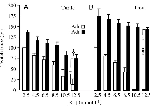
Ventricular preparations from turtle and trout were subjected to increments in extracellular [K+]. For both species, twitch force decreased as [K+] was elevated. At 12.5 mmol l−1K+, the force was close to zero, representing a reduction relative to the force at 2.5 mmol l−1 K+ of 87±14 % for turtle (Fig. 1A) and approximately 100 % for trout (Fig. 1B). These reductions were due to the elevated [K+], as shown by an extrapolation of previously obtained results (Hartmund and Gesser, 1996), which provide the change in force for preparations run under similar conditions and for similar periods but at a constant [K+] of 2.5 mmol l−1. In the presence of 10µmol l−1 adrenaline, force increased as expected (Fig. 1). Furthermore, the negative effect of high [K+] on twitch force was substantially diminished; the force at 12.5 mmol l−1[K+] compared with that at 2.5 mmol l−1 was reduced by 41±13 % for turtle (Fig. 1A) but by only 12±5 % for trout (Fig. 1B). The effect of adrenaline was greater in trout than in turtle cardiac muscle. In turtle, the force in 12.5 mmol l−1 K+and 10µmol l−1adrenaline did not differ significantly from
the force developed after 60 min in 2.5 mmol l−1 [K+] in the absence of adrenaline, i.e. control conditions (Fig. 1A), whereas it was significantly (P<0.001) greater than the value recorded under control conditions for trout (Fig. 1B).
Adrenaline-dependence
Dose–response experiments (Fig. 2) also showed that trout heart muscle tended to be more sensitive to adrenaline than turtle heart muscle. The stimulation of force by adrenaline did not differ significantly in 2.5 and 10 mmol l−1 K+ for either species, and it was close to maximal at 1µmol l−1adrenaline, although an increase in adrenaline concentration from 5 to 10µmol l−1caused a weak but significant (P<0.05) increase in twitch force for the turtle preparation in 10 mmol l−1K+(Fig. 2).
Anoxia
Elevations of extracellular [K+] occur in situations where oxygen levels are likely to be low. In both turtle and trout preparations, anoxia caused a decrease in twitch force, and this was more marked in the trout than in the turtle (Fig. 3A,B) in accordance with a previous study (Hartmund and Gesser, 1996). Under anoxic conditions, as when oxygen was supplied, force was lower at 10 mmol l−1 than at 2.5 mmol l−1 K+. For trout cardiac muscle in 10 mmol l−1K+, force approached zero after 60 min of anoxia (Fig. 3B). For both species, exposure to adrenaline resulted in a twitch force development that was not significantly different at 10 mmol l−1and 2.5 mmol l−1K+under both oxygenated and anoxic conditions (Fig. 3C,D). In the presence of adrenaline, twitch force decreased for both turtle and
0 25 50 75 100 125 150 175 200
Twitch force (%)
2.5 4.5 6.5 8.5 10.5 12.5 +Adr −Adr
*
Turtle Trout
A
[K+] (mmol l-1)
[image:3.612.51.298.68.242.2] [image:3.612.321.562.73.246.2]2.5 4.5 6.5 8.5 10.5 12.5 ‡
B
[Adrenaline] (µmol l-1) 0
50 100 150 200
Force change (%)
[K+] 10 mmol l-1 [K+] 2 mmol l-1
Turtle Trout
**
*
0.1 1 5 10
‡
0
Fig. 1. Effects of increments in extracellular [K+] on twitch force in
ventricular muscle from turtle (A) and trout (B) in the absence and in the presence of 10µmol l−1 adrenaline (Adr). Two preparations of each heart were run in parallel, one with and one without exposure to adrenaline. After stabilisation of force at 2.5 mmol l−1K+, [K+] was
increased in steps of 2 mmol l−1 with stabilisation of force at each step. This sequence of [K+] increases lasted 75 min. The open
diamonds signify twitch force for preparations maintained at 2.5 mmol l−1K+and with no adrenaline after a similar time (60 min).
Values are means + S.E.M. N=6 for both turtle and trout. An asterisk
indicates a significant difference between turtle ventricular twitch force at 12.5 mmol l−1 in the presence and in the absence of adrenaline (*P<0.05), and a double dagger indicates a significant difference between trout ventricular twitch force maintained at 12.5 mmol l−1 in the presence of adrenaline and after 60 min at 2.5 mmol l−1K+in the absence of adrenaline (‡P<0.001).
Fig. 2. Change in twitch force following stepwise increases in adrenaline concentration for ventricular muscle from turtle (N=4) and trout (N=5) treated with either 10 or 2.5 mmol l−1 K+. The
changes in force were normalised to the twitch force at 2.5 mmol l−1 K+recorded just before [K+] was elevated to 10 mmol l−1for one of
trout during the first 20 min of anoxia, and it is noteworthy that the force recorded here was significantly (P<0.01) lower for turtle (Fig. 3C) than for trout (Fig. 3D). Subsequently, however, the twitch force stabilised in the turtle, whereas it continued to fall in the trout over the remaining 40 min of anoxic exposure.
All subsequent results are from turtle heart preparations in which [K+] was changed in one step from 2.5 to 10 mmol l−1.
Action and membrane potential
Table 1 shows that elevation of [K+] was followed by an increase in resting potential to less negative values and a decrease in action potential amplitude and duration. Adrenaline restored the action potential duration in 10 mmol l−1K+ to a value not significantly different from that in 2.5 mmol l−1K+but did not significantly affect action potential duration in 2.5 mmol l−1K+. The changes in resting potential and action potential amplitude following the elevation of [K+] were not significantly influenced by adrenaline. The fact that the elevation of the resting potential in 10 mmol l−1 K+ is maintained in the presence of adrenaline is noteworthy because efflux of Ca2+via Na+/Ca2+exchange appears to be the main mechanism for relaxation in heart muscle of ectothermic vertebrates. According to equation 1 given in the Introduction, diastolic [Ca2+]
i would more than double following the membrane depolarisation of approximately 20 mV that occurs, both in the presence and the absence of adrenaline, when [K+] is changed from 2.5 to 10 mmol l−1.
Extracellular [Ca2+]
The demand on excitation/contraction (E-C) coupling,
[image:4.612.205.561.70.337.2]which includes Na+/Ca2+exchange, should increase following elevation of extracellular [Ca2+]. Preparations were therefore subjected to increments in extracellular [Ca2+] in the presence of adrenaline at high and low [K+] to examine the protection offered by adrenaline at high [K+]. The first increase in Ca2+ caused increases in both peak force and the rate of relaxation. The difference in [K+] had no significant effect on the increase in either twitch force (Fig. 4A) or relaxation rate (Fig. 4B). A Fig. 3. Twitch force under anoxia at
10 mmol l−1 and 2.5 mmol l−1 K+ for (A)
turtle (N=5) and (B) trout (N=8) ventricular muscle in the absence of adrenaline and for (C) turtle (N=5) and (D) trout (N=8) in the presence of 10µmol l−1 adrenaline. For the experimental (Experimental) preparation of the two preparations from each heart run in parallel, [K+] was elevated to 10 mmol l−1
20 min after the onset of anoxia in the experiments run in the absence of adrenaline and 20 min before the onset of anoxia in the experiments in which adrenaline was applied. For the control preparation, [K+]
was maintained at 2.5 mmol l−1. Twitch force was normalised to the twitch force recorded just before any change from control conditions was made. Values are means + S.E.M. Asterisks indicate significant
differences in the twitch force recorded at
10 and 2.5 mmol l−1K+(*P<0.05; **P<0.01; ***P<0.001), and a double dagger indicates a significant difference in the twitch force of trout
preparations recorded after 20 and 60 min of anoxic exposure (‡P<0.001).
0 50 100 150 200 250 0 50 100
Twitch force
(%)
60 20
Control
Turtle
*
Duration of anoxic exposure (min)
***
**
20 40 60
+Adrenaline
0
A
Experimental
*
10 mmol l-1 K+ in experimental samples
***
20 40 60
B
60 20 0
-10 -10
Turtle Trout
C
+Adrenaline
*
D
10 mmol l-1 K+ in experimental samples
Trout
Duration of anoxic exposure (min)
‡
[image:4.612.311.560.424.519.2]Twitch force (%)
Table 1. Effects of increased [K+] on membrane variables in cardiac muscle of the freshwater turtle Trachemys scripta elegans in the presence and absence of 10µmol l−1adrenaline
Action Action Resting potential potential
[K+] potential amplitude duration
(mmol l−1) Adrenaline (mV) (mV) (ms) N
2.5 − −73±2 96±3 990±54 13
2.5 + −75±2 107±8 1105±49 5
10 − −48±4** 67±3* 614±109* 8
10 + −55±3** 67±4* 978±84‡ 5
Values are means ± S.E.M. N is the number of preparations. It should be noted that the control values (2.5 mmol l−1 K+ without adrenaline) include all the preparations used for recording of potentials.
further increase in Ca2+ from 5.3 to 9.1 mmol l−1 had no significant effects.
Sarcoplasmic reticulum
In the presence of adrenaline, elevations of [K+] cause depolarisations that should attenuate extrusion of Ca2+ by Na+/Ca2+exchange and thus delay relaxation. It is possible that such an effect may be compensated for by the sarcoplasmic reticulum, which may be another mechanism for relaxation. This possibility was examined in a series of experiments in which preparations were exposed to ryanodine, a well-established inhibitor of the sarcoplasmic reticulum (e.g. Rousseau et al., 1987; Lindsay et al., 1994). Treatment with ryanodine did not significantly change the effect of elevated [K+] on twitch force (Fig. 5A) and relaxation rate (Fig. 5B), nor did it affect the increase in these variables induced by adrenaline (Fig. 5).
Na+/K+-ATPase
Adrenaline has been shown to stimulate Na+/K+-ATPase in heart muscle cells (Pecker et al., 1986). A stimulation of Na+/K+-ATPase that resulted in a decrease in [Na+]
iby 30 % would cancel the increase in diastolic [Ca2+]
iexpected upon a depolarisation by 20 mV (according to equation 1). It follows that the stimulation of twitch force development and relaxation by adrenaline should be more dependent on Na+/K+-ATPase at a high than at a low extracellular [K+]. The effects of a specific
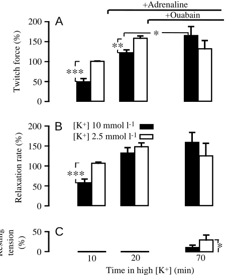
inhibitor of Na+/K+-ATPase, ouabain, on the responses to adrenaline at high and low [K+] were therefore examined. In these experiments, twitch force was significantly lower in high [K+] than in low [K+], even after addition of adrenaline (Fig. 6A). This difference disappeared during subsequent exposure to 0.25 mmol l−1 ouabain, because twitch force increased significantly (P<0.05) in high [K+] but remained unchanged in low [K+]. The relaxation rate after exposure to adrenaline did not differ significantly between high and low [K+] (Fig. 6B). However, relaxation tended to become incomplete in that resting tension tended to increase, although only significantly (P<0.05) so in low [K+] (Fig. 6C).
In another set of experiments, a high dose of ouabain, 3 mmol l−1, was applied. The duration of these experiments was shortened as contractility rapidly deteriorated at this ouabain concentration. As expected, twitch force (Fig. 7A) and relaxation rate (Fig. 7B) were lower in high than in low [K+]. Neither variable changed significantly during the initial 3 min in ouabain, whereas resting tension tended to increase, although the changes were insignificant (Fig. 7C). During the 5 min following the addition of adrenaline, twitch force and relaxation rate increased at high [K+], reaching levels not significantly different from those at low [K+] (Fig. 7A,B). The increases in resting tension became significant, with a tendency, although at the border of significance, to be larger at low than at high [K+] (Fig. 7C).
Stimulation rate
The protection of E-C coupling against the effects of high
0 50 100 150 200 250 300
Relaxation rate (%)
5.3 mmol l-1 9.1 mmol l-1
Ca2+ Ca2+
*
**
*
*
050 100 150 200
250
A
+AdrenalineB
Twitch force (%)
[K+] 2.5 mmol l-1
[K+] 10 mmol l-1 +Rya
−Rya
55
45 70
Time in ryanodine (min)
Relaxation rate (%)
0 50 100 150 200
+Adrenaline
A
Twitch force (%)
10 mmol l-1 K+
0 50 100 150
[image:5.612.61.290.70.327.2]200
B
Fig. 4. Effects of increasing [Ca2+] in the presence of 10µmol l−1
adrenaline at either 10 or 2.5 mmol l−1K+on (A) twitch force and (B)
relaxation rate. The variables were normalised to the values recorded just before [K+] was elevated to 10 mmol l−1 for one of the two
preparations from each heart (N=5) run in parallel. Values are means +S.E.M. Asterisks indicate significant effects of increasing [Ca2+] on
[image:5.612.362.522.70.301.2]twitch force and relaxation rate (*P<0.05; **P<0.01).
Fig. 5. Effects of ryanodine (Rya) (10µmol l−1) on (A) twitch force and (B) relaxation rate in the presence of 10 mmol l−1 K+ and
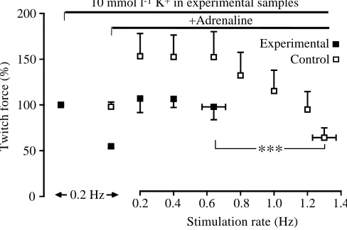
[K+] provided by adrenaline was assessed by examining the development of twitch force at elevated pacing rates. Stimulation frequency was increased in steps for preparations in the presence of adrenaline with either 10 or 2.5 mmol l−1K+. High [K+] was associated with a significant decrease in the maximal frequency at which the preparations were able to respond regularly (Fig. 8).
Discussion
It has been shown (Engstfeld et al., 1961) that increases in extracellular [K+] reduced force development in cardiac preparations from frog and guinea pig in a way that was counteracted by adrenaline.
The present work shows that this can be extended to freshwater turtle and rainbow trout cardiac muscle in which adrenaline not only upregulates twitch force but also diminishes the fraction of force lost following increases in [K+]. In the absence of adrenaline, elevations in [K+] tend to have less effect in inhibiting force development in turtle than in trout cardiac muscle. This is consistent with the fact that
[image:6.612.57.283.72.346.2]extracellular [K+] appears to attain higher values in turtle (Jackson and Ultsch, 1982) than in trout (Nielsen and Lykkeboe, 1992). Adrenaline reverses the situation, however, so that the force reductions following increases in [K+] become smaller for trout than for turtle cardiac muscle. Hence, adrenaline was found to alleviate the inhibitory action of an elevation of [K+] to 12.5 mmol l−1 more efficiently for trout than for turtle myocardium. The dose–response curve (Fig. 2) reveals that adrenaline generally seems to stimulate contractility more potently in trout than in turtle myocardium. Increases in extracellular [K+] are likely to occur during periods of insufficient oxygen supply to the cardiac muscle (Jackson and Ultsch, 1982; Nielsen and Lykkeboe, 1992). The decrease in contractility during extreme oxygen lack, i.e. anoxia, is substantially enhanced by an elevation in [K+]. As previously noted (Hartmund and Gesser, 1996), the maintenance of force development during anoxia is greater for turtle than for trout cardiac muscle. Exposure to adrenaline modifies this situation in several respects. Thus, force development during anoxia is not significantly affected by the Fig. 6. Effects of 0.25 mmol l−1 ouabain, in the presence of
adrenaline (10µmol l−1) and either 10 or 2.5 mmol l−1 K+ on (A)
twitch force, (B) relaxation rate and (C) change in resting tension. Twitch force and relaxation rates were normalised to the control values recorded just before K+ was changed for one of the two
preparations from each heart (N=6) run in parallel. The control value for twitch force was also used to normalise changes in resting tension. Values are means + S.E.M. Asterisks indicate significant changes in twitch force, relaxation rate and resting tension following the increase in [K+] and addition of ouabain (*P<0.05; **P<0.01;
[image:6.612.331.544.74.363.2]***P<0.001).
Fig. 7. Effects of 3 mmol l−1ouabain on the response to 10µmol l−1 adrenaline at either 10 or 2.5 mmol l−1K+for (A) twitch force, (B)
relaxation rate and (C) change in resting tension. Twitch force and relaxation rate were normalised to the control values recorded just before K+was changed for one of the two preparations from each
heart (N=6) run in parallel. The control value for twitch force was also used to normalise changes in resting tension. Values are means + S.E.M. Asterisks indicate significant changes in twitch force, relaxation rate and resting tension following the increase in [K+] and
addition of ouabain and adrenaline (*P<0.05; ***P<0.001).
0 50
+Ouabain
20 70
10
Time in high [K+] (min)
*
**
*
050 100 150 200
+Adrenaline
A
B
Twitch force (%)
[K+] 2.5 mmol l-1 [K+] 10 mmol l-1
***
***
C
Relaxation rate (%)
0 50 100 150 200
Resting tension (%)
*
*
***
*
*
*
*
*
040
+Ouabain
13 18
10
Time in high [K+] (min) 0
50 100 150 200
+Adrenaline
A
B
Twitch force (%)
[K+] 2.5 mmol l-1 [K+] 10 mmol l-1
C
Relaxation rate (%)
0 50 100 150 200
80 120
difference in [K+]. Furthermore, it is not higher for turtle than for trout cardiac muscle. In fact, the reverse was true after the first 20 min of anoxia. Again, the higher adrenaline sensitivity of trout cardiac muscle compared with that of freshwater turtle should be emphasised. The upregulation of anoxic performance by adrenaline indicates that anoxia per se does not necessarily elicit the full anaerobic potential. According to previous observations (Nielsen and Gesser, 1983), this anoxic reserve capacity seems to be larger for cardiac muscle from ectothermic than from endothermic vertebrates.
Engstfeld et al. (1961) demonstrated effects of high [K+]on the resting and action potential of cardiac muscle from frog and guinea pig similar to those recorded for turtle cardiac muscle in the present study (Engstfeld et al., 1961). Thus, elevations of extracellular [K+] shift the resting membrane potential to more positive values and shorten the duration of the action potential. This shortening of the action potential may be associated with a decrease in the Ca2+ transient activating contractility, since action potential duration determines the time available for Ca2+to enter the cell through the L-channels. Furthermore, a study on frog myocardial cells suggests that Ca2+enters the cell not only via the L-channels but also via Na+/Ca2+ exchange during the plateau phase of the action potential (Fan et al., 1996). The generality of the latter observation is unclear, however, as the maintenance of the action potential in guinea pig myocardium seems to be supported by an inward current associated with Ca2+extrusion via Na+/Ca2+exchange (e.g. Paterson et al., 1993).
It is conceivable that adrenaline enhances twitch force at high [K+] via the Ca2+ transient activating contractility. Thus, adrenaline augments the inward Ca2+current during the action potential (e.g. Frace et al., 1993). Furthermore, the present results
at high [K+] show that adrenaline prolongs the action potential duration and presumably the time that Ca2+channels are open, inasmuch as the action potential duration is shortened at high [K+] in the absence but not the presence of adrenaline (Table 1). In this respect, turtle cardiac muscle resembles that from frog and guinea pig (Engstfeld et al., 1961). Unlike the situation at high [K+], adrenaline was found not to prolong the action potential of turtle cardiac muscle at normal [K+]. This difference, together with the assumption that the augmentation of the Ca2+current is not influenced by the [K+], suggests that adrenaline should increase the Ca2+transient and twitch force more at high [K+] than at normal [K+]. However, the situation may be more complicated since the predicted effect on twitch force was not consistent and did not appear in all of the experimental series.
In many ectothermic vertebrates, Na+/Ca2+ exchange appears to be the predominant mechanism of myocardial relaxation (Driedzic and Gesser, 1994). Considering the inward current accompanying the extrusion of Ca2+ by Na+/Ca2+exchange, the depolarisation following an elevation of [K+] should diminish the electrochemical force driving relaxation. Conceivably, adrenaline will accentuate this effect because the depolarisation remains and the amount of activator Ca2+ should be increased in its presence. However, this suggestion was not supported by our experiments. In the presence of adrenaline, similar contractile responses occurred irrespective of K+ levels when extracellular [Ca2+ ] was elevated to challenge E-C coupling including relaxation.
This protection of contractility by adrenaline at high [K+] may involve the sarcoplasmic reticulum. Previous results on trout cardiac muscle suggest that the sarcoplasmic reticulum becomes more important to E-C coupling at high than at low [K+] (El-Sayed and Gesser, 1989). However, ryanodine, a well-established inhibitor of the sarcoplasmic reticulum (e.g. Rousseau et al., 1987; Lindsay et al., 1994), had no significant effect on either peak force or relaxation rate (Fig. 5). The possibility that turtle myocardium has a sarcoplasmic reticulum that is active, but insensitive to ryanodine, seems unlikely because ryanodine interferes with the basic properties of the sarcoplasmic reticulum (e.g. Lindsay et al., 1994). Furthermore, the dose applied (10µmol l−1) gives a maximal effect in trout myocardium (Hove-Madsen, 1992), and ryanodine at concentrations close to 10µmol l−1has clear effects on myocardial tissue from diverse species ranging from rat (Stemmer and Akera, 1986) to octopus (Gesser et al., 1997). Therefore, like many other ectothermic vertebrates (Driedzic and Gesser, 1994), the freshwater turtle appears to possess a myocardium that is not critically dependent on the sarcoplasmic reticulum.
[image:7.612.53.297.75.237.2]Adrenaline may protect E-C coupling by affecting Na+/Ca2+ exchange in a way that counteracts the negative influence of the depolarisation at high [K+]. In skeletal muscle, depression of contractility by increases in extracellular [K+] is counteracted by various means, all involving a stimulation of Na+/K+ -ATPase (e.g. Clausen and Everts, 1991). Adrenaline has been shown to stimulate Na+/K+-ATPase in cardiac muscle (Pecker et al., 1986). Such stimulation could compensate for the depolarisation that occurs in high [K+], since it may lower the Fig. 8. Maximal stimulation rate giving regular twitch force
development in 10 and 2.5 mmol l−1K+in the presence of 10µmol l−1
adrenaline. Stimulation rate was increased in steps until the maximal rate was attained. Two preparations from each heart (N=6) were run in parallel, one in 10 mmol l−1 (Experimental) and one in 2.5 mmol l−1K+(Control). Twitch force was normalised to the twitch
force recorded just before the change in [K+]. Values are means ± S.E.M. Asterisks indicate a significant difference in maximal stimulation rate at 10 and 2.5 mmol l−1K+(***P<0.001).
0.2 0.4 0.6 0.8 1.0 1.2 1.4
Stimulation rate (Hz) 0
50 100 150 200
Twitch force (%)
0.2 Hz
***
Control 10 mmol l-1 K+ in experimental samplescytoplasmic Na+activity and increase the inward Na+gradient and thereby the electrochemical force driving the extrusion of Ca2+. However, this hypothesis was not supported by the present study in which the Na+/K+-ATPase inhibitor ouabain was applied at two concentrations. The lower concentration provided a clearly submaximal inhibition, while the higher concentration should give an almost maximal inhibition of Na+/K+-ATPase. At both concentrations, adrenaline stimulated both twitch force development and relaxation as efficiently in high as in normal [K+]. Thus, Na+/K+-ATPase does not seem to be of critical importance for the stimulation of contractility by adrenaline at high [K+] in turtle heart muscle. A similar conclusion was reached in a study of frog heart muscle (Ryan and Paterson, 1994). This conclusion and the finding that the sarcoplasmic reticulum seems to be without significant importance for contractility might suggest that the capacity of the Na+/Ca2+exchanger (V
max) is such that its function is not attenuated by the elevations of [K+] applied.
The Na+ channels are another target for increases in [K+], which decrease their opening probability and, thus, membrane excitability. This effect seems to be counteracted by adrenaline, as shown in guinea pig cardiac muscle (Paterson et al., 1993). Conceivably, such an effect on the Na+channels and excitability contributed to the finding that the maximal pacing rate eliciting regular force development was substantially lower in 10 than in 2.5 mmol l−1 K+. If this is the case, catecholamines seem to provide a far from complete restoration of Na+ channel activity in turtle cardiac muscle because the decrease in maximal pacing rate at high [K+] occurred in the presence of adrenaline.
In conclusion, adrenaline efficiently counteracts reductions in twitch force development and relaxation at high [K+] under oxygenated and anoxic conditions. This effect of adrenaline was more pronounced for rainbow trout than for freshwater turtle cardiac muscle. The action of adrenaline may depend on a prolongation of the action potential in addition to enhancement of Ca2+current through the L-channels. Despite a partial membrane depolarisation at high [K+], the stimulation of twitch force development and relaxation by adrenaline does not to be seem critically dependent on either the sarcoplasmic reticulum or Na+/K+-ATPase. In contrast to the efficient protection of twitch force at a low pacing rate, the ability to respond to high pacing rates was substantially reduced at high [K+] despite the presence of adrenaline.
The development of the PC-based recordings of force and membrane potential by engineer Einer Larsen and the skilful assistance of technician Dorte Olsson are highly appreciated. This study was supported by the Danish Natural Science Research Council, Center for Respiratory Adaptation.
References
Blaustein, M. P. (1999). Sodium/calcium exchange: Its physiological
implications. Physiol. Rev. 79, 763–854.
Clausen, T. and Everts, M. E. (1991). K-induced inhibition of
contractile force in rat skeletal muscle: role of active Na–K transport. Am. J. Physiol. 261, C799–C807.
Driedzic, W. R. and Gesser, H. (1994). Energy metabolism and
contractility in ectothermic vertebrate hearts: Hypoxia, acidosis and low temperature. Physiol. Rev. 74, 221–258.
El-Sayed, M. F. and Gesser, H. (1989). Sarcoplasmic reticulum,
potassium and cardiac force in rainbow trout and plaice. Am. J.
Physiol. 257, R599–R604.
Engstfeld, G., Antoni, H., Fleckenstein, A., Nast, A. and Hattingberg, M. V. (1961). Die Restitution der Erregungsfortleitung
und Kontraktionskraft des K+-gelähmten Frosch- und
Säugetiermyokards durch Adrenalin. Pflügers Arch. 273, 145–163.
Fan, J., Yaroslav, M. S. and Morad, M. (1996). Regulation of
cardiac sodium–calcium exchanger by β-adrenergic agonists. Proc.
Natl. Acad. Sci. USA 93, 5527–5532.
Frace, A. M., Mery, P. F., Fishmeister, R. and Hartzell, H. C.
(1993). Rate-limiting steps in the β-adrenergic stimulation of cardiac calcium current. J. Gen. Physiol. 101, 227–353.
Gesser, H., Driedzic, W. R., Rantin, F. T. and De Freitas, J. C.
(1997). Ca2+regulation of heart contractility in Octopus. J. Comp.
Physiol. B 167, 474–480.
Hartmund, T. and Gesser, H. (1996). Cardiac force and high-energy
phosphates under metabolic inhibition in four ectothermic vertebrates. Am. J. Physiol. 271, R946–R954.
Hove-Madsen, L. (1992). The influence of temperature on ryanodine
sensitivity and the force–frequency relationship in the myocardium of rainbow trout. J. Exp. Biol. 167, 47–60.
Jackson, D. C. and Ultsch, G. R. (1982). Long-term submergence
at 3 °C of the turtle, Chrysemys picta belli, in normoxic and severely hypoxic water. II. Extracellular ionic responses to extreme lactic acidosis. J. Exp. Biol. 96, 29–43.
Lindsay, A. R. G., Tinker, A. and Williams, A. J. (1994). How does
ryanodine modify ion handling in the sheep cardiac sarcoplasmic reticulum Ca-release channel? J. Gen. Physiol. 104, 425–447.
Møller-Nielsen, T. and Gesser, H. (1992). Sarcoplasmic reticulum
and excitation–contraction coupling at 20 and 10 °C in rainbow trout myocardium. J. Comp. Physiol. B 162, 526–534.
Nielsen, K. E. and Gesser, H. (1983). Effects of [Ca2+]o on
contractility in the anoxic cardiac muscle of mammal and fish. Life
Sci. 32, 1437–1442.
Nielsen, O. B. and Lykkeboe, G. (1992). Changes in plasma and
erythrocyte K+during hypercapnia and different grades of exercise
in trout. J. Appl. Physiol. 72, 1285–1296.
Paterson, D. J., Rogers, J., Powell, T. and Brown, H. F. (1993).
Effect of catecholamines on the ventricular myocyte action potential in raised extracellular potassium. Acta Physiol. Scand. 148, 177–186.
Pecker, M. S., Wook, B. I., Sonn, J. K. and Lee, C. H. (1986).
Effect of norepinephrine and cyclic AMP on intracellular sodium ion activity and contractile force in canine cardiac Purkinje fibers.
Circ. Res. 59, 390–397.
Rousseau, E., Smith, J. S. and Meissner, G. (1987). Ryanodine
modifies conductance and gating behavior of single Ca release channel. Am. J. Physiol. 253, C364–C368.
Ryan, D. M. and Paterson, D. J. (1994). Effect of ouabain and
verapamil on restoration of contraction by adrenaline during high K+in the frog heart. Acta Physiol Scand. 151, 417–419.

![Fig. 4. Effects of increasing [Ca2+relaxation rate. The variables were normalised to the values recordedjust before [Kpreparations from each heart (+twitch force and relaxation rate (*] in the presence of 10 µmol l−1adrenaline at either 10 or 2.5mmoll−1 K+](https://thumb-us.123doks.com/thumbv2/123dok_us/1142312.635090/5.612.61.290.70.327/increasing-relaxation-variables-normalised-recordedjust-kpreparations-relaxation-adrenaline.webp)