Almost all cells possess the ability to regulate their volume upon osmotic perturbations originating from changes in the osmolality of the extracellular medium, imbalances in the influx and efflux rates of osmotically active solutes across the plasma membrane or variations in intracellular metabolism. To maintain both function and volume relatively undisturbed, cells have developed mechanisms that sense and oppose volume changes. It has been shown in proximal renal tubules from rabbit that, if the osmotic changes occur relatively slowly, the
cells are able to maintain a fairly constant volume over a wide osmotic range (Lohr and Grantham, 1986). When subjected to faster and larger osmotic changes, the cells are unable to withstand the perturbation. When the cell volumes depart from the ‘set-point’ by a significant degree, regulatory volume changes are activated and the cell volume is restored by compensatory fluxes of osmotically active particles.
Many cells respond to swelling with an efflux of KCl and an osmotically obligated water efflux (Davis and Finn, 1987; Printed in Great Britain © The Company of Biologists Limited 1998
JEB1415
Regulatory volume decrease (RVD) following hypo-osmotic stimulation was studied using videometric methods in isolated proximal renal tubules from trout (Salmo trutta). The relative tubule diameter increased by 132.0±4.8 % (maximum swelling within 1 min at 15 and 25 °C and within 4 min at 10 °C) following a change from iso-osmotic (290 mosmol kg−1) to hypo-osmotic (160 mosmol kg−1) Ringer’s solution. The tubule diameter subsequently decreased to approximately one-quarter of the maximal value. Ouabain (1 mmol l−1) reduced cell swelling and inhibited the RVD response by 28.0±10.5 %. Furthermore, increasing the bath K+concentration by 30 mmol l−1inhibited RVD by 76.5±3.6 %. The K+channel blocker quinine, but not Ba2+ (1 and 2 mmol l−1), significantly decreased the RVD response (by 25.0±5.4 and 72.3±5.1 % at 0.1 and 0.5 mmol l−1, respectively). Similarly, increasing the Cl− concentration in the bath from 47 to 102 mmol l−1 induced a significant reduction (45.2±7.9 %) in RVD. The RVD response was also markedly reduced (by 54.7±5.3 %) by the Cl− channel blocker indacrinone (MK-196; 0.5 mmol l−1), but only marginally by 5-nitro-2-(3-phenylpropylamino)benzoic acid (NPPB; 1, 5, 8 and 10µmol l−1). Addition of the K+/Cl− symport inhibitor furosemide (0.1 mmol l−1) resulted in a 39.8±3.9 % inhibition of RVD. This inhibition could be completely overcome by simultaneous administration of 1µmol l−1tributyltin (anion exchanger) and furosemide.
Chelation of either extracellular (1 mmol l−1EGTA) or both extra- and intracellular Ca2+ (1 mmol l−1 EGTA, 10µmol l−1 A23187) had no effect on this RVD process.
Furthermore, as measured using the fluorescent dye Fura-2/AM, there was no increase in the intracellular free Ca2+ concentration upon hypo-osmotic stimulation. Administration of the 5-lipoxygenase antagonist ETH 615-139 (20µmol l−1), however, induced a 60 % inhibition of RVD. Simultaneous addition of ETH-615 and either the K+ ionophore gramicidin (0.5 mmol l−1) or the anion exchanger tributyltin (1µmol l−1) could not reverse the ETH 615-139 inhibition. Finally, administration of the cycloxygenase inhibitor indomethacin had only a small, but significant, effect on RVD.
We conclude that RVD following hypo-osmotic swelling is in these cells a temperature- and ouabain-sensitive process that appears to be the result of K+efflux through quinine-sensitive, Ba2+-insensitive K+ channels and Cl− efflux through an MK-196- and furosemide-sensitive Cl−conductance that is relatively unaffected by NPPB. This KCl efflux seems to be regulated by eicosanoids produced by the 5-lipoxygenase. Arachidonic acid metabolites from the cycloxygenase pathway are not involved in this process. Similarly, neither extra- nor intracellular Ca2+ appears to be important for the signalling of RVD.
Key words: proximal renal tubule, teleost, trout, cell volume regulation, KCl efflux, K+ channel, Cl−channel, quinine, barium,
MK-196, NPPB, Ca2+, Fura-2, arachidonic acid, eicosanoid,
5-lipoxygenase, leukotriene, Salmo trutta.
Summary
Introduction
CELL VOLUME REGULATION IN PROXIMAL RENAL TUBULES FROM TROUT
(SALMO TRUTTA)
HILDE KANLI* ANDELI NORDERHUS
Department of Biology, Division of General Physiology, University of Oslo, Norway
*Present address: Department of General Physiology, PO Box 1051 Blindern, University of Oslo, N-0316 Oslo, Norway (e-mail: hilde.kanli@bio.uio.no)
Dellasega and Grantham, 1973; Grinstein et al. 1983; Hoffmann et al. 1984; Terreros et al. 1990; Kristensen and Folke, 1984). Since the relatively high intracellular K+
concentration found in most cells is dependent on the activity of Na+/K+-ATPase, it is not surprising that the osmoregulatory
KCl efflux in many tissues seems to depend on this enzyme (Gagnon et al. 1982; Linshaw and Grantham, 1980). None the less, the manner in which the regulatory volume decrease (RVD)-associated KCl efflux occurs varies among species and among tissues. In some cells, K+and Cl−efflux occur through
separate conductive pathways (Hoffmann et al. 1984; Welling and O’Neil, 1990; Macri et al. 1993; De Smet et al. 1995; Banderali and Roy, 1992). There is emerging evidence that the Cl−conductance could be a rather non-specific anion channel that is also permeable to amino acids (Kirk et al. 1992; Sanchez-Olea et al. 1991; see also Kirk, 1997), and it has recently been found that upon osmotic swelling a Cl− -independent channel opens through which both K+and amino
acids may pass (Bursell and Kirk, 1996). In other cells, RVD is due to furosemide-sensitive K+/Cl−symporters, K+/H+ and
Cl−/HCO3− exchangers or K+ channels and Cl−/HCO3−
exchangers (Lauf, 1985; Cala, 1980; Terreros et al. 1990). The signalling mechanism for this RVD response is not completely understood. Early experiments in several systems indicated that RVD was activated following Ca2+ influx
(Neufield et al. 1983; McCarty and O’Neil, 1991). More recent work has shown that an increase in cytosolic [Ca2+] parallels
hypo-osmotic cell swelling (Beck et al. 1991; Raat et al. 1995; Sardini et al. 1995; Ehrenfeld et al. 1994); this increase may be partly or wholly dependent on the availability of extracellular Ca2+ (McCarty and O’Neil, 1991; Ross and
Cahalan, 1995; Rothstein and Mack, 1990; Raat et al. 1995; Sardini et al. 1995). However, other studies have shown that volume regulation can operate in opossum, rabbit and MDCK renal cells incubated in Ca2+-free medium (Ubl et al. 1988a;
Breton et al. 1992; Roy and Sauve, 1987). A similar relative or complete extracellular Ca2+-independence has also been
observed in hepatocytes, lymphocytes and other non-renal cells (Corasanti et al. 1990; Grinstein et al. 1982; Hoffmann et al. 1984; Margalit et al. 1993; vom Dahl et al. 1991).
Moreover, studies in Ehrlich ascites tumour cells and human lymphocytes indicate that neither intra- nor extracellular Ca2+
is required for RVD (Harbak and Simonsen, 1995; Grinstein and Smith, 1990); other work has suggested that loss of K+
during RVD is not affected by Ca2+, nor does Ca2+affect
RVD-associated ion currents (Harbak and Simonsen, 1995; Beck et
al. 1991; Best et al. 1996).
Metabolites of arachidonic acid and other polyunsaturated fatty acids (i.e. eicosanoids) have, during the last few years, received attention as possible second messengers for the activation of RVD-associated KCl and taurine efflux (for a review, see Lambert, 1994). Of these eicosanoids, it has been found that the synthesis of leukotrienes is stimulated following hypo-osmotic cell swelling (Lambert et al. 1987). Specifically, leukotriene-D4(LTD4) accelerated RVD by stimulation of K+,
Cl− and taurine efflux. Furthermore, decreasing the
intracellular content of LTD4 with 5-lipoxygenase inhibitors
(inhibitors of arachidonic acid metabolism) blocked RVD (Lambert et al. 1987). Findings from other tissues also indicate a role for arachidonic acid metabolites in RVD signalling. For example, inhibition of phospholipase A2 (which catalyzes
phospholipid metabolization to arachidonic acids), 5-lipoxygenase or cycloxygenase decreases the osmoregulatory response associated with RVD in several cell types (Fatherazi
et al. 1994; Ling et al. 1992; Fugelli et al. 1995; Thoroed and
Fugelli, 1994). (Eicosanoids are also produced in fish; see Rowley, 1991.)
In the present study, RVD following hypo-osmotic stimulation of proximal renal tubules from trout (Salmo trutta) was studied using videometric techniques and the fluorescent dye Fura-2/AM. It was observed that RVD in these cells is a temperature- and ouabain-sensitive KCl efflux process. Following hypo-osmotic swelling, osmoregulatory shrinkage seemed to be due to K+ efflux through quinine-sensitive K+
channels that were relatively insensitive to Ba2+. Cl− efflux
appeared to occur via MK-196- and furosemide-sensitive but NPPB-insensitive Cl−conductive pathways. The RVD process was independent of extra- and intracellular Ca2+. Likewise, no
increase in the intracellular Ca2+activity was found upon
hypo-osmotic stimulation. Administration of the 5-lipoxygenase antagonist ETH 615-139, but not the cycloxygenase inhibitor indomethacin, however, inhibited RVD to a major degree. This inhibition probably occurred at the level of both the K+and the
Cl− conductance. Thus, in these trout proximal renal tubules, it appears that KCl efflux associated with hypo-osmotically stimulated RVD is a Ca2+-independent process that is regulated
by arachidonic acid metabolites from the 5-lipoxygenase pathway.
Materials and methods General methodology
Trout (Salmo trutta, 50–100 g, presmolt) obtained from Ims Biological Station, Norwegian Institute for Nature Research, Stavanger, Norway, were kept at 12 °C in standard freshwater aquarium conditions. At the time of the experiments, the fish were decapitated and their renal tissue removed and placed in an iso-osmotic (290 mosmol kg−1) fish Ringer’s solution,
attached to both the optical port of the microscope and a video recorder.
The tubules were incubated for 3–5 min in the iso-osmotic Ringer’s solution with or without test substances (see below). The bath solution was then rapidly exchanged with hypo-osmotic (160 mosmol kg−1) Ringer’s solution (Table 1) and
stimulated for 15 min with or without test substances. The solutions were made hypo-osmotic by omission of 70 mmol l−1
NaCl. For each experiment with a test substance, a control experiment was performed in parallel. Tubular diameters were measured on the screen of a television monitor by replaying the video-taped experiments. The diameters were used as a cell volume indicator, and the data were expressed as relative diameter change (compared with the diameter measured at the time of solution change, i.e. time 0).
Effects of temperature and Na+/K+-ATPase on regulatory volume decrease
To test the effects of temperature on RVD and to find both a functional and practical temperature for the experiments, proximal renal tubules were exposed to iso-osmotic then hypo-osmotic control solutions (as explained above) at 10, 15 or 25 °C. These temperatures range from 2–3 °C below aquarium temperature up to room temperature. Five experiments were performed at each temperature. All other experiments in this study were performed at 15 °C. Further, to determine the role of the Na+/K+-ATPase in RVD in these tubules, 1 mmol l−1
(final concentration) ouabain was added to both the iso-osmotic and hypo-osmotic solutions (seven experiments).
Role of K+efflux during regulatory volume decrease
To determine the role of K+ efflux during RVD in trout
proximal renal tubules, the K+ efflux gradient in the
hypo-osmotic solution was reduced by increasing the K+
concentration from 3 to 33 mmol l−1(iso-osmotic replacement
of mannitol for KCl; Table 1). In the iso-osmotic solutions of both the control experiments (N=5) and the experiments with an increased K+ concentration (N=5), 30 mmol l−1 NaCl was
exchanged with 60 mmol l−1mannitol (KCl concentration was
kept normal, Table 1). To study whether a potential RVD-associated K+ efflux occurred through K+ channels, the K+
channel inhibitors BaCl2 (1 and 2 mmol l−1, three and five
experiments, respectively), quinine (0.1 and 0.5 mmol l−1, three
and five experiments, respectively) or BaCl2(2 mmol l−1) and
quinine (0.5 mmol l−1, five experiments) were added to the
iso-osmotic and hypo-iso-osmotic solutions. In the experiments with BaCl2, the MgSO4 in the iso-osmotic and hypo-osmotic
solutions was substituted with MgCl2.
Role of Cl−efflux during regulatory volume decrease Similar experiments were performed in which the Cl− concentration of the hypo-osmotic solution was increased from 47 mmol l−1 to 77 and 102 mmol l−1 by increasing the NaCl
concentration (five experiments each; Table 1). In these experiments, 60 mmol l−1mannitol (substituted for 30 mmol l−1
NaCl) of the control hypo-osmotic solution was replaced by 70 and 95 mmol l−1 NaCl, respectively. The osmolality of the
high-[Cl−] (102 mmol l−1) hypo-osmotic solution was
increased to 200 mosmol kg−1, while the other hypo-osmotic
osmolalities remained as in the control experiments (160 mosmol kg−1). The iso-osmotic solutions in all these
experiments contained 60 mmol l−1 mannitol substituted for
30 mmol l−1 NaCl (Table 1). To determine whether a
potential Cl− efflux during RVD could occur through Cl− channels, the Cl− channel inhibitors 5-nitro-2-(3-phenylpropylamino)benzoic acid (NPPB) (1, 5, 8 and 10µmol l−1; N=5, 5, 2 and 3, respectively) or MK-196
(indacrinone) (0.5 mmol l−1, five experiments) were added to
[image:3.609.50.567.89.257.2]both the iso-osmotic and hypo-osmotic solutions. Following the demonstration that MK-196 could inhibit RVD, proximal Table 1. Composition of solutions
Iso-osmotic Hypo-osmotic
Iso-osmotic Hypo-osmotic Iso-osmotic Hypo-osmotic control for control for Hypo-osmotic Hypo-osmotic control control high-[K+] high-[K+] high-[Cl−] high-[Cl−] 77 mmol l−1Cl− 102 mmol l−1Cl−
NaCl 140 70 110 40 110 40 70 95
KCl 3 3 3 33 3 3 3 3
NaHCO3 7 7 7 7 7 7 7 7
MgSO4* 5 5 5 5 5 5 5 5
Glucose 5 5 5 5 5 5 5 5
CaCl2 2† 2† 2 2 2 2 2 2
NaH2PO4 1 1 1 1 1 1 1 1
Mannitol 0 0 60 0 60 60 0 0
pH 7.8 7.8 7.8 7.8 7.8 7.8 7.8 7.8
Osmolality 290 160 290 160 290 160 160 200
All solutions were bubbled with compressed 1% CO2in air.
*Substituted with MgCl2 in experiments with BaCl2.
†0.5 mmol l−1in experiments with EGTA or EGTA and A23187.
renal tubules were exposed to both iso-osmotic and hypo-osmotic solutions containing both MK-196 (0.5 mmol l−1; five
experiments) and the anion exchanger tributyltin (TBT; 1µmol l−1) (Wieth and Tosteson, 1979; Wulf and Byington, 1975).
In addition, furosemide (0.1 mmol l−1) was added to both the
iso-osmotic and hypo-osmotic Ringer’s solutions to investigate
whether part of the RVD-associated KCl efflux could occur through K+/Cl− symporters (five experiments). Five
experiments were also performed to determine whether a potential furosemide inhibition of RVD occurred at the level of Cl− efflux only. In these experiments, furosemide (0.1 mmol l−1) and TBT (1µmol l−1) were administered
simultaneously to the iso-osmotic and hypo-osmotic solutions.
Role of Ca2+and arachidonic acid metabolites in regulatory volume decrease
To determine whether extracellular Ca2+ could be an
important signalling factor for RVD, trout proximal renal tubules were exposed to iso-osmotic (290 mosmol kg−1) then
hypo-osmotic (160 mosmol kg−1) Ringer’s solutions in which
2 mmol l−1CaCl
2had been replaced by 1 mmol l−1EGTA and
0.1 mmol l−1 CaCl
2 (five experiments). Control experiments
were performed with similar solutions except for the omission of EGTA. Other experiments were performed in which both the extracellular and intracellular Ca2+ concentrations were
reduced to zero by the addition of 1 mmol l−1 EGTA and
10µmol l−1 A23187 (0.1 mmol l−1 CaCl
2) to both the
iso-osmotic and hypo-iso-osmotic solutions (five experiments). The possible participation of arachidonic acid metabolites in the activation of RVD was tested by using the pharmacological agent ETH 615-139 (20µmol l−1, five experiments) to inhibit
the 5-lipoxygenase pathway of arachidonic acid metabolism. To determine which of the RVD fluxes (K+or Cl−) had been
inhibited by this agent, ETH 615-139 (20µmol l−1) was added
to both the iso-osmotic and hypo-osmotic solutions simultaneously with either the K+ ionophore gramicidin
(0.5 mmol l−1) or the anion exchanger tributyltin (TBT;
1µmol l−1) (five experiments each). In five experiments, the
cycloxygenase inhibitor indomethacin (10µmol l−1; an
antagonist of the metabolism of arachidonic acid into, among other products, the prostaglandins) was added to both the iso-osmotic and hypo-iso-osmotic solutions.
Measurements of variations in intracellular [Ca2+]
To confirm the results with Ca2+ chelation, experiments
were performed using the acetoxymethyl ester form (AM) of the fluorescent intracellular Ca2+ indicator Fura-2
(Grynkiewicz et al. 1985; Sanna et al. 1994). In these
experiments, proximal renal tubules were placed on pieces of coverslip coated with CellTak to improve tissue adhesion to the glass. The coverslips were placed in a glass-bottomed chamber filled with iso-osmotic (290 mosmol kg−1) Ringer’s
solution to which 10µmol l−1Fura-2/AM was added to a final
concentration of 1µmol l−1. Tubules were incubated in this
[image:4.609.43.295.73.549.2]solution for approximately 40 min in the dark at room temperature (25 °C). It had previously been determined that RVD at this temperature is identical to that at 15 °C. Following incubation, excess Fura-2/AM was removed by rinsing the chamber three times with Fura-free iso-osmotic Ringer’s solution. The chambers containing the proximal tubules were then transferred to the stage of a Nikon Diaphot 300 inverted microscope fitted with EPI-fluorescence attachments and a Fig. 1. Electron micrograph of a proximal renal tubule cell from trout
colour-chilled CCD camera and controller (C5310-11, Hamamatsu, Japan). The intensity of the fluorescence signal was measured in the iso-osmotic solutions at excitation wavelengths of 340 and 380 nm (emission wavelength 509 nm). The solution in the chamber was rapidly exchanged with the hypo-osmotic (160 mosmol kg−1) Ringer’s solution,
and the fluorescence intensity was measured as with the iso-osmotic solution. The ratio (R) of the fluorescence intensity (F) at 340 and 380 nm (R=F340/F380) was used as a measure of the
relative Ca2+ concentration in the cells (determined using
Image Pro Plus software, version 2.0, Media Cybernetics, MD, USA). The value of R of the Ca2+response in the hypo-osmotic
solution was expressed as a percentage of that in the iso-osmotic solution (iso-iso-osmotic R set to 100 %).
Chemicals
MK-196 was a gift from MSD, Oslo, Norway. Tributyltin chloride was purchased from Aldrich Chemicals, Germany, and NPPB was purchased from the SMS group, Hørsholm, Denmark. ETH 615-139 was kindly donated by Dr E. Petersen, Leo Pharmaceutical Products Ltd. All other chemicals were purchased from Sigma Chemical Co., St Louis, MO, USA.
Statistics
All data are expressed as means ± standard error (S.E.M.). Significance was established using Student’s t-tests. Differences were considered significant if P<0.05. One-way analysis of variance (ANOVA) and post-hoc comparisons of means (Duncan test) were performed using the Statistica software program (version 5.1, StatSoft, Inc., Tulsa, OK, USA).
Results
Role of temperature and Na+/K+-ATPase in regulatory volume decrease
To determine whether the RVD response in trout proximal renal tubules was temperature-sensitive, tubules were incubated with iso-osmotic solutions and stimulated with hypo-osmotic solutions at 10, 15 or 25 °C. The relative diameter change (and relative volume change) did not vary significantly in the iso-osmotic solutions (Fig. 2A). Following a change to hypo-osmotic solution (time 0), the cells swelled rapidly as the result of water influx, and at 15 and 25 °C the cells reached a maximum relative tubule diameter of 132.0±4.8 % within 1 min. At 10 °C, the cell volumes did not reach their maximum until 4 min after hypo-osmotic stimulation. Following the initial swelling, cell diameters at all temperatures decreased gradually (RVD phase) by approximately 65–75 % as measured 15 min after hypo-osmotic stimulation (Fig. 2A; percentage relative diameter decrease from maximum cell swelling: 74.3±6.3, 74.5±4.5 and 74.5±4.8 at 10, 15 and 25 °C, respectively). All other experiments in this study were performed at 15 °C (see the Discussion), and percentage relative volume regulation for all the controls was set to 100 %.
In the presence of 1 mmol l−1ouabain, cell volumes reached
a maximum at approximately the same time as in the control
experiments (Fig. 2B). Both the amount of cell swelling and the RVD response were, however, reduced (P<0.05) by this Na+/K+-ATPase antagonist (percentage relative diameter
decrease: 72.0±10.5, controls: 100; Table 2; Fig. 2B).
K+efflux during regulatory volume decrease
To determine whether K+ efflux could play a role during
the RVD phase in proximal renal tubules the K+ efflux
−5 0 5 10 15
−0.25 0 0.25 0.50 0.75 1.00 1.25 1.50 1.75
2.00 290 160 mosmol kg−1
10°C 15°C 25°C A
B
*
Time (min)
−3 0 3 6 9 12 15
Relative diameter change
0 0.25 0.50 0.75 1.00 1.25 1.50 1.75 2.00 2.25
Control
1 mmol l−1 ouabain
Fig. 2. (A,B) Effects of temperature (A) and ouabain (B) on the regulatory volume decrease (RVD) in trout proximal renal tubules. Relative diameter change is used as a measure of relative volume change. Isolated tubules were exposed to iso-osmotic (290 mosmol kg−1) trout Ringer’s solution for 5 min prior to
hypo-osmotic (160 mosmol kg−1) stimulation (time 0). Experiments were
performed at solution temperatures of 10, 15 and 25 °C (N=5). In a different set of experiments, tubules were exposed to 1 mmol l−1
ouabain for 3 min in iso-osmotic solution and for 15 min in hypo-osmotic solution (15 °C, N=7). Parallel control experiments were performed without ouabain (N=7). Values are means ± S.E.M. An
[image:5.609.332.543.187.589.2]gradient was decreased by increasing the K+concentration in
the hypo-osmotic solution from 3 to 33 mmol l−1(iso-osmotic
exchange of mannitol for KCl). Increased K+ concentration
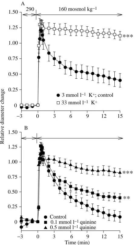
resulted in a significant (P<0.001) inhibition of RVD (Fig. 3A; percentage relative volume change 23.5±3.6; Table 2). To determine whether this K+ efflux could occur
through K+ channels, the K+ channel inhibitor quinine was
added to both the iso-osmotic and hypo-osmotic solutions (Fig. 3B). This inhibitor reduced RVD at 0.1 mmol l−1
(P<0.05; percentage relative volume regulation: 75.0±5.4; Table 2) and more strongly at 0.5 mmol l−1 (P<0.001;
percentage relative volume regulation: 27.7±5.1; Table 2). In contrast, no significant inhibition was found in the presence of either 1 or 2 mmol l−1BaCl
2(data not shown; percentage
relative volume regulation: 74.7±10.0 and 95.1±4.3, respectively). To determine whether Ba2+ and quinine could
exert an additive inhibition of RVD, 2 mmol l−1 BaCl 2 and
0.5 mmol l−1quinine were added simultaneously to both the
iso-osmotic and hypo-osmotic Ringer’s solutions. It was found (data not shown) that the inhibitory effect on RVD was no greater with these two compounds administered together than with quinine alone (percentage relative volume decrease: 49.8±3.9; Table 2; P<0.01).
Cl−efflux during regulatory volume decrease
[image:6.609.320.543.72.477.2]To determine whether Cl−efflux was important for RVD, the Cl−efflux gradient was decreased (Table 1) by increasing the Cl− concentration in the hypo-osmotic solution from 47 to Table 2. Summary of results
Percentage volume regulation Experiment (compared with controls)
Control 100
Ouabain 72.0±10.5*
High [K+] 23.5±3.6***
Quinine (0.1 mmol l−1) 75.0±5.4*
Quinine (0.5 mmol l−1) 27.7±5.1***
Ba2++ quinine 49.8±3.9**
High [Cl−](102 mmol l−1) 54.8±7.9*
NPPB (8µmol l−1) 71.8±8.3*
MK-196 45.3±5.3***
MK-196 + TBT 74.7±9.5*
Furosemide 60.2±3.9***
ETH 615-139 (20µmol l−1) 40.6±4.3***
ETH 615-139 + gramicidin 35.0±7.5*** (0.5 mmol l−1)
ETH 615-139 + TBT 58.2±5.2* (1µmol l−1)
Indomethacin (10µmol l−1) 84.2±4.2*
Temperature experiments are not included since these were used to establish the control value (100 %).
The magnitude of regulatory volume decrease was almost identical at 10, 15 and 25 °C (see text).
Only results that are significantly different from controls are shown: *P<0.05; **P<0.01; ***P<0.001.
NPPB [5-nitro-2-(3-phenylpropylamino)benzoic acid] is a Cl− channel inhibitor; MK-196 (indacrinone) is a Cl−channel inhibitor; ETH 615-139 is a 5-lipoxygenase inhibitor; TBT (tributyltin) is an anion exchanger.
Values are means ±S.E.M., N=3–10.
***
−3 0 3 6 9 12 15 0
0.25 0.50 0.75 1.00 1.25 1.50
3 mmol l−1 K+; control
33 mmol l−1 K+
A
B
Time (min)
−3 0 3 6 9 12 15
Relative diameter change
0 0.25 0.50 0.75 1.00 1.25 1.50
Control
0.1 mmol l−1 quinine
0.5 mmol l−1 quinine
***
**
290 160 mosmol kg−1
Fig. 3. (A,B) Effects of a high [K+] in the bath (A) or the K+channel
blocker quinine (B) on regulatory volume decrease (RVD) in trout proximal renal tubules. Tubules were exposed for 3 min to an iso-osmotic (290 mosmol kg−1) Ringer’s solution with normal K+
concentration (3 mmol l−1). At time 0, the solution was exchanged
with a hypo-osmotic (160 mosmol kg−1) solution in which
60 mmol l−1mannitol had been replaced by 30 mmol l−1 KCl (final
K+concentration 33 mmol l−1) (N=5). Iso-osmotic and hypo-osmotic
solutions with normal K+concentration (3 mmol l−1) were used in the
control experiments (N=5). In a different set of experiments, tubules were preincubated for 3 min in the iso-osmotic Ringer’s solution containing either 0.1 mmol l−1(N=5) or 0.5 mmol l−1 (N=3) quinine
chloride. At time 0, the bath solution was exchanged with hypo-osmotic control Ringer’s solution also containing either 0.1 or 0.5 mmol l−1 quinine. Control experiments (N=7) were performed
without quinine. Values are means ±S.E.M.. An asterisk indicates a
[image:6.609.42.295.84.310.2]77 mmol l−1 (iso-osmotic addition of NaCl for mannitol; final
osmolality 160 mosmol kg−1) or 102 mmol l−1(addition of NaCl
for mannitol; final osmolality 200 mosmol kg−1). It was observed
that increasing the Cl−concentration of the hypo-osmotic solution by 30 mmol l−1had no significant influence on RVD (Fig. 4A)
(percentage relative volume change: 79.4±8.7). Increasing the Cl− concentration by 55 mmol l−1, however, resulted in a
significant inhibition (P<0.05) of RVD (Fig. 4A) (percentage relative volume regulation: 54.8±7.9; Table 2). Since, in the latter experiments, the osmolality of the hypo-osmotic solution was increased by 40 mosmol kg−1compared with the control
hypo-osmotic solution, the hypo-osmotic difference between the iso-hypo-osmotic and the hypo-osmotic solutions was smaller and, therefore, the amount of water influx and cell swelling were reduced compared with the control experiments. The magnitude of RVD in these experiments, thus, cannot be directly compared with the control experiments without introducing some error. Therefore, to study further the possible participation of Cl−in RVD, the Cl−channel inhibitor MK-196 (0.5 mmol l−1) was added to the iso-osmotic
and hypo-osmotic solutions. MK-196 significantly (P<0.001) decreased RVD (Fig. 4B; percentage relative volume decrease: 45.3±5.3; Table 2). To confirm that this effect was due to a decreased Cl−efflux, experiments were performed in which MK-196 (0.5 mmol l−1) was added to the iso-osmotic and
hypo-osmotic solutions together with the anion exchanger TBT. It was found that the presence of TBT resulted in a partial recovery of RVD (Fig. 4B; percentage relative volume regulation: 74.7±9.5; Table 2). In contrast, the Cl−channel antagonist NPPB (1, 5, 8 and 10µmol l−1) had no such negative effect on RVD (data not
shown). In these experiments, it was observed that RVD was slightly, though significantly (P<0.05), inhibited only at 8µmol l−1 NPPB (percentage relative volume regulation:
71.8±8.3; Table 2).
−3 0 3 6 9 12 15
Relative diameter change
0 0.25 0.50 0.75 1.00 1.25
1.50 290 160/200 mosmol kg−1
47 mmol l−1 Cl−; Control
77 mmol l−1 Cl−
102 mmol l−1 Cl−*
A
Time (min)
−3 0 3 6 9 12 15
Relative diameter change
0 0.25 0.50 0.75 1.00 1.25
***
290 160 mosmol kg−1
Control
0.1 mmol l−1 furosemide
C
−3 0 3 6 9 12 15
Relative diameter change
0 0.25 0.50 0.75 1.00 1.25 1.50
1.75 290 160 mosmol kg−1
Control
0.5 mmol l−1 MK-196+
***
*
0.5 mmol l−1 MK-196
1 µmol l−1 TBT
B
Fig. 4. (A–C) Effects of increasing bath Cl− concentration (A) or adding the Cl− channel blocker MK-196 (B) or the K+/Cl−
cotransport inhibitor furosemide (C) on regulatory volume decrease (RVD). Proximal renal tubules from trout were exposed for 3 min to iso-osmotic solutions (290 mosmol kg−1) in which 30 mmol l−1NaCl
had been substituted with 60 mmol l−1mannitol. At time 0, the bath
solution was exchanged with a solution similar to the control hypo-osmotic solution but in which 30 mmol l−1NaCl had been exchanged
with mannitol (160 mosmol kg−1; total Cl−concentration 47 mmol l−1,
N=10), a control hypo-osmotic solution (160 mosmol kg−1; total Cl−
concentration 77 mmol l−1) or a control hypo-osmotic solution to
which 25 mmol l−1NaCl had been added (200 mosmol kg−1; total Cl−
concentration 102 mmol l−1). In a different set of experiments,
proximal renal tubules were exposed to 0.5 mmol l−1MK-196 added
to both the iso-osmotic (290 mosmol kg−1) and hypo-osmotic
(160 mosmol kg−1) solutions. To determine whether the effect of
MK-196 could be related to Cl−efflux, proximal renal tubules in another set of experiments were exposed to iso-osmotic and hypo-osmotic solutions to which both 0.5 mmol l−1MK-196 and the anion
exchanger tributyltin (TBT, 1µmol l−1) had been added. Control
experiments were performed without MK-196. In yet other experiments, tubules were exposed to 0.1 mmol l−1 furosemide for
3 min in iso-osmotic solution (290 mosmol kg−1). At this time, the
solution was exchanged (time 0) with a hypo-osmotic control Ringer’s solution containing 0.1 mmol l−1 furosemide. Values are
means ± S.E.M. Unless otherwise indicated, N=5. An asterisk
[image:7.609.327.550.133.760.2]Finally, to study whether some of the KCl efflux associated with the RVD process could occur through the K+/Cl−
symporter, 0.1 mmol l−1furosemide was added to both the
iso-osmotic and hypo-iso-osmotic solutions. This inhibitor caused a
significant (P<0.001) inhibition of the volume regulatory response (Fig. 4C; percentage relative volume regulation: 60.2±3.9; Table 2). This inhibition could be reversed by the addition of 1µmol l−1 TBT to the furosemide-containing
solutions (data not shown, percentage relative volume regulation: 89.4±6.4).
Signalling of regulatory volume decrease
To study whether Ca2+influx from the extracellular medium
was necessary for activation of RVD, proximal renal tubules were incubated in iso-osmotic (290 mosmol kg−1, 3 min) and
stimulated with hypo-osmotic (160 mosmol kg−1) Ringer’s
solutions in which [Ca2+] had been chelated towards zero by
the addition of 1 mmol l−1 EGTA (0.1 mmol l−1 Ca2+). No
significant change in RVD was induced by this chelation (percentage relative volume regulation: 92.1±8.2; Fig. 5A). Similarly, chelation of both intra- and extracellular Ca2+
(10µmol l−1 A23187, 1 mmol l−1 EGTA, 0.1 mmol l−1 Ca2+)
had no significant effect on RVD (percentage relative volume regulation: 86.8±8.0; Fig. 5A). These results were confirmed using the intracellular Ca2+indicator Fura-2/AM. No increase
in intracellular Ca2+ activity was observed following
hypo-osmotic stimulation (Fig. 5B).
The role of arachidonic acid metabolites in the signalling of Time (min)
Relative diameter change
0 0.25 0.50 0.75 1.00 1.25
1.50 290 160 mosmol kg−1
10 µmol l−1 A23187 +
1 mmol l−1 EGTA + 0.1 mmol l−1 Ca2+
Control
1 mmol l−1 EGTA + 0.1 mmol l−1 Ca2+
A
% Change in relative [Ca
2+
]i
0 10 20 30 40 50 60 70 80 90 100
Iso-osmotic Hypo-osmotic B
Time (min)
Relative diameter change
0 0.25 0.50 0.75 1.00 1.25 1.50 1.75
***
Control
20 µmol l−1 ETH 615-139
290 160 mosmol kg−1
C
−3 0 3 6 9 12 15
−3 0 3 6 9 12 15
Fig. 5. (A–C) Effect of reducing extracellular and both extra- and intracellular Ca2+activity on regulatory volume decrease (RVD) (A)
and the role of intracellular Ca2+(B) and the 5-lipoxygenase pathway
(C) in RVD in proximal renal tubules from trout. Tubules were exposed to an iso-osmotic (290 mosmol kg−1) Ringer’s solution
containing either 1 mmol l−1EGTA and 0.1 mmol l−1Ca2+or 10µmol
l−1 A23187, 1 mmol l−1 EGTA and 0.1 mmol l−1 Ca2+. After 3 min
(time 0), the solution was exchanged with hypo-osmotic Ringer’s containing similar concentrations of A23187, EGTA and Ca2+.
Parallel control experiments were performed without A23187 and EGTA. In additional experiments, changes in intracellular free Ca2+
concentration were measured upon hypo-osmotic stimulation (B). Proximal renal tubules of trout were exposed to iso-osmotic (290 mosmol kg−1) Ringer’s solution preloaded with the fluorescent
intracellular Ca2+indicator Fura-2/AM (N=3). The bath solution was
then exchanged with hypo-osmotic Ringer’s solution (160 mosmol kg−1). The intensity of the fluorescence signal was
measured at excitation wavelengths of 340 and 380 nm (emission wavelength 509 nm). The ratio (R) of the fluorescence intensity (F) at 340 and 380 nm (R=F340/F380) was used as a measure of the relative
Ca2+concentration in the cells. The value of R of the Ca2+response
in the hypo-osmotic solution is expressed as a percentage of that in the iso-osmotic solution (iso-osmotic R set to 100 %). Finally, to test whether arachidonic acid metabolites from the 5-lipoxygenase pathway could be involved in the signalling of RVD in these tubules, the 5-lipoxygenase inhibitor ETH 615-139 (20µmol l−1) was added
to iso-osmotic (290 mosmol kg−1) and hypo-osmotic
(160 mosmol kg−1) Ringer’s solutions. Control experiments were
[image:8.609.56.282.136.746.2]RVD in these tubules was then examined. Inhibition of 5-lipoxygenase (the enzyme that catalyzes the production of leukotrienes) with 20µmol l−1ETH 615-139 induced a strong
inhibition (P<0.001) of RVD (Fig. 5C; percentage relative volume regulation: 40.6±4.3; Table 2). This inhibition could not be overcome using either the K+ ionophore gramicidin
(0.5 mmol l−1; percentage relative volume regulation: 35.0±7.5;
Table 2; data not shown) or the Cl−/OH−exchanger tributyltin (1µmol l−1; percentage relative volume regulation: 58.2±5.2;
Table 2; data not shown). Inhibition of cycloxygenase (the enzyme that catalyzes arachidonic acid breakdown into prostaglandins) by 10µmol l−1 indomethacin induced only a
small, though significant (P<0.05), decrease in RVD (percentage relative volume regulation: 84.2±4.2; data not shown).
Discussion
In terrestrial species, cell volume regulatory mechanisms have developed mainly in response to variations in the internal environment. For aquatic species, however, cell volume regulation is critical in dealing both with variations in the external environment and with changes in the composition of extracellular body fluids. In fish, both the kidneys and the gills are important in maintaining the composition of extracellular body fluid relatively constant. Most saltwater teleosts are exposed to salt loading and water loss and, therefore, potential cell shrinkage. These fish excrete excess salt through the gills and faeces, while urine output is kept to a minimum. In contrast, the typical freshwater teleost is threatened by dilution of extracellular body fluid and cell swelling due to water influx. In such fish, excess water is excreted as hypo-osmotic urine. Thus, for the trout used in the present study, the physiological meaning of the regulatory volume decrease response is probably in maintaining a relatively constant cell volume during osmotic variations induced both by changes occurring within the animal itself (for example, changes in metabolism) and by the external environment (fresh water).
The role of temperature and the Na+/K+-ATPase in regulatory volume decrease
The cell volumes of the proximal renal tubules remained relatively constant in iso-osmotic (290 mosmol kg−1) solutions
(Fig. 2A). This lack of change is to be expected as there should be no significant difference in osmolality between the intra-and extracellular media. Upon hypo-osmotic (160 mosmol kg−1) stimulation, these water-permeable cells
swelled rapidly due to water influx. This swelling was followed by an RVD phase in which the cell diameters decreased by approximately 65–75 %. The rate of cell swelling appeared to be nearly the same at both 15 and 25 °C. At 10 °C, however, the time from hypo-osmotic stimulation to maximum cell swelling was longer than that at 15 and 25 °C (4 min at 10 °C compared with 40 s at 15 °C and 50 s and 25 °C). Thus, the time of onset of the RVD phase was delayed by approximately 3 min at 10 °C compared with the higher temperatures, resulting in an elevated maximum cell volume. These results may be explained by the observations that the plasma membrane
fluidity and the activity of transport molecules, e.g. the Na+/K+-ATPases (Charnock et al. 1971; Esmann and Skou,
1988), are temperature-sensitive in many tissues. Furthermore, it is also possible that, as observed in isolated rabbit proximal renal tubules, hypothermia inhibits RVD through an increase in Na+accumulation (Grantham et al. 1977). The remainder of
the experiments in the present study were performed at 15 °C, a temperature at which the RVD response was rapid. In addition, this temperature is near that of the aquarium.
Since Na+/K+-ATPase, which is necessary to maintain the
intracellular K+ concentration, is important in cell volume
regulation in iso-osmotic solutions (Gagnon et al. 1982; Grantham et al. 1977; Linshaw and Grantham, 1980), the role of this transporter in hypo-osmotically induced volume regulation was studied by the addition of the cardiac glycoside ouabain. Ouabain was found both to reduce the amount of cell swelling upon hypo-osmotic stimulation and to inhibit RVD (Fig. 2B). Volume regulation in proximal renal tubules has been found to be inhibited by ouabain in other studies (Gagnon et al. 1982; Kirk et al. 1987; Linshaw and Grantham, 1980). Some researchers have attributed this effect to the importance of Na+
and water efflux during RVD (Gagnon et al. 1982). It has, however, been noted in proximal straight tubules that the cells initially shrink upon exposure to ouabain (Kirk et al. 1987; Linshaw et al. 1977). This shrinkage is followed by cell swelling to a steady-state volume. As a consequence of the high K+ permeability of the basolateral membrane, the initial
ouabain-induced shrinking was thought to be because K+efflux
exceeded Na+ influx (Grantham et al. 1977; Welling et al.
1985). This theory corresponds well with the observation that the intracellular K+ activity is quickly reduced upon ouabain
administration, the effect occurring within minutes after addition (Biagi et al. 1981). In the present study, cell shrinkage in the iso-osmotic solution, but no subsequent swelling, was also observed upon ouabain administration (Fig. 2B). The lack of cell swelling in the iso-osmotic solution is probably due to the short exposure to ouabain, only 3 min, prior to hypo-osmotic stimulation. In comparison, Gagnon et al. (1982) observed cell swelling only after approximately 20 min of exposure to ouabain. Therefore, if indeed ouabain induces K+efflux in
iso-osmotic solutions, it is to be expected that a short (minutes) incubation in ouabain followed by hypo-osmotic stimulation would lead to (i) a cell swelling that is less than that in the control preparation due to the pre-shrunken state of the cells (result of K+ efflux), and (ii) a compromised RVD due to a
reduction in the intracellular concentration of the osmoeffector K+(discussed below). In addition, as has been proposed by Kirk et al. (1987), it is likely that, owing to the coupling between the
pump and the K+ leak, the K+ permeability might have been
decreased indirectly upon inhibition of Na+/K+-ATPase.
Mechanism of osmoregulatory KCl efflux during regulatory volume decrease
K+efflux
As observed in tissues of a variety of species, K+ and Cl−
also appear to be important for RVD in trout proximal renal tubules (Hoffmann et al. 1984; Macri et al. 1993; Terreros et
al. 1990). As others have found in renal cells (Kirk et al. 1987;
Rothstein and Mack, 1990), we found that RVD was reduced when the efflux gradient for K+ was decreased by increasing
the peritubular K+ concentration (Fig. 3A). It appears that
much of this K+ efflux occurs through K+ channels since
administration of the K+ channel inhibitor quinine (Cook,
1988) inhibited the trout proximal tubule RVD response by 70 % (Fig. 3B). Interestingly, it was found that the K+channel
inhibitor Ba2+ (Cook, 1988) had little effect on RVD. This
result was initially surprising since Ba2+-sensitive K+ loss
during RVD has been described in other tissues (Haddad and Graf, 1989; Lambert et al. 1984; Lau et al. 1984; Terreros et
al. 1990; Welling et al. 1985). A similarly slight effect of Ba2+
has, however, been documented in hepatocytes where quinidine blocked RVD-associated K+efflux more effectively
than Ba2+ (Haussinger et al. 1990). Furthermore,
hypo-osmotically stimulated K+ loss that is more sensitive to
quinine/quinidine than to Ba2+has been documented in other
cells (De Smet et al. 1995; Illek et al. 1992). In MDCK cells, Ba2+is unable to inhibit RVD-associated K+loss significantly
(Simmons, 1991). Contrary to observations in proximal renal tubules from rabbit (Lapointe and Duplain, 1991), in the present study addition of both Ba2+ (2 mmol l−1) and quinine
(0.5 mmol l−1) induced no additional inhibitory effect on
volume regulation compared with each of the inhibitors alone.
Cl−efflux
It appears that Cl−efflux is important for RVD in the trout proximal renal tubules used in the present study. Increases in extracellular Cl− concentration (from 47 to 77 and 102 mmol l−1), to reduce the electrochemical gradient for Cl−
efflux, induced a slight inhibition of RVD (Fig. 4A). Administration of the Cl−channel blocker MK-196, however, resulted in a 55 % inhibition of RVD (Fig. 4B). It was confirmed that this effect was due to inhibition of the Cl− conductance because simultaneous addition of MK-196 and the anion carrier tributyltin almost completely reversed this repression (Fig. 4B). Interestingly, the Cl− channel blocker NPPB (Wangemann et al. 1986), which has been reported to inhibit swelling-activated Cl− efflux (Best et al. 1996; Grunewald et al. 1993; Kubo and Okada, 1992), had only minor effects on RVD in the present study. Of the four concentrations of NPPB tested (1, 5, 8 and 10µmol l−1), only
8µmol l−1significantly inhibited RVD. Although the IC 50 of
Cl− channel inhibition by NPPB is 8×10⁻⁸mol l−1 in thick
ascending limb sections of rabbit nephrons (Wangemann et al. 1986), and 1µmol l−1 NPPB has been used in cell volume
regulation studies in other proximal renal tubules (Völkl and Lang, 1988), the NPPB concentration used for various tissues in several different studies varies a great deal and is frequently much higher than the IC50noted above. Thus, even though a
slight effect was noted at 8µmol l−1in the present study, it may
still be that this inhibitor could have been more effective at another concentration. At any rate, if NPPB-inhibitable Cl−
channels are involved to a significant degree in RVD in these trout proximal renal tubules, these channels do not belong to the most NPPB-sensitive group. Furthermore, it should be noted that NPPB at high concentrations inhibits swelling-activated K+ channels in HT-29/B6 cells (human colon
adenocarcinoma origin; Illek et al. 1992). The concentrations necessary to induce this effect (IC50114µmol l−1) were much
higher than those used in the present study; however, it has been suggested that as ‘little’ as 10µmol l−1 NPPB may
interfere with the activity of other types of K+channels (Illek et al. 1992). Thus, it is not known whether some of the slight
effect of 8µmol l−1NPPB in the present study may have been
due to inhibition of RVD-activated K+channels.
Finally, hypo-osmotic stimulation was performed in the presence of 0.1 mmol l−1furosemide to exclude the possibility
that K+ and Cl− efflux during RVD in these proximal renal
tubules may occur through a furosemide-sensitive K+/Cl−
symporter, which has been localized to the basolateral cell membrane in mammalian (Greger and Schlatter, 1983) and
Amphiuma means (Guggino, 1986) renal cells. It was observed
that furosemide inhibited RVD by approximately 40 %; thus, it is possible that the K+/Cl−symporter functions in conjunction
with osmoregulatory K+and Cl−channels (Fig. 4C). However,
it could be that most of this effect is due to inhibition of the Cl− conductance only, since simultaneous addition of TBT and furosemide resulted in an almost complete restoration of RVD. Indeed, Cl−channels that are directly or indirectly furosemide-sensitive have been observed in a variety of tissues (Uchida et
al. 1995; Ishikawa and Cook, 1993; Evans et al. 1986).
Signalling of regulatory volume decrease Role of extra- and intracellular Ca2+
It appears that the RVD process in proximal renal tubules is not dependent on Ca2+ influx from the extracellular medium
since chelation of bath Ca2+with EGTA (0.1 mmol l−1Ca2+)
had no effect on hypo-osmotically induced volume recovery (Fig. 5A). The tubules were exposed to EGTA for only 3 min in the iso-osmotic solution before hypo-osmotic stimulation (also in the presence of EGTA); thus, the treatment should not have interfered with intracellular Ca2+ stores to a significant
degree (Ross and Cahalan, 1995). This result is in agreement with findings from both renal (Ubl et al. 1988a; Breton et al. 1992; Roy and Sauve, 1987) and non-renal (Corasanti et al. 1990; Hoffmann et al. 1984; Margalit et al. 1993; vom Dahl
et al. 1991) cells. Nevertheless, cells apparently vary in their
need for extracellular Ca2+influx during RVD. For example,
in the absence of extracellular Ca2+, a complete regulatory
volume decrease following hypo-osmotic stimulation does not occur in some renal cells in culture (Ehrenfeld et al. 1994; Suzuki et al. 1990).
However, it appears that the signal for RVD in the renal tubules used in the present study is not Ca2+ released from
intracellular stores either, since intracellular Ca2+ chelation
with the Ca2+ ionophore A23187 and extracellular EGTA
(0.1 mmol l−1 Ca2+) did not change the RVD response
the intracellular free Ca2+concentration as observed with the
Ca2+ indicator Fura-2/AM (Fig. 5B). These findings were
initially surprising since they are in contrast with several studies in which chelation of intracellular Ca2+or inhibition of
Ca2+ release from intracellular stores results in a reduced or
completely inhibited RVD (Hoffmann et al. 1984; Grinstein et
al. 1982; Terreros et al. 1990). Similarly, the results are not in
agreement with the participation of Ca2+-activated K+channels
proposed to be activated during the RVD process (Kawahara
et al. 1991; Neufield et al. 1983; Terreros et al. 1990). In
addition, increases in the intracellular Ca2+activity paralleling
cell swelling have been documented using fluorescent dyes in several renal cell types (Rothstein and Mack, 1990; Beck et al. 1991; McCarty and O’Neil, 1991; Raat et al. 1995; Ishii et al. 1996) and in some non-renal cells (Sardini et al. 1995; Ross and Cahalan, 1995). There are, however, some studies reporting no apparent increase in intracellular free [Ca2+] upon
hypo-osmotic stimulation (Harbak and Simonsen, 1995; Thomas-Young et al. 1993; Grinstein and Smith, 1990; Rink
et al. 1983). In addition to these observations, no effect of Ca2+
on RVD-associated ion currents has been observed in some cells (Beck et al. 1991; Best et al. 1996) and, moreover, it has been found that the RVD-associated K+loss known to occur
through a K+ channel could not be blocked by several
inhibitors of Ca2+-activated K+ channels (Harbak and
Simonsen, 1995; Thomas-Young et al. 1993). Thus, emerging evidence indicates that intracellular Ca2+ may not be a
requirement for RVD in all cells. In interpreting these results, it should be kept in mind that it is possible that increases in [Ca2+]
iare too localized to be registered by our apparatus (as
discussed by Foskett, 1994). In addition, although the Fura-2/AM concentration used in the present study is within the range normally used to study increases in intracellular [Ca2+],
the presence of the intracellular dye may itself buffer Ca2+to
such an extent that normal cell function is disrupted (Kanli et
al. 1992; Rink et al. 1983). Furthermore, it cannot be excluded
that the volume expansion itself may induce changes in the fluorescence signals. These artefacts seem mostly to be noted as increases in the intracellular free Ca2+ concentration
(Botchkin and Matthews, 1993; Ishii et al. 1996) and are, therefore, of no consequence for the results with trout proximal tubules. In fact, in the present study, a decrease in the relative amount of free intracellular Ca2+ was observed upon
hypo-osmotic stimulation (Fig. 5B), which is consistent with the expected result of cytoplasmic dilution. It seems relatively certain that, owing to the lack of effect of intracellular Ca2+
chelation on volume recovery, increases in the free intracellular Ca2+concentration, either due to influx from the
extracellular medium or due to release from intracellular stores, is not responsible for the signalling of RVD in these cells.
Role of arachidonic acid metabolites
Application of the 5-lipoxygenase inhibitor ETH 615-139 induced a 60 % inhibition of RVD in trout proximal renal tubules (Fig. 5C). This inhibitor reduces the amount of
arachidonic acid that is metabolized into the hydroperoxy eicosatetraenoic acids, which give rise to the signal molecules of the leukotriene family. Only arachidonic acid is discussed here as being the origin of the eicosanoids; however, several fatty acids, e.g. eicosatrienoic acid and eicosapentaenoic acid, may also give rise to several of the prostaglandins and leukotrienes (Rowley, 1991). We are aware that direct evidence for the importance of eicosatetraenoic acids in fish is lacking; however, clear evidence exists for an eicosanoid-generating potential in many trout organs (Knight et al. 1995). It should be noted, however, that it is not known whether the 5-lipoxygenase inhibitor used in the present study is as specific in trout proximal renal tubules as it reportedly is in other tissues. It has been found in other studies that this lipoxygenase is important for RVD (Lambert et al. 1987). Furthermore, of the eicosanoids produced, LTD4 was shown to be able to
stimulate KCl efflux. In human platelets, the lipoxygenase product hepoxilin A3 has been identified as a signalling
molecule for RVD-associated K+fluxes (Margalit et al. 1993).
In addition, RVD in Carassius auratus proximal renal tubules can be inhibited by 5-lipoxygenase antagonists (Fugelli et al. 1995), and it has been found that the RVD-associated taurine efflux in fish red blood cells is greatly reduced by similar lipoxygenase inhibitors (Thoroed and Fugelli, 1994). Thus, it appears that there is a role for arachidonic acid metabolites as signals for volume recovery following cell swelling in cell types from both mammalian and non-mammalian vertebrates. It is not surprising, therefore, that arachidonic acid metabolites have been found to regulate both mammalian (Ando and Asano, 1995; Besseghir, 1985; Kinoshita et al. 1989) and fish (Gupta et al. 1985; Wales and Gaunt, 1986) renal function.
To determine whether the effect of 5-lipoxygenase inhibition on RVD was a result of a decrease in K+efflux, trout
proximal renal tubules were exposed to iso-osmotic and hypo-osmotic solutions to which both ETH 615-139 and the K+
ionophore gramicidin had been added. No partial or full recovery of RVD could be observed in the presence of gramicidin. ETH 615-139 and the anion exchanger TBT were then added to the Ringer’s solutions to determine whether the 5-lipoxygenase effect could be attributed to an inhibition of Cl− efflux. Since no recovery of osmoregulatory shrinkage was found in this instance either, it appears that arachidonic acid metabolites from the 5-lipoxygenase pathway are important for the activation of both K+and Cl−efflux during RVD. A similar
result has been obtained in Ehrlich ascites tumour cells, where it has been found that LTD4can activate both the K+and the
Cl−conductances (Lambert, 1994). It is not known at present which of the 5-lipoxygenase products regulates the osmoregulatory KCl efflux in these proximal renal tubules.
Arachidonic acid metabolic products from the cycloxygenase pathway have also been reported to play a role in RVD. In some renal tissue, cycloxygenase inhibition slightly reduces hypo-osmotically induced efflux of the osmoeffector taurine (Fugelli et al. 1995), while a similar treatment reduces activation of hypo-osmotically induced K+ channels in
RVD was observed upon cycloxygenase inhibition in trout proximal renal tubules, which is in accordance with studies on Ehrlich cells (Lambert, 1994).
The signal for swelling-induced increased breakdown of membrane phospholipids into arachidonic acid and its metabolites is not yet understood. One could speculate that the enzymes responsible for metabolization of membrane phospholipids are stretch-sensitive and, thus, activated upon plasma membrane stretch secondary to cell swelling (Lambert, 1994). In human platelets, it has been suggested that mechanical stress activates a G-protein localized in the plasma membrane (Margalit et al. 1993). The dissociated G-protein will, in turn, activate phospholipase A2, and thus induce an
increase in arachidonic acid production. Furthermore, it has been determined that membrane stretch can activate the production of 1,4,5-trisphosphate and tetrakisphosphate in cardiomyocytes (Dassouli et al. 1993). The possibility also
exists that the channels/transporters responsible for RVD are themselves directly stretch-activated (Filipovic and Sackin, 1992; Ubl et al. 1988b; Christensen, 1987). However, the exact nature of the sensing mechanism of cell swelling remains to be resolved.
In conclusion, a tentative model of RVD in trout proximal renal tubules can be proposed (Fig. 6). As a result of cell swelling, a temperature- and ouabain-sensitive regulatory volume decrease mechanism is activated, possibly in response to stretch of the plasma membrane, resulting in increased K+
and Cl− efflux through separate conductances. (Na+ entering
from the lumen is transported out of the cell via basolateral Na+/K+-ATPases.) Osmotically obligated water follows. The
K+channel is inhibited by quinine, but is relatively insensitive
to Ba2+. The Cl−conductance, in contrast, is sensitive to the
Cl− channel blocker MK-196 but is not very responsive to NPPB inhibition. It also appears that the Cl− conductance is furosemide-inhibitable. A minor participation of the furosemide-sensitive K+/Cl−cotransporter during RVD cannot
be excluded. Arachidonic acid metabolites from the 5-lipoxygenase pathway seem to activate both the K+and the Cl−
conductances during RVD, while arachidonic acid metabolites from the cycloxygenase pathway appear to play a minor role in this KCl efflux. It cannot be ruled out, however, that in this system other fatty acids may have given rise to the eicosanoids. Neither extra- nor intracellular Ca2+appears to regulate RVD.
Thanks are extended to Mrs Tove Bakar at the Electron Microscopy Laboratory, Department of Biology, University of Oslo, for technical assistance with the electron microscopy work and to Mr Gunnar Kinn at the Norwegian Radiation Protection Authority for his assistance with the Fura-2/AM measurements. We are also indebted to Mr Erik Mørk for his help with the computer-assisted drawing and to Jørn Olsen and Torstein Kvernstuen for designing and manufacturing the cooling system. We wish to thank Professor Daniel A. Terreros for introducing us to the videometric techniques, Professor Kjell Fugelli, Division of General Physiology, University of Oslo, for helpful discussions in preparing this paper and Kristin Kardel M.S. and Ellen Roll M.S. for critically reviewing the manuscript. This work was supported by the Norwegian Research Council and the Nansen Foundation.
References
ANDO, Y. AND ASANO, Y. (1995). Luminal prostaglandin E2 modulates sodium and water transport in rabbit cortical collecting ducts. Am. J. Physiol. 268, F1093–F1101.
BANDERALI, U. AND ROY, G. (1992). Activation of K+ and Cl−
channels in MDCK cells during volume regulation in hypotonic media. J. Membr. Biol. 126, 219–234.
BECK, J. S., BRETON, S., LAPRADE, R. AND GIEBISCH, G. (1991). Volume regulation and intracellular calcium in the rabbit proximal convoluted tubule. Am. J. Physiol. 260, F861–F867.
BESSEGHIR, K. (1985). Renal tubular action of prostaglandin E2 on
water and electrolyte excretion in the nonanesthetized chicken. J. Pharmac. exp. Ther. 233, 823–829.
Luminal Basolateral
Na+ Na
+
K+
Quinine
X
AAP +
+
H2O
K+
Cl− −
K+
Cl−
Furosemide AA
5-Lipox.
MK-196 Furosemide? ?
−
[image:12.609.56.287.74.285.2]−
Fig. 6. Speculative model of signalling of regulatory volume decrease (RVD) in trout proximal renal tubule cells. Na+,
cotransported with other substrates (X) into the cell from the lumen, is transported out of the cell via the basolateral Na+/K+-ATPase.
Upon cell swelling, KCl efflux occurs through quinine-inhibitable K+ channels and MK-196-sensitive (a Cl− channel blocker) and
possibly furosemide-sensitive Cl− conductances. Osmotically obligated water follows. A minor component of the osmoregulatory KCl efflux may occur through furosemide-sensitive K+/Cl−
symporters. Both the K+ and the Cl− conductances appear to be
regulated by arachidonic acid metabolic products from the 5-lipoxygenase pathway. It is possible that plasma membrane stretch during cell swelling somehow activates the phospholipase A2 that
produces arachidonic acid from membrane phospholipids. Arachidonic acid metabolites from the cycloxygenase pathway (i.e. prostaglandins) appear not to be involved in the RVD signalling process. Furthermore, neither extra- nor intracellular Ca2+appears to
BEST, L., SHEADER, E. A. AND BROWN, P. D. (1996). A volume-activated anion conductance in insulin-secreting cells. Pflügers Arch. 431, 363–370.
BIAGI, B., KUBOTA, T., SOHTELL, M. AND GIEBISCH, G. (1981).
Intracellular potentials in rabbit proximal tubules perfused in vitro. Am. J. Physiol. 240, F200–F210.
BOTCHKIN, L. M. ANDMATTHEWS, G. (1993). Cell volume changes
are accompanied by artifactual changes in internal calcium as measured by fura-2 fluorescence. Biophys. J. 64, A78.
BRETON, S., BECK, J. S., CARDINAL, J., GIEBISCH, G. ANDLAPRADE, R.
(1992). Involvement and source of calcium in volume regulatory decrease of collapsed proximal convoluted tubule. Am. J. Physiol.
263, F656–F664.
BURSELL, J. D. H. AND KIRK, K. (1996). Swelling-activated K
transport via two functionally distinct pathways in eel erythrocytes. Am. J. Physiol. 270, R61–R70.
CALA, P. M. (1980). Volume regulation by Amphiuma red blood cells: The membrane potential and its implications regarding the nature of the ion-flux pathways. J. gen. Physiol. 76, 683–708.
CHARNOCK, J. S., DOTY, D. M. ANDRUSSELL, J. C. (1971). The effect
of temperature on the activity of (Na++K+)-ATPase. Archs
Biochem. Biophys. 142, 633–637.
CHRISTENSEN, O. (1987). Mediation of cell volume regulation by Ca2+
influx through stretch-activated channels. Nature 330, 66–68. COOK, N. S. (1988). The pharmacology of potassium channels and
their therapeutic potential. Trends. pharmac. Sci. 9, 21–28.
CORASANTI, J. G., GLEESON, D. ANDBOYER, J. L. (1990). Effects of
osmotic stresses on isolated rat hepatocytes. I. Ionic mechanisms of cell volume regulation. Am. J. Physiol. 258, G290–G298.
DASSOULI, A., SULPICE, J. C., ROUX, S. AND CROZATIER, B. (1993).
Stretch-induced inositol trisphosphate and tetrakisphosphate production in rat cardiomyocytes. J. molec. cell. Cardiol. 25, 973–982.
DAVIS, C. W. AND FINN, A. L. (1987). Interactions of sodium transport, cell volume and calcium in frog urinary bladder. J. gen. Physiol. 89, 687–702.
DELLASEGA, M. AND GRANTHAM, J. J. (1973). Regulation of renal
tubule cell volume in hypotonic media. Am. J. Physiol. 224, 1288–1294.
DESMET, P., SIMAELS, J. ANDVANDRIESSCHE, W. (1995). Regulatory
volume decrease in a renal distal tubular cell line (A6). I. Role of K+and Cl−. Pflügers Arch. 430, 936–944.
EHRENFELD, J., RASCHI, C. AND BROCHIERO, E. (1994). Basolateral
potassium membrane permeability of A6 cells and cell volume regulation. J. Membr. Biol. 138, 181–195.
ESMANN, M. ANDSKOU, J. C. (1988). Temperature-dependencies of
various catalytic activities of membrane-bound Na+/K+-ATPase
from ox brain, ox kidney and shark rectal gland and of C12E8-solubilized shark Na+/K+-ATPase. Biochim. biophys. Acta 944,
344–350.
EVANS, M. G., MARTY, A., TAN, Y. P. ANDTRAUTMANN, A. (1986).
Blockage of Ca-activated Cl conductance by furosemide in rat lacrimal glands. Pflügers Arch. 406, 65–68.
FATHERAZI, S., IZUTSU, K. T., WELLNER, R. B. ANDBELTON, C. M.
(1994). Hypotonically activated chloride current in HSG cells. J. Membr. Biol. 142, 181–193.
FILIPOVIC, D. ANDSACKIN, H. (1992). Stretch- and volume-activated
channels in isolated proximal tubule cells. Am. J. Physiol. 262, F857–F870.
FOSKETT, J. K. (1994). The role of calcium in the control of
volume-regulatory transport pathways. In Cellular and Molecular
Physiology of Cell Volume Regulation (ed. K. Strange), pp. 259–277. Boca Raton, FL: CRC Press.
FUGELLI, K., KANLI, H. ANDTERREROS, D. A. (1995). Taurine efflux
is a cell volume regulatory process in proximal renal tubules from the teleost Carassius auratus. Acta physiol. scand. 155, 223–232.
GAGNON, J., OUIMET, D., NGUYEN, H., LAPRADE, R., LEGRIMELLEC,
C., CARRIERE, S. ANDCARDINAL, J. (1982). Cell volume regulation in the proximal convoluted tubule. Am. J. Physiol. 243, F408–F415.
GRANTHAM, J. J., LOWE, C. M., DELLASEGA, M. AND COLE, B. R.
(1977). Effect of hypotonic medium on K and Na content of proximal renal tubules. Am. J. Physiol. 232, F42–F49.
GREGER, R. ANDSCHLATTER, E. (1983). Properties of the basolateral
membrane of the cortical thick ascending limb of Henle’s loop of rabbit kidney: a model for secondary active chloride transport. Pflügers Arch. 396, 325–334.
GRINSTEIN, S., CLARKE, C. A., ROTHSTEIN, A. ANDGELFAND, E. W.
(1983). Volume-induced anion conductance in human B lymphocytes is cation independent. Am. J. Physiol. 245, C160–C163.
GRINSTEIN, S., DUPRE, A. AND ROTHSTEIN, A. (1982). Volume
regulation by human lymphocytes. Role of calcium. J. gen. Physiol.
79, 849–868.
GRINSTEIN, S. AND SMITH, J. D. (1990). Calcium-independent cell
volume regulation in human lymphocytes. Inhibition by charybdotoxin. J. gen. Physiol. 95, 97–120.
GRUNEWALD, J. M., GRUNEWALD, R. W. ANDKINNE, R. K. (1993). Ion
content and cell volume in isolated collecting duct cells: effect of hypotonicity. Kidney Int. 44, 509–517.
GRYNKIEWICZ, G., POENIE, M. AND TSIEN, R. Y. (1985). A new
generation of Ca2+indicators with greatly improved fluorescence
properties. J. physiol. Chem. 260, 3440–3450.
GUGGINO, W. B. (1986). Functional heterogeneity in the early distal
tubule of the Amphiuma kidney: evidence for two modes of Cl−and K+transport across the basolateral cell membrane. Am. J. Physiol.
250, F430–F440.
GUPTA, O. P., LAHLOU, B., BOTELLA, J. AND PORTHE-NIBELLE, J. (1985). In vivo and in vitro studies on the release of cortisol from interrenal tissue in trout. I. Effects of ACTH and prostaglandins. Exp. Biol. 43, 201–212.
HADDAD, P. ANDGRAF, J. (1989). Volume-regulatory K+fluxes in the
isolated perfused rat liver: characterization by ion transport inhibitors. Am. J. Physiol. 257, G357–G363.
HARBAK, H. ANDSIMONSEN, L. O. (1995). The K+channels activated
during regulatory volume decrease (RVD) are distinct from those activated by Ca2+-mobilizing agonists in Ehrlich mouse ascites
tumor cells. J. Physiol., Lond. 482, 12P (abstract).
HAUSSINGER, D., STEHLE, T. ANDLANG, F. (1990). Volume regulation
in liver: further characterization by inhibitors and ionic substitutions. Hepatology 11, 243–254.
HOFFMANN, E. K., SIMONSEN, L. O. AND LAMBERT, I. H. (1984).
Volume-induced increase of K+and Cl−permeabilities in Ehrlich
ascites tumor cells. Role of internal Ca2+. J. Membr. Biol. 78,
211–222.
ILLEK, B., FISCHER, H., KREUSEL, K. M., HEGEL, U. ANDCLAUSS, W. (1992). Volume-sensitive basolateral K+ channels in HT-29/B6
cells: block by lidocaine, quinidine, NPPB and Ba2+. Am. J.
Physiol. 263, C674–C683.
ISHII, T., HASHIMOTO, T. AND OHMORI, H. (1996). Hypotonic
stimulation induced Ca2+release from IP3-sensitive internal stores