4-Bromo-8-methoxyquinoline
Neil Vasdev,aPadmakar V. Kulkarni,bAlan A. Wilson,a Sylvain Houleaand Alan J. Loughc*
a
PET Centre, Centre for Addiction and Mental Health and Department of Psychiatry, University of Toronto, 250 College Street, Toronto, Ontario, Canada M5T 1R8, bDepartment of Radiology, The University of Texas, Southwestern Medical Center at Dallas, 5323 Harry Hines Blvd, Dallas, Texas 75390, USA, andcDepartment of Chemistry, University of Toronto, 80 St. George Street, Toronto, Ontario, Canada M5S 3H6
Correspondence e-mail: [email protected]
Received 8 May 2008; accepted 14 May 2008
Key indicators: single-crystal X-ray study;T= 150 K; mean(C–C) = 0.004 A˚; Rfactor = 0.028;wRfactor = 0.060; data-to-parameter ratio = 16.9.
The non-H atoms of the title molecule, C10H8BrNO, are
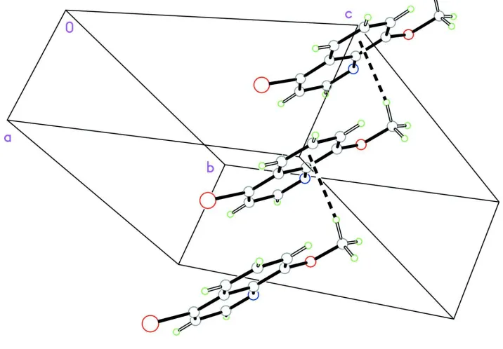
essentially coplanar. In the crystal structure, molecules are linked by weak intermolecular C—H (arene) interactions, forming one-dimensional chains along theaaxis.
Related literature
For related literature, see: Michael (2008); Kulkarni et al. (2006); Irving & Pinnington (1957).
Experimental
Crystal data
C10H8BrNO Mr= 238.08
Orthorhombic,P212121
a= 5.1615 (1) A˚
b= 12.1337 (6) A˚
c= 14.2436 (7) A˚
V= 892.05 (6) A˚3 Z= 4
MoKradiation = 4.56 mm 1
T= 150 (1) K 0.300.120.11 mm
Data collection
Nonius KappaCCD diffractometer Absorption correction: multi-scan
(SORTAV; Blessing 1995)
Tmin= 0.545,Tmax= 0.607
6134 measured reflections 2026 independent reflections 1872 reflections withI> 2(I)
Rint= 0.035
Refinement
R[F2> 2(F2)] = 0.028 wR(F2) = 0.059 S= 1.01 2026 reflections 120 parameters
H-atom parameters constrained
max= 0.38 e A˚ 3 min= 0.40 e A˚ 3
Absolute structure: Flack (1983), 815 Friedel pairs
Flack parameter: 0.017 (11)
Table 1
Hydrogen-bond geometry (A˚ ,).
D—H A D—H H A D A D—H A
C10—H10A Cgi
0.98 2.66 3.531 (3) 148
Symmetry code: (i)x 1;y;z.Cgis the centroid of the C4–C9 ring.
Data collection: COLLECT (Nonius, 2002); cell refinement:
DENZO-SMN (Otwinowski & Minor, 1997); data reduction:
DENZO-SMN; program(s) used to solve structure:SIR92(Altomare
et al., 1994); program(s) used to refine structure:SHELXTL (Shel-drick, 2008); molecular graphics: PLATON (Spek, 2003) and
SHELXTL; software used to prepare material for publication:
SHELXTL.
We thank Dr Peter P. Antich and Dr Frederick J. Bonte for helpful discussions and support. Financial support for this work was provided by the Natural Sciences and Engineering Research Council of Canada (NSERC).
Supplementary data and figures for this paper are available from the IUCr electronic archives (Reference: PV2082).
References
Altomare, A., Cascarano, G., Giacovazzo, C., Guagliardi, A., Burla, M. C., Polidori, G. & Camalli, M. (1994).J. Appl. Cryst.27, 435.
Blessing, R. H. (1995).Acta Cryst.A51, 33–38. Flack, H. D. (1983).Acta Cryst.A39, 876–881.
Irving, H. & Pinnington, A. R. (1957).J. Chem. Soc.pp. 285–290.
Kulkarni, P., Arora, V., Bennett, M., Roney, C., Partridge, K., Lewis, M., Antich, P. & Bonte, F. (2006).J. Nucl. Med.47, 509P–510P.
Michael, J. P. (2008).Nat. Prod. Rep.25, 166–187.
Nonius (2002).COLLECT. Nonius BV, Delft, The Netherlands.
Otwinowski, Z. & Minor, W. (1997). Methods in Enzymology, Vol. 276,
Macromolecular Crystallography, Part A edited by C. W. Carter Jr & R. M. Sweet pp. 307–326. London: Academic press.
Sheldrick, G. M. (2008).Acta Cryst.A64, 112–122. Spek, A. L. (2003).J. Appl. Cryst.36, 7–13. Acta Crystallographica Section E
Structure Reports Online
supporting information
Acta Cryst. (2008). E64, o1117 [doi:10.1107/S1600536808014591]
4-Bromo-8-methoxyquinoline
Neil Vasdev, Padmakar V. Kulkarni, Alan A. Wilson, Sylvain Houle and Alan J. Lough
S1. Comment
Quinoline derivatatives are established chelating agents and also have applications as precursors for pesticides and
pharmaceuticals (Michael, 2008). Our laboratories are pursuing the development of radiohalogenated 8-hydroxyquinoline
derivatives for positron emission tomography (PET) and single photon emission computed tomography (SPECT),
specifically to image extracellular glial deposition of amyloid plaque protein in Alzheimer's disease and matrix
metalloproteinases in tumours (Kulkarni et al., 2006). 4-Bromo-8-methoxyquinoline, first reported by Irving &
Pinnington (1957) may be used as a precursor for radiohalogenation reactions to prepare labelled
8-hydroxyquinoline-based PET or SPECT radiopharmaceuticals. To our surprise, neutral compounds bearing a 4-halogen substituted,
8-phen-oxyquinoline core have not yet been studied by single-crystal X-ray crystallography. In the present study we report the
crystal structure of the title compound at 150 K.
The non-hydrogen atoms of title molecule (Fig. 1), C10H8BrNO, are essentially co-planar (r.m.s. deviation of all non-H
atoms = 0.0242 Å). In the crystal structure, molecules are linked by weak intermolecuar C—H···π(arene) interactions to
form one-dimensional chains along the a axis (Fig. 2). There are no other hydrogen bonds or π···π stacking interactions.
S2. Experimental
X-ray quality crystals were obtained by evaporation of a solution of the title compound (ECA International Corporation,
Palatine, Illinois, USA) in chloroform.
S3. Refinement
H atoms were placed in calculated positions with C—H = 0.95Å (aryl) and 0.98Å (methyl) and were included in the
Figure 1
Molecular structure showing 30% probability displacement ellipsoids (arbitrary spheres for H atoms).
Figure 2
[image:3.610.126.484.379.628.2]4-Bromo-8-methoxyquinoline
Crystal data
C10H8BrNO Mr = 238.08
Orthorhombic, P212121 Hall symbol: P 2ac 2ab a = 5.1615 (1) Å b = 12.1337 (6) Å c = 14.2436 (7) Å V = 892.05 (6) Å3 Z = 4
F(000) = 472 Dx = 1.773 Mg m−3
Mo Kα radiation, λ = 0.71073 Å Cell parameters from 6134 reflections θ = 2.9–27.5°
µ = 4.56 mm−1 T = 150 K Needle, colourless 0.30 × 0.12 × 0.11 mm
Data collection
Nonius KappaCCD diffractometer
Radiation source: fine-focus sealed tube Graphite monochromator
Detector resolution: 9 pixels mm-1 φ scans and ω scans with κ offsets Absorption correction: multi-scan
(SORTAV; Blessing 1995) Tmin = 0.545, Tmax = 0.607
6134 measured reflections 2026 independent reflections 1872 reflections with I > 2σ(I) Rint = 0.036
θmax = 27.5°, θmin = 2.9° h = −5→6
k = −14→15 l = −18→18
Refinement
Refinement on F2 Least-squares matrix: full R[F2 > 2σ(F2)] = 0.028 wR(F2) = 0.059 S = 1.01 2026 reflections 120 parameters 0 restraints
Primary atom site location: structure-invariant direct methods
Secondary atom site location: difference Fourier map
Hydrogen site location: inferred from neighbouring sites
H-atom parameters constrained w = 1/[σ2(F
o2) + (0.0306P)2 + 0.0333P] where P = (Fo2 + 2Fc2)/3
(Δ/σ)max = 0.001 Δρmax = 0.38 e Å−3 Δρmin = −0.40 e Å−3
Extinction correction: SHELXTL (Sheldrick, 2008), Fc*=kFc[1+0.001xFc2λ3/sin(2θ)]-1/4 Extinction coefficient: 0.0062 (8)
Absolute structure: Flack (1983), 815 Friedel pairs
Absolute structure parameter: −0.017 (11)
Special details
Geometry. All e.s.d.'s (except the e.s.d. in the dihedral angle between two l.s. planes) are estimated using the full
covariance matrix. The cell e.s.d.'s are taken into account individually in the estimation of e.s.d.'s in distances, angles and torsion angles; correlations between e.s.d.'s in cell parameters are only used when they are defined by crystal symmetry. An approximate (isotropic) treatment of cell e.s.d.'s is used for estimating e.s.d.'s involving l.s. planes.
Refinement. Refinement of F2 against ALL reflections. The weighted R-factor wR and goodness of fit S are based on F2,
conventional R-factors R are based on F, with F set to zero for negative F2. The threshold expression of F2 > σ(F2) is used only for calculating R-factors(gt) etc. and is not relevant to the choice of reflections for refinement. R-factors based on F2 are statistically about twice as large as those based on F, and R- factors based on ALL data will be even larger.
Fractional atomic coordinates and isotropic or equivalent isotropic displacement parameters (Å2)
x y z Uiso*/Ueq
O1 0.1007 (3) 0.62674 (16) 0.72708 (14) 0.0259 (4)
N1 0.4260 (4) 0.64140 (17) 0.58326 (14) 0.0233 (4)
C1 0.5884 (5) 0.6481 (2) 0.51287 (18) 0.0278 (6)
H1A 0.5819 0.7126 0.4752 0.033*
C2 0.7717 (5) 0.5671 (2) 0.48878 (19) 0.0268 (6)
H2A 0.8842 0.5768 0.4366 0.032*
C3 0.7833 (5) 0.4746 (2) 0.54217 (19) 0.0239 (6)
C4 0.6190 (5) 0.4611 (2) 0.62176 (17) 0.0188 (5)
C5 0.6239 (5) 0.3694 (2) 0.68244 (18) 0.0232 (6)
H5A 0.7429 0.3109 0.6718 0.028*
C6 0.4556 (5) 0.3650 (2) 0.75705 (17) 0.0243 (6)
H6A 0.4624 0.3038 0.7986 0.029*
C7 0.2739 (5) 0.4487 (2) 0.77325 (18) 0.0235 (6)
H7A 0.1558 0.4424 0.8241 0.028*
C8 0.2656 (5) 0.5400 (2) 0.71586 (17) 0.0195 (5)
C9 0.4408 (5) 0.54916 (19) 0.63810 (16) 0.0199 (5)
C10 −0.0731 (5) 0.6201 (2) 0.80535 (18) 0.0271 (7)
H10C −0.1784 0.6872 0.8083 0.041*
H10D 0.0269 0.6126 0.8635 0.041*
H10A −0.1865 0.5559 0.7978 0.041*
Atomic displacement parameters (Å2)
U11 U22 U33 U12 U13 U23
Br1 0.02472 (15) 0.03108 (16) 0.03239 (16) 0.00303 (10) 0.00306 (11) −0.00756 (11) O1 0.0264 (10) 0.0255 (11) 0.0259 (10) 0.0030 (8) 0.0047 (7) 0.0004 (8) N1 0.0281 (10) 0.0212 (11) 0.0206 (11) −0.0009 (9) −0.0021 (8) 0.0001 (10) C1 0.0339 (13) 0.0259 (14) 0.0237 (14) −0.0019 (10) 0.0030 (10) 0.0087 (14) C2 0.0265 (12) 0.0299 (14) 0.0240 (14) −0.0065 (10) 0.0049 (12) 0.0001 (13) C3 0.0215 (13) 0.0257 (14) 0.0246 (15) −0.0015 (10) −0.0025 (10) −0.0078 (12) C4 0.0210 (13) 0.0184 (13) 0.0169 (13) −0.0037 (9) −0.0018 (9) −0.0016 (11) C5 0.0224 (12) 0.0215 (14) 0.0257 (14) 0.0016 (11) −0.0064 (10) −0.0019 (12) C6 0.0328 (15) 0.0176 (13) 0.0225 (13) −0.0037 (13) −0.0074 (11) 0.0035 (11) C7 0.0257 (14) 0.0258 (15) 0.0191 (14) −0.0088 (11) 0.0018 (11) −0.0010 (11) C8 0.0208 (13) 0.0185 (13) 0.0193 (13) −0.0003 (10) −0.0021 (10) −0.0014 (11) C9 0.0207 (12) 0.0204 (13) 0.0186 (12) −0.0042 (10) −0.0046 (10) 0.0010 (10) C10 0.0240 (14) 0.0314 (17) 0.0258 (15) 0.0015 (12) 0.0047 (11) −0.0028 (12)
Geometric parameters (Å, º)
Br1—C3 1.895 (2) C4—C9 1.429 (3)
O1—C8 1.362 (3) C5—C6 1.374 (4)
O1—C10 1.433 (3) C5—H5A 0.9500
N1—C1 1.309 (3) C6—C7 1.402 (4)
N1—C9 1.367 (3) C6—H6A 0.9500
C1—C2 1.407 (4) C7—C8 1.378 (4)
C1—H1A 0.9500 C7—H7A 0.9500
C2—H2A 0.9500 C10—H10C 0.9800
C3—C4 1.425 (3) C10—H10D 0.9800
C4—C5 1.409 (4) C10—H10A 0.9800
C8—O1—C10 116.0 (2) C5—C6—H6A 119.3
C1—N1—C9 116.9 (2) C7—C6—H6A 119.3
N1—C1—C2 125.0 (2) C8—C7—C6 120.4 (2)
N1—C1—H1A 117.5 C8—C7—H7A 119.8
C2—C1—H1A 117.5 C6—C7—H7A 119.8
C3—C2—C1 118.1 (2) O1—C8—C7 124.8 (2)
C3—C2—H2A 121.0 O1—C8—C9 115.1 (2)
C1—C2—H2A 121.0 C7—C8—C9 120.0 (2)
C2—C3—C4 121.0 (2) N1—C9—C4 123.7 (2)
C2—C3—Br1 119.4 (2) N1—C9—C8 118.0 (2)
C4—C3—Br1 119.58 (19) C4—C9—C8 118.3 (2)
C5—C4—C3 124.6 (2) O1—C10—H10C 109.5
C5—C4—C9 120.1 (2) O1—C10—H10D 109.5
C3—C4—C9 115.3 (2) H10C—C10—H10D 109.5
C6—C5—C4 119.6 (3) O1—C10—H10A 109.5
C6—C5—H5A 120.2 H10C—C10—H10A 109.5
C4—C5—H5A 120.2 H10D—C10—H10A 109.5
C5—C6—C7 121.5 (2)
Hydrogen-bond geometry (Å, º)
D—H···A D—H H···A D···A D—H···A
C10—H10A···Cgi 0.98 2.66 3.531 (3) 148