Rietveld refinement of Sr
5(AsO
4)
3Cl from
high-resolution synchrotron data
Anthony M. T. Bell,a* C. Michael B. Henderson,b Richard F. Wendlandtcand Wendy J. Harrisonc
aSynchrotron Radiation Source, STFC Daresbury Laboratory, Daresbury, Warrington, Cheshire WA4 4AD, England,bSchool of Earth, Atmospheric and Environmental Sciences, University of Manchester, Manchester, M13 9PL, England, and cDepartment of Geology and Geological Engineering, Colorado School of Mines, Golden, CO 80401, USA
Correspondence e-mail: [email protected]
Received 6 February 2009; accepted 11 February 2009
Key indicators: powder synchrotron study;T= 298 K; mean(As–O) = 0.020 A˚; Rfactor = 0.052;wRfactor = 0.066; data-to-parameter ratio = 14.1.
The apatite-type compound, pentastrontium tris[arsenate(V)] chloride, Sr5(AsO4)3Cl, has been synthesized by ion exchange at high temperature from a synthetic sample of mimetite [Pb5(AsO4)3Cl] with SrCO3as a by-product. The results of the Rietveld refinement, based on high resolution synchrotron X-ray powder diffraction data, show that the title compound crystallizes in the same structure as other halogenoapatites with general formula A5(YO4)3X (A = divalent cation, Y= pentavalent cation, andX= F, Cl or Br) in the space group
P63/m. The structure consists of isolated tetrahedral AsO43
anions (the As atom and two O atoms have msymmetry),
separated by two crystallographically independent Sr2+ cations that are located on mirror planes and threefold rotation axes, respectively. One Sr atom is coordinated by nine O atoms and the other by six. The chloride anions (site symmetry 3) are at the 2asites and are located in the channels of the structure.
Related literature
For crystal chemistry of apatites, see: Mercier et al. (2005); White & ZhiLi (2003); Wuet al.(2003). For powder diffraction data on Sr As-apatite, see: Kreidler & Hummel (1970). Atomic coordinates as starting parameters for the Rietveld (Rietveld, 1969) refinement of the present phases were taken from Bellet al.(2008); Daiet al.(1991); de Villierset al.(1971). For related Sr—Cl-apatites, see: Ðordevic´ et al. (2008); Sudarsanan & Young, (1974, 1980); Becket al.(2006); Noet-zold et al. (1995); Noetzold & Wulff (1996, 1997, 1998); Swafford & Holt (2002); Wardojo & Hwu (1996). For synthetic work, see: Baker (1966); Essington (1988); Harrison et al.
(2002).
Experimental
Crystal data
Sr5(AsO4)3Cl
Mr= 890.31
Hexagonal,P63=m
a= 10.1969 (1) A˚
c= 7.28108 (9) A˚
V= 655.63 (2) A˚3
Z= 2
Synchrotron radiation
= 0.998043 A˚
T= 298 K
Specimen shape: cylinder 400.70.7 mm
Specimen prepared at 100 kPa Specimen prepared at 1258 K Particle morphology: powder, white
Data collection
In-house design diffractometer Specimen mounting: capillary Specimen mounted in transmission
mode
Scan method: step
Absorption correction: fixed at 0 2min= 2, 2max= 60
Increment in 2= 0.01 Refinement
Rp= 0.052
Rwp= 0.066
Rexp= 0.047
RB= 0.090
S= 2.00
Excluded region(s): 2-62
Profile function: Pseudo Voigt 225 reflections
16 parameters
Preferred orientation correction: none
Table 1
Selected geometric parameters (A˚ ,).
Sr1—O1 2.49 (2) Sr1—O2i 2.59 (2) Sr1—O3i 3.01 (1) Sr2—O2ii 2.53 (2) Sr2—O3iii 2.44 (1) Sr2—O3iv 2.94 (1) Sr2—O1v 3.02 (2) Sr2—Cl1iv 3.156 (3) As1—O3 1.57 (1) As1—O1 1.72 (2) As1—O2 1.70 (2)
O3—As1—O3vi
121 (1) O3—As1—O1 105.8 (7)
O3—As1—O2 106.3 (6) O1—As1—O2 112 (1)
Symmetry codes: (i)xy;x;z; (ii)yþ1;xyþ1;z; (iii)y;xþyþ1;z; (iv)
x;yþ1;z; (v)xþy;xþ1;z; (vi)x;y;zþ1 2.
Data collection: local software; cell refinement:CELREF(Laugier
& Bochu, 2003) and GSAS (Larson & Von Dreele (2004); data
reduction: local software; method used to solve structure: coordinates taken from a related compound; program(s) used to refine structure:
GSAS and EXPGUI (Toby, 2001); molecular graphics: VESTA
(Momma & Izumi, 2008); software used to prepare material for publication:publCIF(Westrip, 2009).
AMTB acknowledges the use of the EPSRC’s Chemical Database Service at Daresbury (Fletcheret al., 1996).
Supplementary data and figures for this paper are available from the IUCr electronic archives (Reference: BR2096).
References
Baker, W. E. (1966).Am. Mineral.51, 1712–1721.
Beck, H. P., Douiheche, M., Haberkorn, R. & Kohlmann, H. (2006).Solid State Sci.8, 64–70.
Bell, A. M. T., Henderson, C. M. B., Wendlandt, R. F. & Harrison, W. J. (2008).
Acta Cryst.E64, i63–i64.
Dai, Y.-S., Hughes, J. M. & Moore, P. B. (1991).Can. Mineral.29, 369–376. Ðordevic´, T., Sˇutovic´, S., Stojanovic´, J. & Karanovic´, Lj. (2008).Acta Cryst.
C64, i82–i86.
Essington, M. E. (1988).Soil Sci. Soc. Am. J.52, 1566–1570.
Fletcher, D. A., McMeeking, R. F. & Parkin, D. J. (1996).Chem. Inf. Comput. Sci.36, 746–749.
inorganic compounds
i16
Bellet al. doi:10.1107/S1600536809005054 Acta Cryst.(2009). E65, i16–i17Acta Crystallographica Section E Structure Reports
Online
Mineralogical Association 18th General Meeting, Sept 1–6, 2002, Edin-burgh, Scotland. Abstract A18–10, meeting program with abstracts, page 185.
Kreidler, E. R. & Hummel, F. A. (1970).Am. Mineral.55, 170–184. Larson, A. C. & Von Dreele, R. B. (2004). General Structure Analysis System
(GSAS). Report LAUR 86-748. Los Alamos National Laboratory, New Mexico, USA.
Laugier, J. & Bochu, B. (2003).CELREF. http://www.CCP14.ac.uk/tutorial/ lmgp/CELREF.htm.
Mercier, P. H. J., Le Page, Y., Whitfield, P. S., Mitchell, L. D., Davidson, I. J. & White, T. J. (2005).Acta Cryst.B61, 635–655.
Momma, K. & Izumi, F. (2008).J. Appl. Cryst.41, 653–658.
Noetzold, D. & Wulff, H. (1996).Phys. Status Solidi Sect. A,158303–311.
Noetzold, D. & Wulff, H. (1998).Phys. Status Solidi Sect. B,207271–281. Noetzold, D., Wulff, H. & Herzog, G. (1995).Phys. Status Solidi Sect. B,191
21–30.
Rietveld, H. M. (1969).J. Appl. Cryst.2, 65–71.
Sudarsanan, K. & Young, R. A. (1974).Acta Cryst.B30, 1381–1386. Sudarsanan, K. & Young, R. A. (1980).Acta Cryst.B36, 1525–1530. Swafford, S. H. & Holt, E. M. (2002).Solid State Sci.4, 807–812. Toby, B. H. (2001).J. Appl. Cryst.34, 210–213.
Villiers, J. P. R. de (1971).Am. Mineral.56, 758–766.
Wardojo, T. A. & Hwu, S.-J. (1996).Acta Cryst.C52, 2959–2960. Westrip, S. P. (2009).publCIF. In preparation.
White, T. J. & ZhiLi, D. (2003).Acta Cryst.B59, 1–16.
supporting information
sup-1
Acta Cryst. (2009). E65, i16–i17supporting information
Acta Cryst. (2009). E65, i16–i17 [doi:10.1107/S1600536809005054]
Rietveld refinement of Sr
5(AsO
4)
3Cl from high-resolution synchrotron data
Anthony M. T. Bell, C. Michael B. Henderson, Richard F. Wendlandt and Wendy J. Harrison
S1. Comment
Apatites are minerals and synthetic compounds with general formula A5(YO4)3X, containing tetrahedrally coordinated
YO43- anions (Y = pentavalent cation) and a monovalent anion X such as F-, Cl- or OH-. The divalent cations frequently belong to the alkaline earth group, but other cations like Pb2+ are also known. For a review of the structures and
crystal-chemistry of these materials, see Mercier et al. (2005), White & ZhiLi (2003) and Wu et al., (2003). Apatites containing
arsenic (As-apatites) are of interest as hosts for storage of arsenic removed from contaminated water (Harrison et al.,
2002). Powder diffraction data for the Sr containing As-apatite Sr5(AsO4)3Cl (Kreidler & Hummel, 1970) was indexed in
space group P63/m. Related crystal structures have also been reported for Ca5(AsO4)3Cl (Wardojo and Hwu, 1996) and for
Sr5(AsO4)3F and (Sr1.66Ba0.34)(Ba2.61Sr0.39)(AsO4)3Cl (Đordević et al., 2008). The crystal structure of Sr5(AsO4)3Cl
in space group P63/m is reported in the present communication. We recently reported the related crystal structure of
Ba5(AsO4)3Cl (Bell et al., 2008).
Table 1 shows refined interatomic distances and angles for the Sr5(AsO4)3Cl structure. The averaged Sr1—O and Sr2—
O distances of respectively 2.70 Å and 2.72 Å, compare with Sr1—O and Sr2—O distances in: Sr5(AsO4)3F
(Đordević et al. 2008) of 2.71 Å and 2.62 Å; 2.71 Å and 2.63 Å for Sr5(VO4)3Cl (Beck et al., 2006); 2.67 Å and
2.62 Å for Sr5(PO4)3Cl (Sudarsanan and Young, 1974); and 2.67 Å and 2.59 Å for Sr5(PO4)3F (Swafford and Holt, 2002).
The As—O distances are characteristic for tetrahedral AsO4 units. The O—As—O angles deviate significantly from the
ideal tetrahedral angle of 109.5°, indicating a strong distortion.
The refined lattice parameters for Sr5(AsO4)3Cl are similar to the previously published parameters of a = 10.18 Å, c =
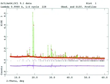
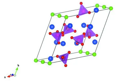
7.28 Å given by Kreidler & Hummel (1970). Fig. 1 shows the Rietveld difference plot for the present refinement. The
crystal structure of Sr5(AsO4)3Cl, showing the isolated tetrahedral AsO43- anions separated by Sr2+ cations and Cl- anions,
is displayed in Fig. 2.
S2. Experimental
This work was part of an attempt to synthesize analogues of Pb5(AsO4)3Cl (mimetite) with Pb2+ substituted by alkaline
earth cations. All starting materials were well crystallized solids. Pb5(AsO4)3Cl was precipitated by titration of 0.1M
Na2HAsO4 into a well stirred, saturated PbCl2 solution at room temperature (procedure modified from methods of Baker
(1966) and Essington (1988)). The molar ratio of Pb:As was slightly greater than 5:3, allowing for excess PbCl2 during
the precipitation. A very fine-grained pure solid formed immediately, which was then separated, washed, and dried.
Typically, five de-ionized water washes were needed to reduce the conductivity of the wash water to < 50 µS.cm-1.
Sr5(AsO4)3Cl was successfully synthesized by ion exchange of Pb5(AsO4)3Cl with molten SrCl2 at 1258 K (modified from
the method given by Kreidler & Hummel (1970)). Two fusions were required to completely eliminate formation of Pb
containing solid solutions and to yield the Pb free title compound. Excess metal in the form of SrCl2 was removed from
S3. Refinement
The main Bragg reflections of the high resolution synchrotron X-ray powder diffraction pattern could be indexed in space
group P63/m with similar lattice parameters to those of the published powder diffraction data (Kreidler & Hummel,
1970). Some broad and weak Bragg reflections were matched by the pattern of SrCO3 in space group Pmcn.
Initial lattice parameters for the two phases were refined using CELREF (Laugier & Bochu, 2003). The P63/m crystal
structure of Ba5(AsO4)3Cl (Bell et al., 2008) was used as a starting model for the Rietveld (Rietveld, 1969) refinement of
the structure of Sr5(AsO4)3Cl. The crystal structure of strontianite (de Villiers et al., 1971) was used as a starting model
for refinement of the structure of SrCO3. Isotropic atomic displacement parameters were used for both phases. For the
Sr5(AsO4)3Cl phase soft constraints were used for the As—O distances in the AsO4 tetrahedral units. These distances were
restrained to those for mimetite (Dai et al., 1991). For the SrCO3 phase only the coordinates and the atomic displacement
parameters for Sr were refined, the C and O coordinates were fixed to those in the starting model and the C and O atomic
displacement parameters were fixed at zero. Proportions of the two phases were refined as 76.6 (1) wt.% Sr5(AsO4)3Cl
[image:4.610.127.486.295.568.2]and 23.4 (1) wt.% SrCO3.
Figure 1
Rietveld difference plot for the multi-phase refinement of Sr5(AsO4)3Cl and SrCO3. The red crosses, and green and pink
lines show respectively the observed, calculated and difference plots. Calculated Bragg reflection positions are indicated
supporting information
[image:5.610.118.490.74.337.2]sup-3
Acta Cryst. (2009). E65, i16–i17Figure 2
The crystal structure of Sr5(AsO4)3Cl. Pink tetrahedra show AsO4 units with As5+ cations as orange spheres and O2- anions
as red spheres. Large blue spheres represent Sr2+ cations and small green spheres Cl- anions.
pentastrontium tris[arsenate(V)] chloride
Crystal data
Sr5(AsO4)3Cl
Mr = 890.31
Hexagonal, P63/m
a = 10.1969 (1) Å
c = 7.28108 (9) Å
V = 655.63 (2) Å3
Z = 2
Dx = 4.510 (1) Mg m−3
Synchrotron radiation, λ = 0.998043 Å
T = 298 K
Particle morphology: powder white
cylinder, 40 × 0.7 mm
Specimen preparation: Prepared at 1258 K and 100 kPa
Data collection
In-house design diffractometer
Radiation source: Synchrotron
Si(111) channel-cut crystal monochromator
Specimen mounting: capillary Data collection mode: transmission Scan method: step
2θmin = 6°, 2θmax = 60°, 2θstep = 0.01°
Refinement Rp = 0.052
Rwp = 0.066
Rexp = 0.047
RBragg = 0.090
R(F2) = 0.33148
χ2 = 3.992 5801 data points
Excluded region(s): 2-6° 2θ
Profile function: Pseudo Voigt 16 parameters
0 restraints 4 constraints (Δ/σ)max = 0.001
Experimental. Absorption correction fixed at zero, all attempts to refine this term in GSAS were unsuccessful so this term was fixed at zero. CELREF was used for initial lattice parameter determinations before Rietveld refinement. Lattice parameters from GSAS refinement are quoted in the paper.
Fractional atomic coordinates and isotropic or equivalent isotropic displacement parameters (Å2)
x y z Uiso*/Ueq
Sr1 0.33333 0.66667 0.008 (1) 0.0246 (9)
Sr2 0.2496 (5) 0.9936 (6) 0.25 0.0246 (9)
As1 0.4057 (5) 0.3718 (5) 0.25 0.029 (2)
O1 0.337 (3) 0.496 (2) 0.25 0.015 (4)
O2 0.598 (2) 0.464 (2) 0.25 0.015 (4)
O3 0.354 (2) 0.284 (2) 0.063 (2) 0.015 (4)
Cl1 0.0000 0.0000 0.0000 0.031 (5)
Geometric parameters (Å, º)
Sr1—O1i 2.49 (2) Sr2—O3vi 2.44 (1)
Sr1—O1ii 2.49 (2) Sr2—O3vii 2.94 (1)
Sr1—O1 2.49 (2) Sr2—O3viii 2.94 (1)
Sr1—O2iii 2.59 (2) Sr2—O1ii 3.02 (2)
Sr1—O2iv 2.59 (2) Sr2—Cl1viii 3.156 (3)
Sr1—O2v 2.59 (2) Sr2—Cl1ix 3.156 (3)
Sr1—O3iv 3.01 (1) As1—O3 1.57 (1)
Sr1—O3iii 3.01 (1) As1—O3x 1.57 (1)
Sr1—O3v 3.01 (1) As1—O1 1.72 (2)
Sr2—O2i 2.53 (2) As1—O2 1.70 (2)
Sr2—O3iv 2.44 (1)
O3—As1—O3x 121 (1) O3—As1—O2 106.3 (6)
O3—As1—O1 105.8 (7) O3x—As1—O2 106.3 (6)
O3x—As1—O1 105.8 (7) O1—As1—O2 112 (1)