2,2
000,5,5
000-Tetrachlorobenzidine
Onome Ugono, Marcel Douglas Jr, Nigam P. Rath and Alicia M. Beatty*
Department of Chemistry and Biochemistry, University of Missouri–St Louis, St Louis, Missouri, USA
Correspondence e-mail: [email protected]
Received 17 May 2010; accepted 2 August 2010
Key indicators: single-crystal X-ray study;T= 100 K; mean(C–C) = 0.001 A˚;
Rfactor = 0.025;wRfactor = 0.075; data-to-parameter ratio = 36.1.
In the crystal structure of the title compound, C12H8Cl4N2, molecules lie on crystallographic twofold axes at the centre of the C—C bonds linking the benzene rings, such that the asymmetric unit consists of a half-molecule. The individual molecules participate in intermolecular N—H N, N— H Cl, C—H Cl and Cl Cl [3.4503 (3) A˚ ] interactions.
Related literature
For studies involving the use of benzidines in organic synth-eses, see: Schwenecke & Mayer (2005). For studies on 2,20,5,50 -tetrachlorobenzidine in crystal engineering, see: Dobrzycki & Wozniak (2007, 2008). For our studies on related structures, see: Beattyet al.(2002a, 2002b); Ugonoet al.(2009).
Experimental
Crystal data
C12H8Cl4N2
Mr= 322.00 Monoclinic,I2=a a= 17.2346 (11) A˚
b= 3.8767 (2) A˚
c= 18.1573 (19) A˚ = 94.872 (3)
V= 1208.77 (16) A˚3
Z= 4
MoKradiation = 0.96 mm1
T= 100 K
0.230.220.14 mm
Data collection
Bruker APEXII CCD diffractometer
Absorption correction: multi-scan (SADABS; Sheldrick, 1996)
Tmin= 0.806,Tmax= 0.879
11878 measured reflections 2961 independent reflections 2595 reflections withI> 2(I)
Rint= 0.025
Refinement
R[F2> 2(F2)] = 0.025
wR(F2) = 0.075
S= 1.07 2961 reflections
82 parameters
H-atom parameters constrained
max= 0.62 e A˚ 3
min=0.67 e A˚ 3
Table 1
Hydrogen-bond geometry (A˚ ,).
D—H A D—H H A D A D—H A
N1—H1A Cl2i 0.88 2.79 3.3650 (8) 124
C3—H3 Cl1ii
0.95 2.71 3.5013 (9) 141
N1—H1B N1i
0.88 2.90 3.2159 (12) 103
Symmetry codes: (i)xþ1;yþ1 2;zþ
1 2; (ii)xþ
1 2;y1;z.
Data collection:APEX2(Bruker, 2007); cell refinement:SAINT
(Bruker, 2007); data reduction:SAINT; program(s) used to solve structure:SHELXS97(Sheldrick, 2008); program(s) used to refine structure: SHELXL97 (Sheldrick, 2008); molecular graphics:
SHELXTL(Sheldrick, 2008); software used to prepare material for publication:SHELXTLandPLATON(Spek, 2009).
The authors are grateful to the University of Missouri–St. Louis for generous support and instrumentation. MD is grateful to the ACS Project SEED for a summer research fellowship. AMB acknowledges support from the NSF-CAREER # 0645758.
Supplementary data and figures for this paper are available from the IUCr electronic archives (Reference: FJ2305).
References
Beatty, A. M., Grange, K. E. & Simpson, A. E. (2002a).Chem. Eur. J.8, 3254– 3259.
Beatty, A. M., Schneider, C. L., Simpson, A. E. & Zaher, J. L. (2002b).
CrystEngComm,4, 282–287.
Bruker (2007).APEX2andSAINT. Bruker AXS Inc., Madison, Wisconsin, USA.
Dobrzycki, L. & Wozniak, K. (2007).CrystEngComm,9, 1029–1041. Dobrzycki, L. & Wozniak, K. (2008).CrystEngComm,9, 525–533.
Schwenecke, H. & Mayer, D. (2005).Benzidine and Benzidine Derivatives.In
Ullmann’s Encyclopedia of Industrial Chemistry. Weinheim: Wiley–VCH. Sheldrick, G. M. (1996).SADABS. University of Go¨ttingen, Germany. Sheldrick, G. M. (2008).Acta Cryst.A64, 112–122.
Spek, A. L. (2009).Acta Cryst.D65, 148–155.
Ugono, O., Rath, N. P. & Beatty, A. M. (2009).Cryst. Growth Des.9, 4595– 4598.
Acta Crystallographica Section E Structure Reports Online
supporting information
Acta Cryst. (2010). E66, o2285 [https://doi.org/10.1107/S1600536810030886]
2,2
′
,5,5
′
-Tetrachlorobenzidine
Onome Ugono, Marcel Douglas Jr, Nigam P. Rath and Alicia M. Beatty
S1. Comment
Benzidines are of great importance in the pigments industry, as their derivatives are employed in the syntheses of a
variety of azo-dyes (Schwenecke and Mayer 2005). Furthermore, the presence of two amine functionalities renders this
class of molecules attractive to crystal engineers, who may wish to incorporate this class of rigid linear molecule in larger
extended networks via hydrogen bonds (Dobrzycki and Wozniak 2008 a, b). During the course of experiments aimed at
reacting the title compound with pyrazole-3,5-dicarboxylic acid, a crystalline phase was obtained and shown to be
composed solely of 2,2′,5,5′-tetrachlorobenzidine. A search of the Cambridge crystallographic structural database showed
that this phase has not previously been reported. This molecule packs in monoclinic I2/a, a non-standard setting of C2/c,
with one half of the molecule in the asymmetric unit. Very weak hydrogen bonding interactions exist in the structure; the
increased lengths are probably due to the bulky chlorine atoms ortho to the amine functionalities.
S2. Experimental
A 20 ml scintillation vial was charged with 52.0 mg (0.30 mmol) of 3,5-pyrazole dicarboxylic acid monohydrate, which
was dissolved in 5.0 ml of a 3:2 MeOH:H2O mixture affording a homogenous solution. To this solution was then added
48.3 mg (0.15 mmol) of 2,2′,5,5′-tetrachlorobenzidine. The mixture obtained was filtered and the filtrate was allowed to
slowly evaporate to yield colorless single crystals of 2,2′,5,5′-tetrachlorobenzidine after a week.
S3. Refinement
All non hydrogen atoms were refined anisotropically. Phenyl hydrogen atoms were placed in calculated positions and
treated with a riding model C–H= 0.95 Å, Uiso(Haryl)= 1.2Ueq(C) for aromatic carbons. The amine hydrogen atoms were
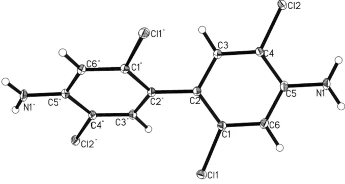
Figure 1
Molecular structure showing 50% probability displacement ellipsoid.
2,2′,5,5′-Tetrachlorobenzidine
Crystal data
C12H8Cl4N2
Mr = 322.00 Monoclinic, I2/a Hall symbol: -I 2ya a = 17.2346 (11) Å b = 3.8767 (2) Å c = 18.1573 (19) Å β = 94.872 (3)° V = 1208.77 (16) Å3
Z = 4
F(000) = 648 Dx = 1.769 Mg m−3 Melting point = 309–311 K Mo Kα radiation, λ = 0.71073 Å Cell parameters from 5249 reflections θ = 4.5–36.4°
µ = 0.96 mm−1
T = 100 K Blocks, colorless 0.23 × 0.22 × 0.14 mm
Data collection
Bruker APEXII CCD diffractometer
Radiation source: fine-focus sealed tube Graphite monochromator
φ and ω scans
Absorption correction: multi-scan (SADABS; Sheldrick, 1996) Tmin = 0.806, Tmax = 0.879
11878 measured reflections 2961 independent reflections 2595 reflections with I > 2σ(I) Rint = 0.025
θmax = 36.4°, θmin = 3.1°
h = −28→28 k = −3→6 l = −30→26
Refinement
Refinement on F2 Least-squares matrix: full R[F2 > 2σ(F2)] = 0.025
wR(F2) = 0.075
S = 1.07 2961 reflections 82 parameters
0 restraints
H-atom parameters constrained w = 1/[σ2(F
o2) + (0.0404P)2 + 0.5838P] where P = (Fo2 + 2Fc2)/3
Special details
Experimental. All H atoms were added in their calculated positions and were treated using appropriate riding models.
Geometry. All e.s.d.'s (except the e.s.d. in the dihedral angle between two l.s. planes) are estimated using the full covariance matrix. The cell e.s.d.'s are taken into account individually in the estimation of e.s.d.'s in distances, angles and torsion angles; correlations between e.s.d.'s in cell parameters are only used when they are defined by crystal symmetry. An approximate (isotropic) treatment of cell e.s.d.'s is used for estimating e.s.d.'s involving l.s. planes.
Refinement. Refinement of F2 against ALL reflections. The weighted R-factor wR and goodness of fit S are based on F2, conventional R-factors R are based on F, with F set to zero for negative F2. The threshold expression of F2 > σ(F2) is used only for calculating R-factors(gt) etc. and is not relevant to the choice of reflections for refinement. R-factors based on F2 are statistically about twice as large as those based on F, and R- factors based on ALL data will be even larger.
Fractional atomic coordinates and isotropic or equivalent isotropic displacement parameters (Å2)
x y z Uiso*/Ueq
Cl1 0.160889 (12) 0.57866 (5) 0.112011 (11) 0.01179 (5)
Cl2 0.490222 (11) −0.04833 (5) 0.098542 (11) 0.01279 (6)
N1 0.42631 (4) 0.2547 (2) 0.23242 (4) 0.01399 (13)
H1A 0.4109 0.3405 0.2736 0.017*
H1B 0.4733 0.1666 0.2320 0.017*
C1 0.25315 (5) 0.4094 (2) 0.10312 (5) 0.00977 (13)
C2 0.27558 (5) 0.2934 (2) 0.03511 (4) 0.00940 (12)
C3 0.35064 (5) 0.1556 (2) 0.03688 (4) 0.01012 (13)
H3 0.3688 0.0715 −0.0077 0.012*
C4 0.39925 (5) 0.1373 (2) 0.10113 (5) 0.00990 (13)
C5 0.37670 (5) 0.2584 (2) 0.16844 (4) 0.01036 (13)
C6 0.30195 (5) 0.3964 (2) 0.16774 (5) 0.01065 (13)
H6 0.2842 0.4830 0.2123 0.013*
Atomic displacement parameters (Å2)
U11 U22 U33 U12 U13 U23
Cl1 0.00978 (9) 0.01507 (9) 0.01067 (9) 0.00235 (6) 0.00183 (6) 0.00083 (6)
Cl2 0.00832 (9) 0.01894 (10) 0.01088 (10) 0.00207 (6) −0.00065 (6) 0.00105 (6)
N1 0.0119 (3) 0.0218 (3) 0.0076 (3) 0.0000 (3) −0.0027 (2) −0.0006 (2)
C1 0.0089 (3) 0.0118 (3) 0.0086 (3) 0.0002 (2) 0.0006 (2) 0.0005 (2)
C2 0.0084 (3) 0.0118 (3) 0.0079 (3) −0.0002 (2) 0.0000 (2) 0.0000 (2)
C3 0.0093 (3) 0.0127 (3) 0.0081 (3) 0.0001 (2) −0.0002 (2) −0.0006 (2)
C4 0.0076 (3) 0.0130 (3) 0.0088 (3) 0.0001 (2) −0.0004 (2) 0.0003 (2)
C5 0.0098 (3) 0.0128 (3) 0.0082 (3) −0.0021 (2) −0.0009 (2) 0.0005 (2)
C6 0.0110 (3) 0.0135 (3) 0.0074 (3) −0.0007 (2) 0.0005 (2) −0.0005 (2)
Geometric parameters (Å, º)
Cl1—C1 1.7402 (8) C2—C3 1.3974 (11)
Cl2—C4 1.7295 (8) C2—C2i 1.4872 (16)
N1—C5 1.3827 (11) C3—C4 1.3791 (11)
C1—C2 1.3993 (12) C6—H6 0.9500
C5—N1—H1A 120.0 C2—C3—H3 118.9
C5—N1—H1B 120.0 C3—C4—C5 121.97 (7)
H1A—N1—H1B 120.0 C3—C4—Cl2 119.03 (6)
C6—C1—C2 122.84 (8) C5—C4—Cl2 118.98 (6)
C6—C1—Cl1 115.40 (6) N1—C5—C6 121.10 (8)
C2—C1—Cl1 121.75 (6) N1—C5—C4 122.30 (8)
C3—C2—C1 115.29 (7) C6—C5—C4 116.56 (7)
C3—C2—C2i 120.00 (8) C1—C6—C5 121.05 (8)
C1—C2—C2i 124.63 (8) C1—C6—H6 119.5
C4—C3—C2 122.26 (8) C5—C6—H6 119.5
C4—C3—H3 118.9
C6—C1—C2—C3 1.26 (12) C3—C4—C5—N1 −177.11 (8)
Cl1—C1—C2—C3 −178.33 (6) Cl2—C4—C5—N1 4.10 (11)
C6—C1—C2—C2i 178.20 (6) C3—C4—C5—C6 0.59 (12)
Cl1—C1—C2—C2i −1.39 (9) Cl2—C4—C5—C6 −178.20 (6)
C1—C2—C3—C4 −0.36 (12) C2—C1—C6—C5 −1.26 (12)
C2i—C2—C3—C4 −177.46 (6) Cl1—C1—C6—C5 178.35 (6)
C2—C3—C4—C5 −0.56 (13) N1—C5—C6—C1 178.02 (8)
C2—C3—C4—Cl2 178.23 (6) C4—C5—C6—C1 0.29 (12)
Symmetry code: (i) −x+1/2, y, −z.
Hydrogen-bond geometry (Å, º)
D—H···A D—H H···A D···A D—H···A
N1—H1A···Cl2ii 0.88 2.79 3.3650 (8) 124
C3—H3···Cl1iii 0.95 2.71 3.5013 (9) 141
N1—H1B···N1ii 0.88 2.90 3.2159 (12) 103