The Canine J wave
C.F. Agudelo, P. Schanilec
Faculty of Veterinary Medicine, University of Veterinary and Pharmaceutical Sciences, Brno, Czech Republic
ABSTRACT: The J wave is a deflection immediately following the QRS complex of the surface ECG. The J wave has been observed in humans under physiological and pathophysiological conditions. We describe in this paper the ratio of incidence of this phenomenon in healthy dogs and dogs with pathological disease and the effect of exercise on its size and shape. At rest, a J wave was observed at the R-ST junction of the ECG in 11 of 34 adult dogs, usually in leads I, II, III, aVR, and aVF and left lateral precordial leads. After a submaximal exercise test there were no variations in the shape or the size of the J wave.
Keywords: electrocardiography; exercise test; repolarisation; dogs
List of abbreviations
ACVIM = American College of Veterinary Internal Medicine, DMVD = degenerative mitral valve disease, ECG = electrocardiographic, ISACHC = International Small Animal Cardiac Health Council
Partially supported by the Ministry of Education, Youth and Sports of the Czech Republic. The J wave or Osborn wave is an
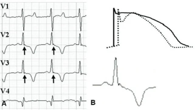
electrocardio-graphic (ECG) finding at the R-ST junction (J point) that has a characteristic protuberant morphology (Figure 1A). This finding can often be confused with a small secondary R wave (R’). Although the J wave was first described in 1920 in a patient with hyper-calcaemia, Osborn (1953) studied the effect of hy-pothermia on the respiratory and cardiac function in dogs and defined the appearance of the J wave.
The J wave has been observed under physiological conditions in the ECG of humans and some other species, such as baboons and dogs (Osborn 1953; Detweiler 2010). This wave can be partially or com-pletely buried in the QRS complex and can be seen as a normal variant in 2–5% of the human popula-tion (Figure 1B). On the other hand, the presence of J waves can represent a “current of injury” and seem to be indicative of severe ischaemia (Yan and Antzelevitch 1996). Several other conditions have been described to occur together with the presence of J waves, such as hypothermia, hypercalcaemia, brain injury, subarachnoid haemorrhage, Chagas disease, sepsis, vasospastic angina, Brugada
syn-drome, acute myocardial ischaemia, Prinzmetal angina, hypertrophy of the left ventricle, cocaine or haloperidol overdosing and idiopathic ventricular fibrillation associated with J waves in inferior leads (Rituparna et al. 2007). Typically, the J waves should have an amplitude and duration of at least 1 mm and 10 milliseconds, respectively, in at least two consec-utive beats (Hlaing et al. 2005). In humans they occur frequently in leads II, III, and aVF and the precor-dial leads: V5–V6 (Shinde et al. 2007; Borggrefe and Schimpf 2010). There is a lack of information about the incidence and morphological characteristics of J waves in other species. The spike-and-dome mor-phology of the epicardial action potentialis absent in canine neonates, gradually appearing over the firstfew months of life (Yan and Antzelevitch 1996).
a prominent transient outward current (Ito) in the ventricular epicardium that produces a transmu-ral voltage gradient during the early ventricular repolarisation which can be accountable for the presence of the J wave (Yan and Antzelevitch 1996; Hlaing et al. 2005; Rituparna et al. 2007; Shinde et al. 2007). Moreover, it is possible to distinguish a J wave coincident with the epicardial action poten-tial notch in the ECG when the electrical activation originates in the endocardium and spreads to the epicardium. In contrast, if activation starts from the epicardium and travels to the endocardium, the J wave disappears from the ECG because it is buried within the QRS complex (Rituparna et al. 2007). However, a spike-and-dome morphology of the action potential is clearlypresent in cells other than epicardial cells (Yan and Antzelevitch 1996). Cells displaying a prominentnotched configura-tion have been described in the deep layersof the endocardial structures of the ventricles of the heart, as well as incells residing in the deep structures of the free wall, particularlyin the M region. Cells in the M regioncould contribute to the J wave with an endocardial-to-epicardial transmuralactivation sequence (Yan and Antzelevitch 1996).
In pathological circumstances the presence of J waves can be explained by several mechanisms. The presence of a prominent Ito in the epicardium has been shown to sensitise tissue to the effects of high calcium whereas hypothermia accentuates the
voltage gradient across ventricular myocardium (Rituparna et al. 2007). Intracranial pathology can induce differences in electrophysiological respon-siveness of the epicardium and endocardium to ace-tylcholine and isoproterenol (Hlaing et al. 2005). J waves in coronary artery disease are likely to be transient and may indicate a current of injury in imminent myocardial infarctions (useful in localis-ing the infarct-related arterial territory) or in myo-cardial revascularisation (Rituparna et al. 2007).
Interestingly, in a normal ventricular activation sequence from endocardium to epicardium, an ac-celeration of heart rate reduces Ito due to its slow recovery from inactivation, resulting in a decrease in the J wave size (Hlaing et al. 2005). The aim of this study was to evaluate the overall presence of J waves in a randomised geriatric population of normal dogs and dogs with heart disease and to evaluate the effects of exercise on its size and shape.
MATERIAL AND METHODS
[image:2.595.143.466.93.275.2]The ECGs of 34 dogs were evaluated. Patients were chosen from the canine patient population, who visited the Small Animal Clinic at the University of Veterinary and Pharmaceutical Sciences, Brno, Czech Republic. Dogs were taken from the geriatric age group because of the higher rate of degenerative mitral valve disease (DMVD). DMVD dogs were
Figure 1. A. Surface ECG of a 12-year-old mongrel dog showing, at the J point (R-S junction), an additional characteristic small wave (black arrows) in the left precordial leads (V1 = CV5RL; V2 = CV6UL; V3 = CV6UL; V4 = V10). B. Cellular basis for the early repolarisation syndrome. The figure represents a model of endocardial and epicardial (dashed line) action potentials with a simultaneous surface ECG (below). The J wave is present due to the action potential notch in the epicardium but not the endocardium. Ventricular epicardium displays an action potential with a prominent transient outward K+ current (I
patients diagnosed based on clinical examination, ECG, thoracic radiographs and echocardiography. In addition, dogs were already on medication for the DMVD and belonged to a functional classifi-cation Ib-II according to the International Small Animal Cardiac Health Council (ISACHC) (Fox et al. 1999) and therapeutic classification B2–C2 (American College of Veterinary Internal Medicine – ACVIM) (Atkins et al. 2009). In the control group the average age was 9.7 years. There were three males and seven females including four mongrels, two King Charles Cavalier Spaniels, two French Poodles, one Dachshund and one Czech Terrier. The average weight was 9.3 kg. In the DMVD group the average age was 11.9 years, 13 were male and 11 females, which included seven Dachshunds, six French Poodles, five mongrels, two German Spitz, two Japanese Chins, one Czech Terrier and one Pekinese. Their average weight was 8 kg.
ECG exercise test. Owners were allowed to be with the dogs to minimise the effects of the stress. Standard 10-lead ECG (EKG Praktik Veterinary®, SEIVA, Czech Republic) in standing position was recorded as reported in the literature (Hanton and Rabemampianina 2006). No electronic filters were used during ECG signal acquisition. The ECGs were recorded at 50 mm/s calibrated to display voltage signals at 1 cm/mV. Dogs underwent a graded sub-maximal test over the course of 3 min. Dogs were put on a treadmill (Treadmill 188N, Formerfit®, Czech Republic) and the ECG machine was attached. Dogs were not pre-trained to walk on the treadmill and were not familiar with the equipment. The graded exercise was a modified protocol based on reports from the literature for veterinary patients and was categorised as a submaximal test (Table 1).
The presence and changes of J waves was evaluat-ed at rest and during every stage of exercise. J waves were measured semi-automatically by manually lo-cating callipers on the J wave and the provided ECG software calculated the value in a table. Differences between groups were evaluated using the Mann-Whitney test and during the test in each group with
the Wilcoxon test. A P-value less than 0.05 denoted the presence of a statistically significant difference. Results are indicated as mean ± standard deviation. The present study was approved by the Bioethical Committee of the University of Veterinary and Pharmaceutical Sciences, Brno.
RESULTS
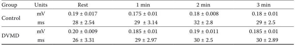
The presence of J waves at rest was detected in four control dogs (40%) and in seven DMVD dogs (29.1%) (Figure 2). J wave amplitude and duration in control dogs was 0.185 ± 0.017 mV and 28 ± 2.4 ms, respec-tively, and in DMVD dogs the values were 0.20 ± 0.009 mV and 26 ± 3.31 ms, respectively (Table 2). J waves were present in the control group in leads I, II, aVL, aVR, aVF and CV6LL. In DMVD dogs the waves were present in leads I, II, aVF, CV6LL and CV6LU. No statistical differences were found between groups and the J wave did not change significantly in any of the groups during exercise.
DISCUSSION
[image:3.595.305.532.113.185.2]In this study, the J wave or Osborne wave was studied in a randomised population of geriatric dogs with and without heart disease. The J wave was found in all leads except for III, CV5RL and V10. Interestingly, the presence of waves was more prominent in the limb leads (mainly in lead aVL) than in precordial leads underlying the impor-tance of evaluating all standard leads in a surface ECG. In dogs and humans the manifestationof the
Table 1. Submaximal exercise treadmill protocol
Stage Speed (m/s) Evaluation Rest 0 ECG 1 min 0.27 ECG 2 min 0.50 ECG 3 min 1.00 ECG
Table 2. Amplitude and duration of J waves during exercise in lead II. Values indicated as average ± SD
Group Units Rest 1 min 2 min 3 min
Control mV 0.19 ± 0.017 0.175 ± 0.01 0.18 ± 0.008 0.18 ± 0.01
ms 28 ± 2.54 29 ± 3.14 32 ± 2.8 29 ± 2.5
DVMD mV 0.20 ± 0.009 0.185 ± 0.01 0.19 ± 0.011 0.185 ± 0.01
[image:3.595.61.532.685.757.2]J wave in ECG leads (II, III, aVR, aVF, and mid to left precordialleads V3 through V6) in which the mean vector axis istransmurally oriented across the left ventricle and septum is most prominent
(Yan and Antzelevitch 1996). In spite of its larger notch, the rightventricular epicardium might be expected to make only a minorcontribution to the J wave under normal conditions, becauseof a thinner ventricular wall and a briefer activation time (Yan and Antzelevitch 1996).
None of the patients in this study exhibited signs of hypothermia, hypercalcaemia, or other conditions leading to the presence of pathological J waves. Thus, the presence of J waves may consti-tute a normal finding in approximately one third of the population of geriatric dogs. Exercise-induced changes in J waves were not observed in any of the groups (Figure 3). The behaviour of the J wave in this study seems to be conservative. It was present at the beginning of exercise in 11 dogs and did not change its amplitude or duration significantly. It is possible that the load of exercise did not produce “currents of injury” that are indicative of severe ischaemia. As we also did not observe changes in DMVD dogs, physical exertion might be recom-mended in dogs with DMVD in categories B2–C2. Also, there is a low possibility that exercise in this group of dogs might protect from arrhythmias since J waves are generally associated with re-entrant ar-rhythmias (Rituparna et al. 2007; Jastrzebski 2009).
CONCLUSIONS
[image:4.595.63.291.98.465.2]Since none of the patients in this study exhibited signs of hypothermia, hypercalcaemia, or any other physical and internal changes, it can be concluded that J waves constitute a normal finding in approxi-mately 30–40% of geriatric subjects. Submaximal testing in dogs with DMVD is feasible and safe. Exertion did not alter the duration, amplitude or shape of the J waves; thus, it is possible that under a
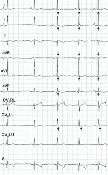
Figure 2. 10-lead surface ECG in a normal eight-year-old female Czech Terrier showing presence of J waves (arrows) in leads I, II, aVR, aVL, aVF and CV6LL at rest. Speed 50 mm/s, amplitude 10 mm = 1 mV
Figure 3. Behaviour of the J wave in an 11 year-old Dachshund with DMVD during submaximal exercise; A = 1 min, B = 2 min, C = 3 min. Note that the J wave did not change significantly Lead I. Paper speed 50 mm/s, amplitude 10 mm = 1 mV
[image:4.595.71.517.601.729.2]submaximal load of exercise dogs do not develop is-chaemic processes detected using the ECG method.
REFERENCES
Atkins RC, Bonagura JD, Ettinger SM, Fox PR, Gordon SG, Haggstrom J, Hamlin RL, Keene B, Luis-Fuentes V, Stepien RL (2009): Guidelines for the diagnosis and treat-ment of canine chronic valvular heart disease. Journal of Veterinary Internal Medicine 23, 1142–1150.
Borggrefe M, Schimpf R (2010): J-Wave syndromes caused by repolarization or depolarization mechanisms: A de-bated issue among experimental and clinical electro-physiologists. Journal of the American College of Cardiology55, 798–800.
Detweiler DK (2010): The dog electrocardiogram. A critical review. In: Macfarlane PW, van Oosterom A, Pahlm O, Kligfield P, Janse K, Camm J (eds.): Comprehensive
Elec-trocardiology. 2nd ed. Pergamon Press, Oxford. 1886–1903.
Fox PR, Sisson D, Moise NS (1999): Appendix A: Recom-mendations for diagnosis of heart disease treatment of heart failure in small animals. International Small Animal Cardiac Health Council. Textbook of Canine and Feline
Cardiology, Principles and Clinical Practice. 2nd ed. W.B.
Saunders, Philadelphia. 883–901.
Hanton G, Rabemampianina Y (2006): The electrocardio-gram of the Beagle dog: reference values and effect of sex, genetic strain, body position and heart rate. Laboratory Animals 40, 123–136.
Hlaing T, DiMino T, Kowey PR, Yan GX (2005): ECG repo-larization waves: Their genesis and clinical implications. Annals of Noninvasive Electrocardiology 10, 211–223. Jastrzebski M (2009): Ischemic J wave: Novel risk marker
for ventricular fibrillation? Heart Rhythm 6, 829–835. Osborn JJ (1953): Experimental hypothermia; respiratory
and blood pH changes in relation to cardiac function. American Journal of Physiolology 175, 389–398. Rituparna S, Suresh S, Chandrashekhar M, Purvez G, Sunil
S, Durairaj M, Yash L, Di Diego JM, Antzlevitch C (2007): Occurrence of “J waves” in 12-lead ECG as a marker of acute ischemia and their cellular basis. Pacing and Clin-ical Electrophysiology 30, 817–819.
Shinde R, Shinde S, Makhale C, Grant P, Sathe S, Durairaj M, Lokhandwala Y, Di Diego J, Antzelevitch C (2007): Occurrence of “J waves” in 12-lead ECG as a marker of acute ischemia and their cellular basis. Pacing and Clin-ical Electrophysiology 30, 817–819.
Yan GX, Antzelevitch C (1996): Cellular basis for the elec-trocardiographic J wave. Circulation 93, 372–379. Yan GX, Lankipalli RS, Burke JF, Musco S, Kowey PR (2003):
Ventricular repolarization components on the electro-cardiogram: cellular basis and clinical significance. Jour-nal of the American College of Cardiology42, 401–409.
Received: 2014–08–07 Accepted after corrections: 2015–03–13
Corresponding Author:
Carlos F. Agudelo, University of Veterinary and Pharmaceutical Sciences Brno, Faculty of Veterinary Medicine, Small Animal Clinic, Palackeho 1/3, 612 42 Brno, Czech Republic