Long-term evaluation of bicipital tenodesis
with T-staple in three dogs: a case report
S. Pinna, A. De Giorgi, G. Spinella
Department of Veterinary Medical Sciences, University of Bologna, Italy
ABSTRACT: The purpose of this report is to describe theT-staple tenodesis of biceps brachii in three client-owned dogs and long-term ultrasonographic follow-ups. The orthopaedic examination revealed grade 2/4 (n = 2) and 3/4 (n = 1) lameness, moderate pain on passive movement and positivity to the tendon biceps test with a complete extension of the elbow during the flexion of the shoulder (Cases 1 and 2). Ultrasound examination was crucial for diagnosis of partial or complete rupture and tenosynovitis of biceps tendon. Surgical tenodesis was carried out with a metal T-staple. One-year ultrasound follow-up was performed, confirming the correct integration of the staple on the bicipital fibres and the absence of macroscopic tendon injury or degenerative disease. The results suggest that the metal T-staple could be a good alternative for bicipital tenodesis in dogs.
Keywords: biceps brachii tendon; ultrasonography; staple; dog
Lesions of the biceps tendon have been described as the third most common cause of shoulder lame-ness after shoulder instability and osteochondritis dissecans of the humeral head in dogs (Bardet 1999). In veterinary practice, avulsion of the biceps bra-chii tendon from the supraglenoid tubercle, tendon rupture and bicipital tenosynovitis have been surgi-cally treated with tenodesis or tenotomy (Bruce et al. 2000; Wall and Taylor 2002; Adamiak and Szalecki 2003; Cook et al. 2005). The biceps tenodesis pro-cedure involves cutting the biceps tendon close to its insertion on the supraglenoid tubercle and then anchoring the tendon along its anatomical course more distally along the humerus. Several different anchoring techniques have been described using ar-throtomy or arthroscopy (Piermattei and Flo 1997; Adamiak and Szalecki 2003; Innes and Brown 2004; Cook et al. 2005). Otherwise, the biceps tenotomy procedure involves transecting the tendon and leav-ing it free, usleav-ing arthrotomy, arthroscopy, or in a percutaneous procedures (Piermattei and Flo 1997; Venturini et al. 1998; Denny and Butterworth 2000; Cook et al. 2005; Esterline et al. 2005;Bergenhuyzen et al. 2010).
The purpose of this article is to report the clinical and radiological outcomes as well as the
ultrasono-graphic findings after the execution of the biceps tenodesis procedure on three dogs.
Case description
Three client-owned dogs were presented at the Department of Veterinary Medical Sciences, University of Bologna, for investigation of moder-ate-to-severe forelimb lameness. Clinical exami-nation, diagnostic imaging and surgical treatment were performed as follows.
Ultrasound examination of the right shoulder performed with a 10 MHz linear probe, revealed a partial rupture of the biceps brachii tendon with incomplete disruption of the normal fibrillar pat-tern of the tendon next to its insertion on the su-praglenoid tubercle. The surface of the tubercle appeared rough and bumpy and small hyperecho-ic bone fragments were attached to the tendon. Moderate and inhomogeneous hypoechoic fluid was present in the tendon sheath as well as in the articular space. Distally, the mean echogenicity [scale of 256 grey levels (0 = black; 255 = white)] of the tendon along the intertubercular groove was in the normal range compared with that of the con-tralateral limb (Spinella et al. 2013). This aspect was consistent with the diagnosis of partial biceps tendon avulsion and, according to Kramer’s classi-fication, grade 2 tenosynovitis (Kramer et al. 2001). Case 2. A six-year-old female working Irish Red Setter dog, weighing 21 kg, showed lameness of the left forelimb of one-month duration due to a trau-ma. An anti-inflammatory drug had been adminis-tered without improvement. On initial evaluation the dog showed a grade 2 (out of 4) lameness, mod-erate pain, hypotrophy of the shoulder muscles, and he was positive to the biceps tendon test performed with shoulder flexion and elbow extension.
Radiographic views of the left shoulder revealed a defect of the supraglenoid tubercle which was ir-regular, and a bone fragment on the cranial aspect of the tubercle, as well as a soft mineralisation in the proximal part of the bicipital groove.
During ultrasound of the left shoulder joint, per-formed with a 10 MHz linear probe, a longitudinal
scan revealed complete rupture of the biceps ten-don with complete disruption of the normal fibril-lar pattern in the proximal part. The supraglenoid tubercle appeared rough with a small fragment evi-dent in the articular surface (Figure 1A). Moreover, around the suprascapular tendon insertion the collection of heterogeneous and hypoechoic fluid was observed. The mean echogenicity of the left bicipital tendon along the intertubercular groove was in the normal range compared to that of the contralateral limb. These findings led to the diag-nosis of complete brachii biceps tendon rupture, grade 2 bicipital tenosynovitis and suprascapular hematoma in resolution.
Case 3. A six-year-old male working Belgian Mali- nois, weighing 33 kg, had been treated for left fore-limb lameness for four months previous to pres-entation. Intra-articular corticosteroids had given only a short improvement. The dog showed grade 3 (out of 4) lameness, slight reduction of the shoulder muscle, moderate pain in both flexion and exten-sion of the shoulder, and the elbow did not extend jointly with shoulder flexion.
Radiographic findings of the left shoulder in-cluded signs of moderate osteoarthritis with os-teophytes (range of 2–4 mm) on the caudal rim of the glenoid cavity, caudal humeral head, and smaller osteophytes (range of 1–2 mm) along the bicipital tendon groove. Mild signs of subchondral bone sclerosis were also evident.
[image:2.595.101.488.545.698.2]Ultrasonography of the left scapula-humeral re-gion revealed a severe and chronic grade 3 teno-synovitis of the biceps brachii tendon (Figure 2A), with moderate tendon hypotrophy. Moreover, an
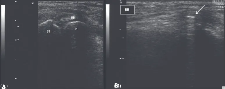
Figure 1. Six-year-old female Irish Red Setter dog. A: Longitudinal scan of left shoulder joint. Complete rupture of the biceps brachii (BB) tendon on its proximal part. A small bone fragment was easily recognised on the supraglenoid tubercle (ST); (H = humerus). B: 12-month follow-up. No inflammatory reactions, mineralised lesions or tendon disruption were observed around the T-staple (white arrow)
irregular hyperechoic profile of the intertubercular groove consistent with osteophytes, was observed. The mean echogenicity of the tendon along the in-tertubercular groove was mildly reduced compared to that of the contralateral limb.
Surgical treatment.In brief, three types of shoul-der diseases (partial and complete brachii biceps tendon rupture, and chronic tenosynovitis) were diagnosed. Each dog was surgically treated using the same tenodesis procedure.
A modified cranio-medial approach to the shoul-der was performed (Piermattei and Johnson 2004). The superficial and deep pectoral muscles were exposed and their insertions were freed from the proximal border of the humerus. The extension synovial sheath of the joint capsule was incised only distally to the transverse humeral ligament. The latter was transected, and the biceps tendon was exposed. Traction on the bicipital tendon in the distal direction was applied and it was later cut as proximally as possible, without exposing the supra-glenoid tubercle. The intertubercular groove was inspected and osteophytes were removed, when present. Furthermore, to identify the correct func-tional point of fixation, a mild traction was applied on the bicipital tendon in order to achieve a flexion of the elbow, as well as to allow its passive exten-sion. The tendon was re-attached to the humerus just distally to the intertubercular groove using a metal T-staple, positioned and fixed in a caudo-lateral direction. The extremity of the tendon, re-sulting in an excess proximally to the staple, was removed at 5–6 mm from the staple.
All other muscular structures were repositioned and subcutaneous and cutaneous layers were sutured.
The metal surgical implant was constructed of AISI 316L stainless steel (Figure 3-inset).
Pre- and postoperative care. Perioperative anal-gesia was achieved through methadone (0.3 mg/kg intramuscularly) in premedication followed by fentanyl (5 mcg/kg/h intravenous) during sur-gery. After surgery, each dog received carprofen (2–4 mg/kg daily subcutaneous) for seven days and tramadol (2–5 mg/kg twice daily orally), if required for supplemental analgesia. The owners were in-structed to limit physical activity and to allow walks on a leash for 30 days before leaving the dogs to resume normal levels of activity.
Follow-up and outcome monitoring. Follow-ups were carried out at 1, 2, 4 and 12 months after surgery in all three cases.
Clinical examination revealed an improvement of the clinical signs and the complete disappearance of pain and lameness at one month after surgery.
Radiographs were taken at the end of the surgery to verify the correct position of the staple in all dogs. Further radiographic exams were performed during the follow-up (1, 2, 4, and 12 months) which did not reveal significant changes of the shoulder image, nor implant migration (Figure 3).
Ultrasonographic follow-ups were performed in order to monitor the healing process and the inflammatory reaction next to the metal T-staple implant. All three clinical cases showed common features with respect to postoperative ultrasono-graphic healing, despite the different initial
pa-Figure 2. Six -year-old male Belgian Malinois dog. A: Longitudinal scan of the biceps brachii tendon along the bicipi-tal groove. A grade 3 tenosynovitis of the biceps brachii tendon was observed. B: 30-day follow-up revealed a moder-ate inflammatory response around the T-staple (white arrow) and imperfect fibre alignment
[image:3.595.98.492.94.265.2]thologies. One month after tenodesis (Figure 2B), a mild-to-moderate hypoechoic inflammatory re-action was observed in the bicipital tendon at the site of fixation close to the staple. Moreover, an imperfect fibre alignment was observed in Case 3. During ultrasound follow-ups at two and four months after surgery, a correct tendon fibre align-ment was observed in all three cases, with a mild anechoic pattern around the staple. At 12 months follow-up, no inflammatory reaction was seen at the T-staple site. Neither mineralised lesions nor tendon disruption were observed in any of the three clinical cases (Figure 1B).
Both imaging exams were also performed on the sound contralateral forelimb and each dog was its own control for pre-surgical and follow-up evalu-ations.
DISCUSSION
The three cases described in this report were affected by the most frequent bicipital lesions. Tenodesis was performed as the patients were working dogs, and the healing process after teno-desis was followed up, using x-ray and ultrasound examinations, for up to one year.
The signalment, age and weight reported in our three cases are consistent with the literature (Rivers et al. 1992; Kramer et al. 2001). Bergenhuyzen et
al. (2010) described a lower mean age of 3.5 years but a wider range from six months to 10 years of age, and the dogs of that clinical communication were represented by medium-large breeds, mainly by Border Collies and Bernese Mountain dogs, but no information was reported about the weight of the animals (Bergenhuyzen et al. 2010). Previously, a case report concerning 15 dogs described wider weight and age ranges, 13–44 kg (mean 28.4 kg; median 29 ± 9.2 kg) and 1–15 years (mean 6.9 years; median 7 ± 3.8 years), respectively (Bruce et al. 2000).
The normal ultrasonographic anatomy of the canine biceps brachii tendon as well as ultrasono-graphic aspects of bicipital tendinitis and tenosy-novitis have been widely described in dogs (Bruce et al. 2000; Kramer et al. 2001; Spinella et al. 2013). The benefits of ultrasonographic examination were proven by other authors whose data run counter to the initial results obtained by Rivers et al. (1992), who observed a low sensitivity of this technique. Ultrasonography provided immediate information on tendon integrity, lesion classification and as-sisted in treatment choice. Moreover, it allowed the monitoring of the healing process after injures or surgical procedures (Rivers et al. 1992; Kramer et al. 2001), providing information on tendon fibre reor-ganisation and alignment, as well as the recovery of normal mean echogenicity of the tendon structure.
In the present report, the efficacy of ultrasonog-raphy as well as its suitability for the diagnosis of complete or partial rupture of the biceps brachii tendon and tenosynovitis, as well as in monitor-ing the healmonitor-ing process and inflammatory reaction when the tendon fixation is achieved by a T-staple, is confirmed and verified. Our findings are con-sistent with the results reported by other authors, confirming that better results are obtained when a linear probe is used for tendon examination in dogs and horses (Long and Nyland 1999; Bruce et al. 2000; Kramer et al. 2001; Agut et al. 2009; Vilar et al. 2011; Spinella et al. 2013). Moreover, ultra-sonographic findings confirmed the biological in-activity and biocompatibility of stainless steel AISI 316L, and revealed minimal tissue reaction of fibre tendon around it, with no chronic or degenerative lesions, up to 12 months after tenodesis (Navarro et al. 2008).
[image:4.595.65.291.97.234.2]In the veterinary literature, viewpoints regard-ing biceps tenodesis versus tenotomy for surgical intervention have been conflicting, and the lack of
papers comparing the long-term results of the two techniques did not enable one to be unequivocally considered better than the other.
The biceps tenotomy may be performed using arthrotomy, arthroscopy (Wall and Taylor 2002), or percutaneously (Venturini et al. 1998; Esterline et al. 2005). Excellent long-term results were re-ported by Bergenhuyzen (2010) who performed the shoulder arthroscopy in a standardised manner using craniolateral and caudolateral portals for a standard compartmental approach as Van Ryssen et al. described in 1993. Moreover, a cadaveric evalu-ation of arthroscopic tenotomy was performed to improve and give more information on this tech-nique (Holsworth et al. 2002). The authors ob-served that the craniolateral camera port provided an optimised tendon visualisation, if a combined moderate shoulder and elbow flexion was applied. Moreover, tenotomy was easier to perform using a blade and could not be carried out with the shaver
(Holsworth et al. 2002).
The biceps tenodesis can be carried out using arthroscopy or arthrotomy (open procedure). Cook stated that since the forelimb bears the majority of the weight in dogs, biceps tenodesis might be advantageous for this species, and he performed the procedure using arthroscopy (Cook et al. 2005). He used two kinds of devices, a cannulated inter-ference screw in four dogs and a screw with tissue washer in two dogs, fixed into the bone of the most distal point in the intertubercular groove which was visible arthroscopically (Cook et al. 2005).
Arthroscopic procedures performed on the shoul-der have been widely documented (Van Ryssen et al. 1993; Martini et al. 2002), but only Cook’s clinical study reported arthroscopic tenodesis (Cook et al. 2005). Excellent long-term results were described by open biceps tenodesis (Stobie et al. 1995), in contrast with the mixed results described in other papers (Bruce et al. 2000; Innes and Brown 2004).
Regarding biceps tenodesis, there are no evi-dence-based recommendations for the ideal level at which to cut and stabilise the tendon.
In the surgical technique described in the present paper, the exposure of the tendon was achieved with low invasiveness, without opening the joint capsule or exposing the supraglenoid tubercle, but only the synovial sheath just beyond the transverse humeral ligament. This procedure provided an easier ap-proach to the tendon distally from its insertion on the supraglenoid tubercle in contrast with the
open approach which involves incision of the joint capsule (Todoroff 1998; Denny and Butterworth 2000; Adamiak and Szalecki 2003; Piermattei and Johnson 2004). Unlike other described procedures, we did not approach the bicipital tendon using os-teotomy of the greater tubercle (Stobie et al. 1995; Piermattei and Johnson 2004).
The arthroscopic approach and the mini-arthrot-omy permit tenotmini-arthrot-omy and tenodesis (if done) close to the proximal insertion of the tendon that could involve an injured part of the tendon in the tenode-sis. In the procedure described here a traction on the bicipital tendon in the distal direction was ap-plied which was later cut as proximally as possible, without exposing the supraglenoid tubercle. The part of the tendon proximal to the site of tenodesis was removed to decrease the risk of inflammation.
Arthroscopically, shoulder inspection is excel-lent in all joint compartments (Van Ryssen et al. 1993; Martini et al. 2002), and the transection of the tendon is made after tenodesis (Cook et al. 2005). To perform the tenodesis using an open procedure it was our preference to cut the tendon before its fixation according to the techniques described in the literature (Piermattei and Flo 1997; Denny and Butterworth 2000; Adamiak and Szalecki 2003). The tendon was fixed with the elbow flexed to achieve the correct tension on the muscle in Cases 1 and 2 (positive in the tendon biceps test), with a com-plete extension of the elbow during the flexion of the shoulder.
Several methods of bicipital tenodesis have been previously described, e.g. suturing of the tendon to the periosteum, using a bone screw and spiked washer, giving a new attachment to the supraspina-tus tendon, transporting the tendon through a hole drilled in the greater tubercle or using a ligament staple to reattach the tendon into the intertuber-cular groove (Piermattei and Flo 1997; Denny and Butterworth 2000).
The rationale for our approach of performing the tenodesis with a metal-T-staple was related to the idea that this kind of staple with a “T” shape pressing uniformly on the transected tendon would facilitate more rapid healing and prevent ischaemic injures, as was then observed during the ultrasono-graphic follow-ups.
described a tendon calcification, visualised after a radiological re-evaluation. In our study we did not observe any dystrophic calcification, both with radiography and ultrasonography exams; further, no major or minor complications such as infection, migration of implant, or seroma occurred.
Seroma, displacement or joint laxity, infection, pain and intermittent lameness can all be poten-tial complications in both tenodesis and tenotomy procedures, but a detailed perusal of the literature indicates that most studies report beneficial out-comes. By contrast, in human medicine, a cosmetic “Popeye deformity” is frequently observed in pa-tients treated with tenotomy (Gurnani et al. 2015).
This is likely due to the different anatomical con-formation of the canine biceps tendon.
A limitation of this report was the small number of patients examined. These studies should there-fore be extended, and accompanied by a histologi-cal tendon evaluation, in order to create a more reliable clinical picture. However, to the authors’ knowledge postoperative ultrasound assessment after tenodesis has not been reported until now. The ultrasound findings reported here suggest that the surgical procedure performed in the three dogs did not weaken the bicipital tendon and preserved elbow flexion and the functional muscle.
Acknowledgement
The authors thank Dr. Ramona Raduc (freelance linguist) for her assistance with English editing.
ReFeReNCeS
Adamiak Z, Szalecki P (2003): Treatment of bicipital teno-synovitis with double tenodesis. Journal of Small Animal Practice 44, 539–540.
Agut A, Martinez ML, Sanchez-Valverde MA, Soler M, Rod-ríguez MJ (2009): Ultrasonographic characteristics (cross-sectional area and relative echogenicity) of the digital flexor tendons and ligaments of the metacarpal region in Pure-bred Spanish horses. Veterinary Journal 180, 377–383. Bardet JF (1999): Lesions of the biceps tendon diagnosis
and classification – a retrospective study of 25 cases in 23 dogs and one cat. Veterinary and Comparative Ortho-paedics and Traumatology 12, 188–195.
Bergenhuyzen ALR, Vermote KAG, Van Bree H, Van Ryssen HB (2010): Long-term follow-up after arthroscopic
ten-otomy for partial rupture of the biceps brachii tendon. Veterinary and Comparative Orthopaedics and Trauma-tology 23, 51–55.
Bruce WJ, Burbidge HM, Bray JP, Broome CJ (2000): Bi-cipital tendinitis and tenosynovitis in the dog: a study of 15 cases. New Zealand Veterinary Journal 48, 44–52. Cook JL, Kenter K, Fox DB (2005): Arthroscopic biceps
tenodesis: technique and results in six dogs. Journal of American Animal Hospital Association 41, 121–127. Denny HR, Butterworth SJ (2000): The shoulder. In: AGuide
to Canine and Feline Orthopaedic Surgery. 4th ed. Black-well Science Ltd. 324–329.
Esterline ML, Armbrust L, Roush JK (2005): A comparison of palpation guided and ultrasound guided percutaneous biceps brachii tenotomy in dogs. Veterinary and Com-parative Orthopaedics and Traumatology 18, 135–139. Gurnani N, van Deurzen DF, Janmaat VT, van den Bekerom
MP (2015): Tenotomy or tenodesis for pathology of the long head of the biceps brachii: a systematic review and meta-analysis. Knee Surgery, Sports Traumatology, Ar-throscopy: official journal of the ESSKA. May 15. [Epub ahead of print] DOI 10.1007/s00167-015-3640-6 Holsworth IG, Schulz KS, Ingel K (2002): Cadaveric
evalu-ation of canine arthroscopic bicipital tenotomy. Veteri-nary and Comparative Orthopaedics and Traumatology 15, 215–222.
Innes JF, Brown G (2004): Rupture of the biceps brachii tendon sheath in two dogs. Journal of Small Animal Prac-tice 45, 25–28.
Kramer M, Gerwing M, Sheppard C, Schimke E (2001): Ultrasonography for the diagnosis of diseases of the ten-don and tenten-don sheath of Biceps Brachii muscle. Veteri-nary Surgery 30, 64–71.
Long CD, Nyland TG (1999): Ultrasonographic evaluation of the canine shoulder. Veterinary Radiology and Ultra-sound 40, 372–379.
Martini MF, Pinna S, Del Bue M (2002): A simplified technique for diagnostic and surgical arthroscopy of the shoulder joint in the dog. Journal of Small Animal Practice 43, 7–11. Navarro M, Michiardi A, Planell JA (2008): Biomaterials in
orthopaedics. Journal of the Royal Society Interface 5, 1137–1158.
Piermattei DL, Flo GL (1997): The shoulder joint. In: Brinker WO, Piermattei DL, Flo GL (eds.): Handbook of Small Animal Orthopedics and Fracture Repair. 3rd ed. Saunders, Philadelphia. 228–260.
Piermattei DL, Johnson KA (2004): An atlas of approaches to the bones and joints of the dog and cat, 4th ed. Saun-ders, Philadelphia. 136–141.
Veterinary and Comparative Orthopaedics and Trauma-tology 5, 1–57.
Spinella G, Loprete G, Musella V, Britti D, Vilar JM (2013): Cross-sectional area and mean echogenicity of shoulder and elbow tendons in adult German Shepherd dogs. Vet-erinary and Comparative Orthopaedics and Traumatol-ogy 26, 366–371.
Stobie D, Wallace LJ, Lipowitz AJ, King V, Lund EM (1995): Chronic bicipital tenosynovitis in dogs: 29 cases (1985– 1992). Journal of American Veterinary Medical Associa-tion 207, 201–207.
Todoroff RJ (1998): Surgical treatment of biceps brachii tendon injury. In: Bojrab MJ (ed.): Current Techniques in Small Animal Surgery. 4th ed. Williams and Wilkins, Philadelphia. 1074.
Van Ryssen B, Van Bree H, Vyt P (1993): Arthroscopy of the shoulder joint in the dog. Journal of the American Animal Hospital Association29, 101–105.
Venturini A, Pinna S, Valentini S, Barilli M (1998): Tenot-omy as treatment of displacement of the biceps and of
the contracture of the infraspinatus muscle in dogs (in Italian). AIVPA Journal. Associazione Italiana Veterinari Piccoli Animali 1, 29–33.
Vilar JM, Santana A, Espinosa J, Spinella G (2011): Cross-sectional area of the tendons of the tarsal region in stan-dardbred trotter horses. Equine Veterinary Journal 43, 235–239.
Wall CR, Taylor R (2002): Arthroscopic biceps brachii te-notomy as a treatment for canine bicipital tenosynovitis. Journal of American Animal Hospital Association 38, 169–175.
Wiemer P, van Ryssen B, Gielen I, Taeymans O, van Bree H (2007): Diagnostic findings in a lame-free dog with com-plete rupture of the biceps brachii tendon. A case report in a unilaterally affected working Labrador Retriever. Veterinary and Comparative Orthopaedics and Trauma-tology 20, 73–77.
Received: 2015–04–02 Accepted after corrections: 2016–02–18
Corresponding Author:
Stefania Pinna, Department of Veterinary Medical Sciences, School of Agriculture and Veterinary Medicine, University of Bologna, Italy