2-(2,4-Dichlorophenyl)acetic acid
Jiang-Sheng Li,a* Qi-Xi Heband Peng-Yu Lia
aSchool of Chemistry and Biological Engineering, Changsha University of Science & Technology, Changsha 410004, People’s Republic of China, andbCollege of Chemistry and Chemical Engineering, Hunan University, Changsha 410082, People’s Republic of China
Correspondence e-mail: js_li@yahoo.com.cn
Received 4 December 2009; accepted 6 December 2009
Key indicators: single-crystal X-ray study;T= 113 K; mean(C–C) = 0.002 A˚; Rfactor = 0.027;wRfactor = 0.075; data-to-parameter ratio = 13.4.
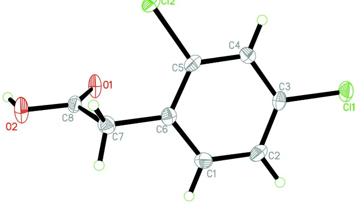
In the title compound, C8H6Cl2O2, the dihedral angle between the C—C( O)—OH carboxyl unit and the benzene ring is 70.70 (4). In the crystal, molecules are linked into inversion
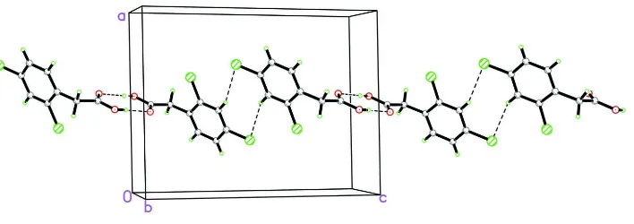
dimers by pairs of O—H O hydrogen bonds. The dimers are linked into chains extending along [001] by weak C—H Cl interactions.
Related literature
For background to carboxylic acids as supramolecular synthons, see: Thalladiet al.(1996). For related structures, see: Hodgson & Asplund (1991); Liet al.(2010).
Experimental
Crystal data
C8H6Cl2O2 Mr= 205.03
Monoclinic,P21=n a= 10.824 (2) A˚
b= 5.6061 (11) A˚
c= 13.820 (3) A˚ = 91.08 (3)
V= 838.4 (3) A˚3 Z= 4
MoKradiation = 0.72 mm1 T= 113 K
0.240.200.12 mm
Data collection
Rigaku Saturn CCD diffractometer Absorption correction: multi-scan
(CrystalClear; Rigaku/MSC, 2005)
Tmin= 0.846,Tmax= 0.918
5321 measured reflections 1484 independent reflections 1237 reflections withI> 2(I)
Rint= 0.037
Refinement
R[F2> 2(F2)] = 0.027 wR(F2) = 0.075 S= 1.10 1484 reflections
111 parameters
H-atom parameters constrained
max= 0.23 e A˚
3 min=0.22 e A˚
3
Table 1
Hydrogen-bond geometry (A˚ ,).
D—H A D—H H A D A D—H A
O2—H2 O1i
0.82 1.85 2.6689 (16) 175 C4—H4 Cl1ii
0.93 2.86 3.731 (2) 156
Symmetry codes: (i)xþ1;yþ1;zþ2; (ii)xþ1;y;zþ1.
Data collection:CrystalClear(Rigaku/MSC, 2005); cell refinement:
CrystalClear; data reduction:CrystalClear; program(s) used to solve structure:SHELXS97(Sheldrick, 2008); program(s) used to refine structure: SHELXL97 (Sheldrick, 2008); molecular graphics:
SHELXTL(Sheldrick, 2008); software used to prepare material for publication:SHELXL97.
Supplementary data and figures for this paper are available from the IUCr electronic archives (Reference: HB5271).
References
Hodgson, D. J. & Asplund, R. O. (1991).Acta Cryst.C47, 1986–1987. Li, J. S., He, Q. X. & Li, P. Y. (2010).Acta Cryst.E66, o39.
Rigaku/MSC (2005).CrystalClear. Rigaku Corporation, Tokyo, Japan. Sheldrick, G. M. (2008).Acta Cryst.A64, 112–122.
Thalladi, V. R., goud, B. S., Hoy, V. J., Allen, F. H., Howard, J. A. K. & Desiraju, G. R. (1996).Chem. Commun.pp. 401–402.
Acta Crystallographica Section E
Structure Reports Online
supporting information
Acta Cryst. (2010). E66, o110 [doi:10.1107/S1600536809052453]
2-(2,4-Dichlorophenyl)acetic acid
Jiang-Sheng Li, Qi-Xi He and Peng-Yu Li
S1. Comment
Carboxylic acid is a supramolecular synthon, widely used to construct supramolecular array with one to three different
dimensions via hydrogen bonds (Thalladi et al., 1996). For our continuous research, we herein report the structure of the
title compound (I).
In the title molecule, (Fig 1), the O1/O2/C7/C8 carboxyl unit forms an angle of 70.70 (4) A with the benzene ring. In the
crystal packing, the molecules are linked into dimers by strong O—H···O H-bonding, which extend down the c axis by
the aid of weak C—H···Cl H-bonding (Table 1 & Fig 2). For related structures, see: Hodgson & Asplund (1991) and Li et
al. (2010).
S2. Experimental
The title compound was available from Hunan institute of Chemical Industry, received without further purification.
Colourless blocks of (I) were obtained by evaporation from its solution of ethyl acetate/petroleum ether 1/2 (v/v).
S3. Refinement
All H atoms were positioned geometrically and constrained to ride on their parent atoms [C—H distances are 0.93 and
[image:2.610.126.482.454.662.2]0.97Å with Uiso(H) = 1.2 Ueq(C) for aromatic and CH2 H atoms, 0.82Å with Uiso = 1.5Ueq (O) for hydroxyl H atom].
Figure 2
The infinite chain formed via alternative O—H···O and C—H···Cl hydrogen bonding down the c axis.
2-(2,4-Dichlorophenyl)acetic acid
Crystal data
C8H6Cl2O2 Mr = 205.03 Monoclinic, P21/n
Hall symbol: -P 2yn
a = 10.824 (2) Å
b = 5.6061 (11) Å
c = 13.820 (3) Å
β = 91.08 (3)°
V = 838.4 (3) Å3 Z = 4
F(000) = 416
Dx = 1.624 Mg m−3
Melting point = 403–405 K Mo Kα radiation, λ = 0.71073 Å Cell parameters from 2684 reflections
θ = 2.4–27.9°
µ = 0.72 mm−1 T = 113 K Block, colourless 0.24 × 0.20 × 0.12 mm
Data collection
Rigaku Saturn CCD diffractometer
Radiation source: rotating anode Confocal monochromator
Detector resolution: 7.31 pixels mm-1 ω and φ scans
Absorption correction: multi-scan (CrystalClear; Rigaku/MSC, 2005)
Tmin = 0.846, Tmax = 0.918
5321 measured reflections 1484 independent reflections 1237 reflections with I > 2σ(I)
Rint = 0.037
θmax = 25.0°, θmin = 2.4°
h = −12→12
k = −6→6
l = −10→16
Refinement
Refinement on F2
Least-squares matrix: full
R[F2 > 2σ(F2)] = 0.027 wR(F2) = 0.075 S = 1.10 1484 reflections 111 parameters 0 restraints
Primary atom site location: structure-invariant direct methods
Secondary atom site location: difference Fourier map
Hydrogen site location: inferred from neighbouring sites
H-atom parameters constrained
w = 1/[σ2(Fo2) + (0.0435P)2]
where P = (Fo2 + 2Fc2)/3
(Δ/σ)max = 0.001
Δρmax = 0.23 e Å−3
Δρmin = −0.22 e Å−3
Extinction correction: SHELXL97 (Sheldrick, 2008), Fc*=kFc[1+0.001xFc2λ3/sin(2θ)]-1/4
Special details
Geometry. All e.s.d.'s (except the e.s.d. in the dihedral angle between two l.s. planes) are estimated using the full covariance matrix. The cell e.s.d.'s are taken into account individually in the estimation of e.s.d.'s in distances, angles and torsion angles; correlations between e.s.d.'s in cell parameters are only used when they are defined by crystal symmetry. An approximate (isotropic) treatment of cell e.s.d.'s is used for estimating e.s.d.'s involving l.s. planes.
Refinement. Refinement of F2 against ALL reflections. The weighted R-factor wR and goodness of fit S are based on F2,
conventional R-factors R are based on F, with F set to zero for negative F2. The threshold expression of F2 > σ(F2) is used
only for calculating R-factors(gt) etc. and is not relevant to the choice of reflections for refinement. R-factors based on F2
are statistically about twice as large as those based on F, and R- factors based on ALL data will be even larger.
Fractional atomic coordinates and isotropic or equivalent isotropic displacement parameters (Å2)
x y z Uiso*/Ueq
Cl1 0.71462 (4) 0.15511 (7) 0.45827 (3) 0.02309 (18)
Cl2 0.35390 (4) 0.34376 (7) 0.70900 (3) 0.02349 (18)
O1 0.55180 (10) 0.4443 (2) 0.88848 (8) 0.0230 (3)
O2 0.46215 (12) 0.7778 (2) 0.94126 (8) 0.0266 (3)
H2 0.4593 0.7022 0.9920 0.040*
C1 0.67428 (16) 0.6663 (3) 0.65565 (12) 0.0196 (4)
H1 0.7191 0.7977 0.6777 0.024*
C2 0.72229 (14) 0.5277 (3) 0.58229 (11) 0.0203 (4)
H2A 0.7977 0.5660 0.5551 0.024*
C3 0.65595 (15) 0.3314 (3) 0.55033 (11) 0.0166 (4)
C4 0.54302 (14) 0.2735 (3) 0.58966 (11) 0.0176 (4)
H4 0.4990 0.1408 0.5680 0.021*
C5 0.49744 (14) 0.4168 (3) 0.66162 (11) 0.0165 (4)
C6 0.56134 (14) 0.6155 (3) 0.69727 (11) 0.0155 (4)
C7 0.51115 (15) 0.7662 (3) 0.77720 (11) 0.0188 (4)
H7A 0.5601 0.9107 0.7826 0.023*
H7B 0.4272 0.8125 0.7601 0.023*
C8 0.51103 (15) 0.6439 (3) 0.87375 (12) 0.0173 (4)
Atomic displacement parameters (Å2)
U11 U22 U33 U12 U13 U23
Cl1 0.0220 (3) 0.0288 (3) 0.0187 (3) 0.00182 (16) 0.00584 (19) −0.00592 (16)
Cl2 0.0160 (2) 0.0273 (3) 0.0274 (3) −0.00391 (15) 0.00862 (19) −0.00387 (17)
O1 0.0288 (6) 0.0256 (7) 0.0146 (6) 0.0115 (5) 0.0033 (5) −0.0024 (5)
O2 0.0387 (8) 0.0245 (7) 0.0168 (6) 0.0114 (6) 0.0075 (6) −0.0012 (5)
C1 0.0210 (9) 0.0183 (9) 0.0195 (9) −0.0041 (6) 0.0008 (8) 0.0001 (7)
C2 0.0158 (8) 0.0250 (9) 0.0203 (9) −0.0023 (7) 0.0046 (7) 0.0027 (7)
C3 0.0194 (8) 0.0192 (9) 0.0114 (8) 0.0035 (7) 0.0018 (7) 0.0015 (6)
C4 0.0179 (8) 0.0182 (8) 0.0168 (8) −0.0013 (7) −0.0004 (7) −0.0024 (7)
C5 0.0130 (8) 0.0213 (8) 0.0153 (8) −0.0004 (6) 0.0017 (6) 0.0047 (7)
C6 0.0193 (8) 0.0161 (8) 0.0111 (8) 0.0013 (6) −0.0003 (7) 0.0028 (6)
Geometric parameters (Å, º)
Cl1—C3 1.7405 (16) C2—H2A 0.9300
Cl2—C5 1.7460 (16) C3—C4 1.386 (2)
O1—C8 1.2185 (19) C4—C5 1.377 (2)
O2—C8 1.316 (2) C4—H4 0.9300
O2—H2 0.8200 C5—C6 1.396 (2)
C1—C2 1.386 (2) C6—C7 1.501 (2)
C1—C6 1.390 (2) C7—C8 1.500 (2)
C1—H1 0.9300 C7—H7A 0.9700
C2—C3 1.382 (2) C7—H7B 0.9700
C8—O2—H2 109.5 C4—C5—Cl2 117.86 (12)
C2—C1—C6 122.16 (15) C6—C5—Cl2 119.55 (13)
C2—C1—H1 118.9 C1—C6—C5 116.81 (15)
C6—C1—H1 118.9 C1—C6—C7 121.50 (14)
C3—C2—C1 118.72 (15) C5—C6—C7 121.68 (15)
C3—C2—H2A 120.6 C8—C7—C6 113.81 (13)
C1—C2—H2A 120.6 C8—C7—H7A 108.8
C2—C3—C4 121.21 (15) C6—C7—H7A 108.8
C2—C3—Cl1 119.43 (13) C8—C7—H7B 108.8
C4—C3—Cl1 119.36 (12) C6—C7—H7B 108.8
C5—C4—C3 118.50 (15) H7A—C7—H7B 107.7
C5—C4—H4 120.7 O1—C8—O2 123.67 (16)
C3—C4—H4 120.7 O1—C8—C7 124.19 (15)
C4—C5—C6 122.59 (15) O2—C8—C7 112.14 (13)
C6—C1—C2—C3 −0.6 (2) C4—C5—C6—C1 0.9 (2)
C1—C2—C3—C4 0.5 (2) Cl2—C5—C6—C1 −179.10 (11)
C1—C2—C3—Cl1 179.98 (12) C4—C5—C6—C7 −178.68 (14)
C2—C3—C4—C5 0.2 (2) Cl2—C5—C6—C7 1.3 (2)
Cl1—C3—C4—C5 −179.22 (11) C1—C6—C7—C8 −109.95 (17)
C3—C4—C5—C6 −1.0 (2) C5—C6—C7—C8 69.59 (19)
C3—C4—C5—Cl2 179.02 (11) C6—C7—C8—O1 2.7 (2)
C2—C1—C6—C5 −0.1 (2) C6—C7—C8—O2 −177.59 (13)
C2—C1—C6—C7 179.49 (14)
Hydrogen-bond geometry (Å, º)
D—H···A D—H H···A D···A D—H···A
O2—H2···O1i 0.82 1.85 2.6689 (16) 175
C4—H4···Cl1ii 0.93 2.86 3.731 (2) 156