6-Iodo-2-methyl-1,3-benzothiazole
Marijana Ðakovic´* and Helena Cˇ icˇak
Department of Chemistry, Faculty of Science, University of Zagreb, Horvatovac 102a, HR-10000 Zagreb, Croatia
Correspondence e-mail: [email protected]
Received 21 January 2011; accepted 7 February 2011
Key indicators: single-crystal X-ray study;T= 296 K; mean(C–C) = 0.004 A˚;
Rfactor = 0.024;wRfactor = 0.064; data-to-parameter ratio = 19.1.
The title compound, C8H6INS, is essentially planar, the largest deviation from the mean plane being for the I atom [0.075 (3) A˚ ]. The crystal structure is mainly stabilized by intermolecular C—I N halogen bonds, forming zigzag supramolecular chains in [101]. Relatively short off-set – contacts [centroid–centroid distance = 3.758 (2) A˚ ] between the thiazole rings of inversion-related molecules link neigh-bouring chains and provide the secondary interactions for building the crystal structure.
Related literature
For the application of benzothiazoles as biologically active compounds, see: Leonget al.(2004); Yildiz-Orenet al.(2004); Lockhartet al.(2005); Shenget al.(2007). For the synthesis of the title compound, see: Racane´et al.(2006, 2011). For related 1,3-benzothiazole structures, see: Matkovic´-Cˇ alogovic´ et al. (2003); Pavlovic´et al.(2009); Ðakovic´et al.(2009); Cˇ icˇaket al. (2010). For graph-set theory, see: Etter (1990); Bernsteinet al. (1995). For a description of the Cambridge Structural Data-base, see: Allen (2002).
Experimental
Crystal data
C8H6INS
Mr= 275.11 Monoclinic,P21=n
˚
V= 884.76 (6) A˚3
Z= 4
MoKradiation
1
Oxford Diffraction Xcalibur diffractometer with a Saphire-3 CCD detector
Absorption correction: multi-scan (CrysAlis PRO; Oxford
Diffraction, 2009) Tmin= 0.253,Tmax= 0.658
13190 measured reflections 1928 independent reflections 1729 reflections withI> 2(I) Rint= 0.027
Refinement
R[F2> 2(F2)] = 0.024 wR(F2) = 0.064
S= 1.06 1928 reflections
101 parameters
H-atom parameters constrained
max= 0.84 e A˚
3 min=0.72 e A˚
3
Table 1
Halogen-bond geometry (A˚ ,).
C4—I1 I1 N1i
C4 N1i
C4—I1 N1i
C4—I1 N1i
2.103 (3) 3.158 (2) 5.257 (4) 175.99 (9)
Symmetry code: (i)1 2þx;
1 2y;
1 2þz.
Data collection: CrysAlis PRO(Oxford Diffraction, 2009); cell refinement: CrysAlis PRO; data reduction: CrysAlis PRO; program(s) used to solve structure: SHELXS97 (Sheldrick, 2008); program(s) used to refine structure:SHELXL97(Sheldrick, 2008); molecular graphics:ORTEP-3(Farrugia, 1997) andMercury(Macrae et al., 2006); software used to prepare material for publication: SHELXL97andPLATON(Spek, 2009).
This research was supported by the Ministry of Science, Education and Sports of the Republic of Croatia, Zagreb (grant Nos. 119–1193079–1332 and 119–1191342–1339).
Supplementary data and figures for this paper are available from the IUCr electronic archives (Reference: FJ2389).
References
Allen, F. H. (2002).Acta Cryst.B58, 380–388.
Bernstein, J., Davies, R. E., Shimoni, L. & Chang, N.-L. (1995).Angew. Chem. Int. Ed. Engl.34, 1555–1573.
Cˇ icˇak, H., Ðakovic´, M., Mihalic´, Z., Pavlovic´, G., Racane´, L. & Tralic´-Kulenovic´, V. (2010).J. Mol. Struct.975, 115–127.
Ðakovic´, M., Cˇ icˇak, H., Soldin, Zˇ. & Tralic´-Kulenovic´, V. (2009).J. Mol. Struct.
938, 125–132.
Etter, M. C. (1990).Acc. Chem. Res.23, 120–126. Farrugia, L. J. (1997).J. Appl. Cryst.30, 565.
Leong, C. O., Suggitt, M., Swaine, D. J., Bibby, M. C., Stevens, M. F. G. & Bradshaw, T. D. (2004).Mol. Cancer Ther.3, 1565–1575.
Lockhart, A., Ye, L., Judd, D. B., Merrittu, A. T., Lowe, P. N., Morgenstern, J. L., Hong, G., Gee, A. D. & Brown, J. (2005).J. Biol. Chem.280, 7677–7684. Macrae, C. F., Edgington, P. R., McCabe, P., Pidcock, E., Shields, G. P., Taylor,
R., Towler, M. & van de Streek, J. (2006).J. Appl. Cryst.39, 453–457. Matkovic´-Cˇ alogovic´, D., Popovic´, Z., Tralic´-Kulenovic´, V., Racane`, L. &
Karminski-Zamola, G. (2003).Acta Cryst.C59, o190–o191.
Oxford Diffraction (2009).CrysAlis PRO. Oxford Diffraction Ltd, Yarnton, England.
Pavlovic´, G., Racane´, L. & Cˇ icˇak, H. (2009).Dyes Pigments,83, 354–362. Racane´, L., Cˇ icˇak, H., Mihalic´, Z., Karminski-Zamola, G. & Tralic´-Kulenovic´,
V. (2011).Tetrahedron Lett.In the press. Structure Reports
Online
supporting information
Acta Cryst. (2011). E67, o620 [doi:10.1107/S1600536811004570]
6-Iodo-2-methyl-1,3-benzothiazole
Marijana
Đ
akovi
ć
and Helena
Č
i
č
ak
S1. Comment
This work was a part of our preparative, structural, mechanistic and computational investigation of a series of substituted
benzothiazoles (bta), which attract considerable interest due to their biological activities.
The molecule is almost ideally planar (r.m.s. deviation = 0.009 Å), with the largest deviation from the plane being that
of atom I1 [0.075 (3) Å] (Fig.1). The geometry of the benzothiazole rings is consistent with other 1,3-benzothiazoles
listed in the CSD base (Allen et al., 2002). The two S—C bonds of the thiazole ring [S1—C1 and S1—C2] differ with
respect to each other, but both are within two bortherline cases, single S—C [1.82 Å] and double S=C [1.56 Å], while the
endocyclic C—N bond is dominantly double in character. The differences in C—C bonds within benzene ring are
common for such fused rings.
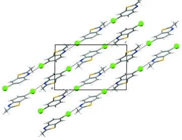
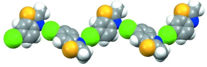
In the crystal structure halogen bonds are the principal specific interactions responsible for the crystal packing. There is
only one short and directional C—I···N contact [C—I = 2.103 (3) Å] (see Table 1) that link the molecules into antiparallel
zigzag C(7) chains (Etter, 1990; Bernstein et al., 1995) in [1 0 - 1] direction (Figs. 2 and 3).
Relatively short off-set π–π contacts [Cg···Cg = 3.758 (2) Å] between the thiazole rings, belonging to the molecules that
are related by an inversion centre, link the neighboring supramolecular chains and provide the secondary interactions for
building the crystal structure.
The structure of the title compound is one more example showing that halogen bonding is also as effective and reliable
tool for assembling molecules into supramolecular architectures.
S2. Experimental
Colourless single crystals of the title compound were obtained by slow evaporation of a dichloromethane solution.
S3. Refinement
All H atoms were placed in geometrically idealized positions and constrained to ride on their parent C atom at distances
of 0.93 or 0.96 Å for aromatic or methyl H atoms, respectively, and with Uiso(H) = 1.2Ueq(C) (for aromatic H) or Uiso(H) =
Figure 1
Molecular structure of the title compound with the atom labeling scheme. Displacement ellipsoids for non-H atoms are
drawn at the 50% probability level.
Figure 2
[image:3.610.126.487.338.616.2]Figure 3
Spacefill representaton of a zigzag halogen bonding chain running in [1 0 - 1].
6-Iodo-2-methyl-1,3-benzothiazole
Crystal data C8H6INS
Mr = 275.11 Monoclinic, P21/n
Hall symbol: -P 2yn a = 8.3255 (3) Å b = 7.6967 (3) Å c = 13.8083 (5) Å β = 90.686 (4)° V = 884.76 (6) Å3
Z = 4
F(000) = 520 Dx = 2.065 Mg m−3
Mo Kα radiation, λ = 0.71073 Å Cell parameters from 10010 reflections θ = 4.4–32.6°
µ = 3.79 mm−1
T = 296 K Plate, colourless 0.47 × 0.38 × 0.14 mm
Data collection
Oxford Diffraction Xcalibur
diffractometer with a Saphire-3 CCD detector Radiation source: Enhance (Mo) X-ray Source Graphite monochromator
Detector resolution: 16.3426 pixels mm-1
CCD scans
Absorption correction: multi-scan
(CrysAlis PRO; Oxford Diffraction, 2009) Tmin = 0.253, Tmax = 0.658
13190 measured reflections 1928 independent reflections 1729 reflections with I > 2σ(I) Rint = 0.027
θmax = 27.0°, θmin = 4.6°
h = −10→10 k = −9→9 l = −17→17
Refinement Refinement on F2
Least-squares matrix: full R[F2 > 2σ(F2)] = 0.024
wR(F2) = 0.064
S = 1.06 1928 reflections 101 parameters 0 restraints
Primary atom site location: structure-invariant direct methods
Secondary atom site location: difference Fourier map
Hydrogen site location: inferred from neighbouring sites
H-atom parameters constrained w = 1/[σ2(F
o2) + (0.0397P)2 + 0.4162P]
where P = (Fo2 + 2Fc2)/3
(Δ/σ)max = 0.001
Δρmax = 0.84 e Å−3
Experimental. Solvent used: CH2Cl2 Crystal mount: glued on a glass fibre Mosaicity (°): 1.1 (1) Frames collected: 892
Seconds exposure per frame: 5 Degree rotation per frame: 1.0 Crystal-Detector distance (mm): 50.0.
Geometry. Bond distances, angles etc. have been calculated using the rounded fractional coordinates. All su's are estimated from the variances of the (full) variance-covariance matrix. The cell e.s.d.'s are taken into account in the estimation of distances, angles and torsion angles
Refinement. Refinement on F2 for ALL reflections except those flagged by the user for potential systematic errors.
Weighted R-factors wR and all goodnesses of fit S are based on F2, conventional R-factors R are based on F, with F set to
zero for negative F2. The observed criterion of F2 > σ(F2) is used only for calculating -R-factor-obs etc. and is not
relevant to the choice of reflections for refinement. R-factors based on F2 are statistically about twice as large as those
based on F, and R-factors based on ALL data will be even larger.
Fractional atomic coordinates and isotropic or equivalent isotropic displacement parameters (Å2)
x y z Uiso*/Ueq
I1 0.96733 (2) 0.14994 (3) 0.30624 (1) 0.0485 (1)
S1 0.66879 (11) 0.75187 (9) 0.49021 (6) 0.0518 (3)
N1 0.6453 (3) 0.5615 (3) 0.64355 (18) 0.0447 (8)
C1 0.6117 (4) 0.7135 (4) 0.6097 (2) 0.0457 (9)
C2 0.7440 (3) 0.5425 (3) 0.4842 (2) 0.0392 (8)
C3 0.8155 (3) 0.4585 (4) 0.40718 (19) 0.0430 (8)
C4 0.8633 (3) 0.2887 (4) 0.42068 (19) 0.0399 (8)
C5 0.8427 (4) 0.2058 (4) 0.5097 (2) 0.0452 (9)
C6 0.7728 (4) 0.2900 (4) 0.5857 (2) 0.0467 (9)
C7 0.7211 (3) 0.4614 (3) 0.5734 (2) 0.0386 (7)
C8 0.5343 (5) 0.8546 (4) 0.6669 (3) 0.0600 (11)
H3 0.83070 0.51470 0.34840 0.0520*
H5 0.87710 0.09160 0.51740 0.0540*
H6 0.75980 0.23380 0.64480 0.0560*
H8A 0.46780 0.80420 0.71580 0.0900*
H8B 0.46950 0.92540 0.62460 0.0900*
H8C 0.61580 0.92510 0.69710 0.0900*
Atomic displacement parameters (Å2)
U11 U22 U33 U12 U13 U23
I1 0.0488 (1) 0.0567 (2) 0.0402 (1) 0.0012 (1) 0.0048 (1) −0.0055 (1)
S1 0.0658 (5) 0.0376 (4) 0.0518 (4) 0.0063 (3) −0.0035 (3) 0.0058 (3)
N1 0.0487 (13) 0.0419 (13) 0.0437 (13) −0.0022 (10) 0.0082 (10) −0.0014 (10)
C1 0.0416 (14) 0.0411 (14) 0.0543 (17) −0.0019 (12) −0.0027 (12) −0.0054 (12)
C2 0.0424 (14) 0.0345 (12) 0.0407 (13) −0.0029 (11) −0.0035 (11) 0.0049 (11)
C3 0.0482 (15) 0.0457 (14) 0.0351 (13) −0.0040 (12) −0.0001 (11) 0.0075 (12)
C4 0.0393 (14) 0.0457 (14) 0.0347 (13) −0.0025 (11) 0.0025 (11) −0.0027 (11)
C5 0.0536 (16) 0.0347 (13) 0.0474 (16) 0.0044 (12) 0.0043 (13) 0.0030 (11)
Geometric parameters (Å, º)
I1—C4 2.103 (3) C4—C5 1.397 (4)
S1—C1 1.748 (3) C5—C6 1.369 (4)
S1—C2 1.731 (2) C6—C7 1.397 (4)
N1—C1 1.289 (4) C3—H3 0.9300
N1—C7 1.395 (4) C5—H5 0.9300
C1—C8 1.494 (5) C6—H6 0.9300
C2—C3 1.385 (4) C8—H8A 0.9600
C2—C7 1.396 (4) C8—H8B 0.9600
C3—C4 1.378 (4) C8—H8C 0.9600
I1···C1i 3.826 (3) H3···H8Avii 2.5800
I1···N1ii 3.158 (2) H5···S1viii 3.1600
I1···H5iii 3.3100 H5···I1iii 3.3100
S1···H5iv 3.1600 H5···H5iii 2.5400
S1···H8Bv 3.1600 H8A···H3ix 2.5800
N1···I1vi 3.158 (2) H8B···S1v 3.1600
C1···I1i 3.826 (3)
C1—S1—C2 89.46 (14) N1—C7—C6 125.3 (2)
C1—N1—C7 110.3 (2) C2—C7—C6 119.0 (2)
S1—C1—N1 115.8 (2) C2—C3—H3 121.00
S1—C1—C8 120.0 (2) C4—C3—H3 121.00
N1—C1—C8 124.1 (3) C4—C5—H5 119.00
S1—C2—C3 129.1 (2) C6—C5—H5 120.00
S1—C2—C7 108.70 (19) C5—C6—H6 120.00
C3—C2—C7 122.2 (2) C7—C6—H6 120.00
C2—C3—C4 117.7 (2) C1—C8—H8A 110.00
I1—C4—C3 120.06 (19) C1—C8—H8B 110.00
I1—C4—C5 119.0 (2) C1—C8—H8C 109.00
C3—C4—C5 120.9 (3) H8A—C8—H8B 109.00
C4—C5—C6 121.1 (3) H8A—C8—H8C 109.00
C5—C6—C7 119.1 (3) H8B—C8—H8C 109.00
N1—C7—C2 115.7 (2)
C2—S1—C1—N1 0.5 (3) S1—C2—C7—C6 180.0 (2)
C2—S1—C1—C8 178.5 (3) C3—C2—C7—N1 −179.1 (2)
C1—S1—C2—C3 179.0 (3) C3—C2—C7—C6 0.4 (4)
C1—S1—C2—C7 −0.6 (2) C2—C3—C4—I1 178.41 (19)
C7—N1—C1—S1 −0.3 (3) C2—C3—C4—C5 −1.1 (4)
C7—N1—C1—C8 −178.2 (3) I1—C4—C5—C6 −178.8 (2)
C1—N1—C7—C2 −0.2 (3) C3—C4—C5—C6 0.8 (5)
C1—N1—C7—C6 −179.6 (3) C4—C5—C6—C7 0.2 (5)
S1—C2—C7—N1 0.5 (3)
Symmetry codes: (i) −x+2, −y+1, −z+1; (ii) x+1/2, −y+1/2, z−1/2; (iii) −x+2, −y, −z+1; (iv) x, y+1, z; (v) −x+1, −y+2, −z+1; (vi) x−1/2, −y+1/2, z+1/2; (vii)