Octaaquabis(
l
2-1
H
-pyrazole-3,5-di-carboxylato)tricopper(II) tetrahydrate
Zhi-Gang Li,aShao-Ai Li,aDe-Quan Liu,aYi-Hua Huangb and Jing-Wei Xuc*
aShenzhen Environmental Monitoring Center, Shenzhen 518008, People’s Republic
of China,bShenzhen Environmental Protecting Bureau, Shenzhen 518008, People’s
Republic of China, andcState Key Laboratory of Electroanalytical Chemistry,
Changchun Institute of Applied Chemistry, Chinese Academy of Sciences, Changchun 130022, People’s Republic of China
Correspondence e-mail: jwxu@ciac.jl.cn
Received 22 November 2009; accepted 13 January 2010
Key indicators: single-crystal X-ray study;T= 293 K; mean(C–C) = 0.005 A˚;
Rfactor = 0.036;wRfactor = 0.095; data-to-parameter ratio = 11.8.
In the trinucler CuIIcomplex molecule of the title compound, [Cu3(C5HN2O4)2(H2O)8]4H2O, the central CuII atom is located on an inversion centre and is coordinated in a distorted octahedral geometry. The equatorial sites are occupied by two N and two O atoms from two pyrazole-3,5-dicarboxylate ligands and the axial positions are occupied by two water molecules. The two other symmetry-related CuII atoms are pentacoordinated and assume a square-pyramidal geometry. In the crystal structure, coordinated and uncoordi-nated water molecules and carboxylate O atoms are linked by O—H O hydrogen bonds.
Related literature
For general background to coordination polymers, see: Yaghi
et al.(2003); Kitagawaet al.(2004). For related structures, see: Kinget al.(2003); Li (2005). For graph-set motifs, see: Bern-steinet al.(1995).
Experimental
Crystal data
[Cu3(C5HN2O4)2(H2O)8]4H2O
Mr= 712.97
a= 8.9455 (6) A˚
b= 9.1018 (7) A˚ ˚
= 90.924 (1)
= 117.505 (1)
V= 633.31 (8) A˚3
Z= 1
= 2.59 mm
T= 293 K
0.170.130.05 mm
Data collection
Bruker SMART APEX CCD area-detector diffractometer Absorption correction: multi-scan
(SADABS; Sheldrick, 1996)
Tmin= 0.666,Tmax= 0.877
3535 measured reflections 2412 independent reflections 2198 reflections withI> 2(I)
Rint= 0.010
Refinement
R[F2> 2(F2)] = 0.036
wR(F2) = 0.095
S= 1.08 2412 reflections 205 parameters 12 restraints
H atoms treated by a mixture of independent and constrained refinement
max= 0.89 e A˚
3
min=0.54 e A˚
[image:1.610.313.565.310.409.2]3
Table 1
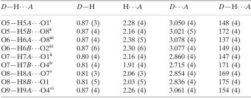
Hydrogen-bond geometry (A˚ ,).
D—H A D—H H A D A D—H A
O5—H5A O1i
0.87 (3) 2.28 (4) 3.050 (4) 148 (4) O5—H5B O8ii
0.87 (4) 2.16 (4) 3.021 (5) 172 (4) O6—H6A O8iii
0.87 (4) 2.38 (5) 3.078 (4) 137 (4) O6—H6B O2iii
0.87 (6) 2.30 (6) 3.077 (4) 149 (4) O7—H7A O1ii
0.80 (4) 2.16 (4) 2.860 (4) 147 (4) O7—H7B O4iv 0.81 (4) 1.91 (4) 2.715 (4) 171 (4) O8—H8A O7v
0.81 (3) 2.06 (3) 2.854 (4) 169 (4) O8—H8B O1 0.81 (5) 2.03 (5) 2.836 (4) 175 (4) O9—H9A O4vi
0.87 (4) 2.26 (4) 3.061 (4) 154 (4)
Symmetry codes: (i)x;y;z1; (ii)xþ1;yþ2;zþ1; (iii)x;yþ1;zþ1; (iv)x1;y;z; (v)x;y;zþ1; (vi)x1;y1;z.
Data collection:SMART(Bruker, 1998); cell refinement: SAINT-Plus(Bruker, 1998); data reduction:SAINT-Plus; program(s) used to solve structure: SHELXS97(Sheldrick, 2008); program(s) used to refine structure:SHELXL97(Sheldrick, 2008); molecular graphics: SHELXTL(Sheldrick, 2008); software used to prepare material for publication:SHELXTL.
This work was supported by the National Analytical Research Center of Electrochemistry and Spectroscopy, Changchun Institute of Applied Chemistry.
Supplementary data and figures for this paper are available from the IUCr electronic archives (Reference: IS2499).
References
Bernstein, J., Davis, R. E., Shimoni, L. & Chang, N.-L. (1995).Angew. Chem. Int. Ed. Engl.34, 1555–1573.
Bruker (1998). SMART and SAINT-Plus. Bruker AXS Inc., Madison, Wisconsin, USA.
King, P., Clerac, R., Anson, C. E., Coulon, C. & Powell, A. K. (2003).Inorg. Chem.423, 705–714.
Kitagawa, S., Kitaura, R. & Noro, S. (2004).Angew. Chem. Int. Ed.43, 2334– 2375.
Li, X.-H. (2005).Acta Cryst.E61, m2405–m2407.
Sheldrick, G. M. (1996).SADABS. University of Go¨ttingen, Germany. Sheldrick, G. M. (2008).Acta Cryst.A64, 112–122.
Yaghi, O. M., O’Keeffe, M., Ockwig, N. W., Chae, H. K., Eddaoudi, M. & Kim, J. (2003).Nature (London),423, 705–714.
Structure Reports Online
supporting information
Acta Cryst. (2010). E66, m216 [https://doi.org/10.1107/S1600536810001595]
Octaaquabis(
µ
2-1
H
-pyrazole-3,5-dicarboxylato)tricopper(II) tetrahydrate
Zhi-Gang Li, Shao-Ai Li, De-Quan Liu, Yi-Hua Huang and Jing-Wei Xu
S1. Comment
The design and synthesis of novel coordination architectures is a fertile field due to the intriguing network topologies and
potential a pplications as new classes of materials (Yaghi et al., 2003; Kitagawa et al., 2004). The ligand of
pyrazole-3,5-dicarboxylic acid has several potential coordination sites involving both two N atoms of the pyrazole ring and four
carboxylate O atoms. These multifunctional coordination sites are highly accessible to metal ions, as such, the ligand can
coordinate as a mono-, bi-, or tetradentate ligand and can act to link together metal centers through a number of bridging
modes (Li, 2005). The divalent copper atoms are easily to precipitate with the OH- when the pyrazole-3,5-dicarboxylic
acids are deprotoned in base water solution, the mixed solution can obtain coordianted polymer single crystals in
hydro-thermal condition (King et al., 2003). Nevertheless, when the ammonia was added to the mixed solution, because of the
complexing action between the copper atoms and NH3, the turbid soltuion became clear. After the ammonia slowly
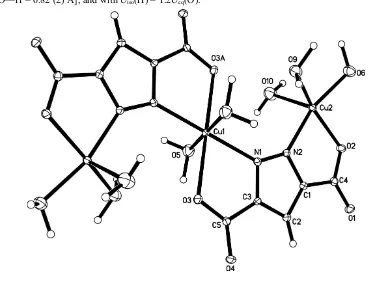
evaporated, we obtained the blue crystals, compound (I), as shown in Fig.1, a copper(II) trimer.
The central copper atom, Cu1, lies on a crystallographic inversion center. The Cu1 atom has a six-coordinate octahedral
geometry, in which two O atoms and two N atoms from two pyrazole-3,5-dicarboxylate ligands occupy the equatorial
plane, and the axial coordination sites are occupied two water molecules; the Cu—N/O bond distances range from
2.003 (2) to 2.437 (3) Å. The other two symmetry-related copper atoms, Cu2, have a pentacoordinate square-pyramidal
geometry, where a pyrazole nitrogen N2 and a carboxylate oxygen O3 from one pyrazole-3,5-dicarboxylate ligand
occupy two coordination sites and the remaining three positions are occupied by water molecules; the Cu—N/O bond
distances range from 1.984 (2) to 2.237 (2) Å. The pyrazole-3,5-dicarboxylate ligand is not strictly planar. Deviation from
the mean plane defined by the pyrazole ring is seen for both carboxylate groups with values ranging from 0.034 (1) to
0.205 (1) Å. The dihedral angle between the two carboxylate mean planes is 11.3 (3)°. It can be seen that the ligand bite
angle at the two different copper centers Cu1 and Cu2 is similar, 74.8 (4) and 80.6 (4)°, respectively. This implies that the
pyrazole-3,5-dicarboxylate ligand is a fairly rigid ligand and retains its integrity on metal chelation.
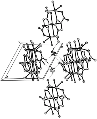
In the asymmetric unit, there are two lattice water molecules, four coordinated water molecules and carboxylate O
atoms, which form complexed hydrogen-bonding interactions. Two lattice water molecules and its symmetric equivalents
together with two carboxylate O atoms from two trimers form a hydrogen-bonded chair conformation, generating an
R46(6) motif (Bernstein et al., 1995). Meanwhile, the four lattice water molecules in each R46(6) motif also bind four
another trimers by O7—H7B···O4 hydrogen bond interaction, and O5—H5B···O8 hydrogen bond interaction. Those
strong hydrogen-bonding interactions as well as some weaker interactions, such as O5—H5A···O1, O6—H6A···O8, O6—
H6B···O2 and O9—H9A···O4, extend the crystal structure into a three-dimensional supramolecular network (Fig. 2).
S2. Experimental
The title complex was prepared by the addition of Cu(BF4)2 (20 mmol) and pyrazole-3,5-dicarboxylic acid (30 mmol) to
(yield 51% based on Cu).
S3. Refinement
Atom H2 was placed geometrically (C—H = 0.93 Å) and refined using a riding model, with Uiso(H) = Ueq(C). The H
atoms bonded to O atoms of water molecules were located in a difference Fourier map and refined, with a bond distance
[image:3.610.109.483.165.446.2]restriction [O—H = 0.82 (2) Å], and with Uiso(H) = 1.2Ueq(O).
Figure 1
A view of (I), with the atom-labeling scheme and 30% probability displacement ellipsoids. [Symmetry code: (A) 1 - x, 1 -
Figure 2
Perspective view of packing structure of (I) along the c axis. For the sake of clarity, H atoms not involved in hydrogen
bonds have been omitted.
Octaaquabis(µ2-1H-pyrazole-3,5-dicarboxylato)tricopper(II) tetrahydrate
Crystal data
[Cu3(C5HN2O4)2(H2O)8]·4H2O Mr = 712.97
Triclinic, P1 Hall symbol: -P 1
a = 8.9455 (6) Å
b = 9.1018 (7) Å
c = 9.1125 (7) Å
α = 103.485 (1)°
β = 90.924 (1)°
γ = 117.505 (1)°
V = 633.31 (8) Å3
Z = 1
F(000) = 361
Dx = 1.869 Mg m−3
Mo Kα radiation, λ = 0.71073 Å Cell parameters from 2128 reflections
θ = 2.3–26.0°
µ = 2.59 mm−1 T = 293 K Tabular, blue
Bruker SMART APEX CCD area-detector diffractometer
Radiation source: fine-focus sealed tube Graphite monochromator
φ and ω scans
Absorption correction: multi-scan (SADABS; Sheldrick, 1996)
Tmin = 0.666, Tmax = 0.877
3535 measured reflections 2412 independent reflections 2198 reflections with I > 2σ(I)
Rint = 0.010
θmax = 26.0°, θmin = 2.3° h = −10→11
k = −11→9
l = −11→9
Refinement
Refinement on F2 Least-squares matrix: full
R[F2 > 2σ(F2)] = 0.036 wR(F2) = 0.095 S = 1.08 2412 reflections 205 parameters 12 restraints
Primary atom site location: structure-invariant direct methods
Secondary atom site location: difference Fourier map
Hydrogen site location: inferred from neighbouring sites
H atoms treated by a mixture of independent and constrained refinement
w = 1/[σ2(F
o2) + (0.0495P)2 + 1.615P] where P = (Fo2 + 2Fc2)/3
(Δ/σ)max = 0.042 Δρmax = 0.89 e Å−3 Δρmin = −0.54 e Å−3
Special details
Geometry. All e.s.d.'s (except the e.s.d. in the dihedral angle between two l.s. planes) are estimated using the full covariance matrix. The cell e.s.d.'s are taken into account individually in the estimation of e.s.d.'s in distances, angles and torsion angles; correlations between e.s.d.'s in cell parameters are only used when they are defined by crystal symmetry. An approximate (isotropic) treatment of cell e.s.d.'s is used for estimating e.s.d.'s involving l.s. planes.
Refinement. Refinement of F2 against ALL reflections. The weighted R-factor wR and goodness of fit S are based on F2, conventional R-factors R are based on F, with F set to zero for negative F2. The threshold expression of F2 > σ(F2) is used only for calculating R-factors(gt) etc. and is not relevant to the choice of reflections for refinement. R-factors based on F2 are statistically about twice as large as those based on F, and R- factors based on ALL data will be even larger.
Fractional atomic coordinates and isotropic or equivalent isotropic displacement parameters (Å2)
x y z Uiso*/Ueq
Cu1 0.5000 0.5000 0.0000 0.01250 (16)
Cu2 0.18693 (5) 0.39943 (5) 0.35269 (4) 0.01365 (14)
O1 0.4661 (3) 0.8149 (3) 0.7106 (3) 0.0205 (5)
O2 0.2463 (3) 0.5801 (3) 0.5590 (3) 0.0180 (5)
O3 0.8061 (3) 0.6833 (3) 0.0762 (3) 0.0187 (5)
O4 0.9786 (3) 0.8716 (3) 0.2903 (3) 0.0198 (5)
O5 0.4555 (4) 0.6959 (4) −0.0015 (3) 0.0319 (7)
H5A 0.420 (6) 0.690 (7) −0.093 (3) 0.038*
H5B 0.541 (5) 0.797 (4) 0.040 (5) 0.038*
O6 −0.0225 (4) 0.2439 (4) 0.4269 (4) 0.0318 (7)
H6A −0.091 (5) 0.148 (4) 0.360 (5) 0.038*
H6B −0.083 (6) 0.291 (6) 0.468 (5) 0.038*
O7 0.2443 (4) 0.8545 (4) 0.1630 (3) 0.0268 (6)
H7A 0.330 (4) 0.923 (5) 0.219 (5) 0.032*
O8 0.2631 (4) 0.9378 (4) 0.8781 (3) 0.0280 (6)
H8A 0.245 (6) 0.913 (6) 0.958 (3) 0.034*
H8B 0.316 (5) 0.899 (6) 0.826 (5) 0.034*
O9 0.1851 (4) 0.2020 (4) 0.1939 (4) 0.0335 (7)
H9A 0.104 (5) 0.102 (4) 0.195 (6) 0.040*
H9B 0.282 (4) 0.202 (7) 0.197 (6) 0.040*
O10 0.0459 (4) 0.4770 (5) 0.2092 (4) 0.0415 (8)
H10A 0.102 (6) 0.580 (4) 0.202 (6) 0.050*
H10B −0.052 (4) 0.452 (7) 0.238 (6) 0.050*
N1 0.5326 (3) 0.5804 (4) 0.2330 (3) 0.0135 (6)
N2 0.4214 (3) 0.5670 (4) 0.3334 (3) 0.0136 (6)
C1 0.5045 (4) 0.6944 (4) 0.4637 (4) 0.0136 (6)
C2 0.6742 (4) 0.7934 (4) 0.4484 (4) 0.0151 (7)
H2 0.7597 0.8884 0.5196 0.015*
C3 0.6852 (4) 0.7162 (4) 0.3012 (4) 0.0125 (6)
C4 0.4011 (4) 0.6993 (4) 0.5882 (4) 0.0153 (7)
C5 0.8344 (4) 0.7607 (4) 0.2152 (4) 0.0136 (7)
Atomic displacement parameters (Å2)
U11 U22 U33 U12 U13 U23
Cu1 0.0145 (3) 0.0118 (3) 0.0090 (3) 0.0055 (2) 0.0011 (2) 0.0007 (2) Cu2 0.0111 (2) 0.0123 (2) 0.0137 (2) 0.00298 (17) 0.00279 (15) 0.00235 (16) O1 0.0194 (12) 0.0210 (13) 0.0134 (12) 0.0060 (11) 0.0028 (10) −0.0012 (10) O2 0.0141 (12) 0.0167 (12) 0.0181 (12) 0.0045 (10) 0.0046 (9) 0.0018 (10) O3 0.0171 (12) 0.0216 (13) 0.0122 (12) 0.0056 (10) 0.0040 (9) 0.0030 (10) O4 0.0116 (11) 0.0210 (13) 0.0172 (12) 0.0012 (10) 0.0018 (9) 0.0026 (10) O5 0.0366 (17) 0.0299 (16) 0.0294 (16) 0.0160 (14) 0.0037 (13) 0.0082 (13) O6 0.0251 (15) 0.0303 (16) 0.0354 (17) 0.0099 (13) 0.0063 (12) 0.0076 (13) O7 0.0209 (14) 0.0209 (14) 0.0335 (16) 0.0080 (12) 0.0098 (12) 0.0025 (12) O8 0.0286 (15) 0.0294 (16) 0.0257 (15) 0.0154 (13) 0.0053 (12) 0.0033 (12) O9 0.0323 (16) 0.0270 (16) 0.0358 (17) 0.0100 (13) 0.0082 (13) 0.0076 (13) O10 0.0351 (18) 0.0381 (19) 0.054 (2) 0.0156 (16) 0.0024 (16) 0.0220 (16) N1 0.0125 (13) 0.0140 (14) 0.0123 (13) 0.0050 (11) 0.0035 (10) 0.0035 (11) N2 0.0119 (13) 0.0146 (14) 0.0112 (13) 0.0044 (11) 0.0020 (10) 0.0018 (11) C1 0.0136 (15) 0.0139 (16) 0.0119 (15) 0.0057 (13) 0.0014 (12) 0.0032 (12) C2 0.0125 (15) 0.0157 (17) 0.0132 (16) 0.0039 (13) 0.0005 (12) 0.0031 (13) C3 0.0127 (15) 0.0126 (16) 0.0103 (15) 0.0047 (13) −0.0010 (12) 0.0027 (12) C4 0.0149 (16) 0.0166 (17) 0.0152 (16) 0.0081 (14) 0.0031 (13) 0.0047 (13) C5 0.0150 (16) 0.0130 (17) 0.0133 (16) 0.0064 (14) 0.0037 (13) 0.0048 (13)
Geometric parameters (Å, º)
Cu1—O5 2.001 (3) O6—H6B 0.87 (6)
Cu1—O5i 2.001 (3) O7—H7A 0.80 (2)
Cu1—N1 2.047 (3) O7—H7B 0.81 (2)
Cu1—N1i 2.047 (3) O8—H8A 0.81 (2)
Cu2—N2 1.985 (3) O9—H9B 0.87 (5)
Cu2—O6 2.002 (3) O10—H10A 0.85 (2)
Cu2—O9 2.021 (3) O10—H10B 0.86 (5)
Cu2—O2 2.059 (2) N1—N2 1.345 (4)
Cu2—O10 2.236 (3) N1—C3 1.354 (4)
O1—C4 1.247 (4) N2—C1 1.357 (4)
O2—C4 1.277 (4) C1—C2 1.392 (5)
O3—C5 1.255 (4) C1—C4 1.481 (5)
O4—C5 1.264 (4) C2—C3 1.390 (5)
O5—H5A 0.87 (2) C2—H2 0.9300
O5—H5B 0.87 (2) C3—C5 1.497 (4)
O6—H6A 0.87 (2)
O5—Cu1—O5i 180.0 Cu2—O6—H6B 116 (3)
O5—Cu1—N1 87.36 (12) H6A—O6—H6B 108 (5)
O5i—Cu1—N1 92.64 (12) H7A—O7—H7B 113 (5)
O5—Cu1—N1i 92.64 (12) H8A—O8—H8B 117 (5)
O5i—Cu1—N1i 87.36 (12) Cu2—O9—H9A 114 (3)
N1—Cu1—N1i 180.00 (18) Cu2—O9—H9B 114 (3)
O5—Cu1—O3i 85.89 (11) H9A—O9—H9B 110 (5)
O5i—Cu1—O3i 94.11 (11) Cu2—O10—H10A 115 (4)
N1—Cu1—O3i 105.11 (9) Cu2—O10—H10B 109 (4)
N1i—Cu1—O3i 74.89 (9) H10A—O10—H10B 114 (6)
O5—Cu1—O3 94.11 (11) N2—N1—C3 107.6 (3)
O5i—Cu1—O3 85.89 (11) N2—N1—Cu1 132.2 (2)
N1—Cu1—O3 74.89 (9) C3—N1—Cu1 116.3 (2)
N1i—Cu1—O3 105.11 (9) N1—N2—C1 108.4 (3)
O3i—Cu1—O3 180.0 N1—N2—Cu2 137.8 (2)
N2—Cu2—O6 165.29 (12) C1—N2—Cu2 113.2 (2)
N2—Cu2—O9 93.98 (12) N2—C1—C2 110.0 (3)
O6—Cu2—O9 92.94 (13) N2—C1—C4 115.9 (3)
N2—Cu2—O2 80.69 (10) C2—C1—C4 134.1 (3)
O6—Cu2—O2 88.18 (11) C3—C2—C1 103.4 (3)
O9—Cu2—O2 158.66 (12) C3—C2—H2 128.3
N2—Cu2—O10 97.78 (12) C1—C2—H2 128.3
O6—Cu2—O10 93.87 (13) N1—C3—C2 110.6 (3)
O9—Cu2—O10 99.41 (14) N1—C3—C5 119.3 (3)
O2—Cu2—O10 101.78 (12) C2—C3—C5 130.1 (3)
C4—O2—Cu2 114.3 (2) O1—C4—O2 124.6 (3)
C5—O3—Cu1 109.0 (2) O1—C4—C1 120.1 (3)
Cu1—O5—H5A 111 (3) O2—C4—C1 115.2 (3)
Cu1—O5—H5B 115 (3) O3—C5—O4 125.8 (3)
H5A—O5—H5B 110 (5) O3—C5—C3 117.4 (3)
Cu2—O6—H6A 115 (3) O4—C5—C3 116.8 (3)
Hydrogen-bond geometry (Å, º)
D—H···A D—H H···A D···A D—H···A
O5—H5A···O1ii 0.87 (3) 2.28 (4) 3.050 (4) 148 (4)
O5—H5B···O8iii 0.87 (4) 2.16 (4) 3.021 (5) 172 (4)
O6—H6A···O8iv 0.87 (4) 2.38 (5) 3.078 (4) 137 (4)
O6—H6B···O2iv 0.87 (6) 2.30 (6) 3.077 (4) 149 (4)
O7—H7A···O1iii 0.80 (4) 2.16 (4) 2.860 (4) 147 (4)
O7—H7B···O4v 0.81 (4) 1.91 (4) 2.715 (4) 171 (4)
O8—H8A···O7vi 0.81 (3) 2.06 (3) 2.854 (4) 169 (4)
O8—H8B···O1 0.81 (5) 2.03 (5) 2.836 (4) 175 (4)
O9—H9A···O4vii 0.87 (4) 2.26 (4) 3.061 (4) 154 (4)