Original Article
A prospective cohort study of negative pressure wound
therapy combined angioplasty for diabetic foot
patients with critical limb ischemia
Tao Wang1*, Rui He2*, Jun Zhao1, Qisheng Gu3, Jiacai Mei1, Mingzhe Shao1, Ye Pan1, Jian Zhang1, Haisheng
Wu1, Min Yu1, Wenchao Yang1, Limei Liu2, Fang Liu2, Weiping Jia2
1Department of Vascular Surgery, Shanghai Jiao-Tong University Affiliated Sixth People’s Hospital of Shanghai,
Shanghai Clinical Medical Center of Diabetes, Multidisciplinary Collaboration Center of Diabetic Foot, 600 Yishan Road, Shanghai 200233, China; 2Department of Endocrinology and Metabolism, Shanghai Key Laboratory of
Diabetes, Shanghai Jiao-Tong University Affiliated Sixth People’s Hospital, Shanghai Clinical Medical Center of Diabetes, Shanghai Key Clinical Center of Metabolic Diseases, Multidisciplinary Collaboration Group of Diabetic Foot, Shanghai Institute for Diabetes, Shanghai 200233, China; 3Shanghai Qisheng Institute for Biological
Material Research, Shanghai 201100, China. *Equal contributors.
Received March 11, 2016; Accepted May 26, 2016; Epub September 15, 2016; Published September 30, 2016
Abstract: To evaluate the clinical efficacy and security of combined negative pressure wound therapy (NPWT) and percutaneous angioplasty (PTA) in the treatment of diabetic foot ulcers with critical limb ischemia (CLI). We en-rolled 97 patients from 142 cases suffered from diabetic foot ulcers into this multi-center prospective cohort study. Patients who were assigned to NPWT-PTA (n=56) or NPWT (n=41) group based on standard off-leading therapy as needed, and were followed-up at 8 weeks and 20 weeks, their ankle-brachial pressure index (ABI), wound area, healing time and adverse events were monitored. A multivariate Cox proportional hazards regression analysis was used to determining the risk factors of wound healing. A greater proportion of wound healing in NPWT-PTA group was observed compared with NPWT group (61.5% vs 40.9%, P=0.004) at 20 weeks. The time of wound healing was earlier in NPWT-PTA group than NPWT group (48.3±32.8 days vs 77.1±27.1 days, P=0.009) at 20 weeks. ABI in NPWT-PTA group was higher than NPWT group at both 8 weeks (0.83±0.19 vs 0.46±0.15, P=0.000) and 20 weeks (0.72±0.17 vs 0.53±0.12, P=0.000). The decrease of ABI (HR=15.000, 95% CI=2.243-100.333, P=0.005) and increase of wound area (HR=0.926, 95% CI=0.866-0.990, P=0.025) were independent risk factors of wound healing. There was no significant difference in adverse events between the two groups. Combined NPWT with PTA therapy is effective and safe for diabetic foot ulcers with CLI by increasing the level of ABI, and results in a higher proportion of wounds closure.
Keywords: Negative pressure wound therapy, percutaneous angioplasty, diabetic ulcer, critical limb ischemia
Introduction
Diabetic foot wounds are one of the most seri-ous and complex sequelae of diabetes mellitus [1, 2]. Non-healing chronic diabetic wounds are often large and deep with compromised wound healing capacity [3]. Various diabetic foot wound treatments have been reported in the literature, including advanced moist wound therapy [4], treatment with growth factors [5], bioengineered tissue or skin substitutes [6], NPWT (negative pressure wound therapy) [7] and so on.
Over the past several years, NPWT has been applied to treat these complex diabetic wound. It is a noninvasive system that creates a local-ized subatmospheric pressure environment,
accelerates wound healing through creating a moist healing environment, preparing the wound bed for closure, reducing edema, and promoting formation of granulation tissue. And it is the delivery of intermittent or continuous subatmospheric pressure by a special pump to keep a closed environment which is connected to an open-celled, resilient and foam-surface dressing covered with an adhesive drape, and
high efficacy of NPWT in treating diabetic foot
wound has been elaborated in several studies [8].
survey (involving more than 1200 diabetic foot patients in 14 highly specialized centers from 10 different European countries) showed that 49% of patients have a preeminent ischemic
component which influences the evolution of
the diabetic foot pathology, leading to a non-healing wound and eventually a major amputa-tion [10]. Meanwhile, some studies have report-ed that the successful revascularization of dia-betes mellitus (DM) patients with critical limb
ischemia (CLI) were beneficial to improve the
healing process of diabetic ischemic ulcers [11]. At present, although bypass surgery still
plays a significant role in the revascularization
of CLI [12], increasing clinical experience over the past two decades shows that endovascular strategies including PTA have low complication rates and high limb salvage rates comparable with bypass surgery [13-16].
To our knowledge, there is little known about
the efficacy of combined NPWT and PTA to treat
diabetic wound with CLI. Therefore, we under-took a prospective cohort study to investigate the effect of NPWT-PTA compared to the thera-py of NPWT in diabetic foot patients accompa-nied by low limb ischemia.
Materials and methods Study design
We did a prospective cohort study to
investi-gate the efficacy of NPWT-PTA therapy in dia
-betic foot wound with CLI. Patients flow through
each stage of this trial including discontinued patients was described in Figure 1. During the
years with ulcer categorized Wagner grade ≥ 2,
presence of CLI (local stenosis > 50% of vessel lumen) according to the criteria of management of Peripheral Arterial Disease in the Trans-Atlantic Inter-Society Consensus Document (TASC II) [12], and evidence of inadequate
per-fusion (defined as ankle brachial index < 0.7).
We excluded patients with wounds resulting
from venous insufficiency, burns, untreated cel -lulitis, untreated osteomyelitis and malignant disease in the wound. A previous PTA in the past months, previous VAC (Vacuum Assisted Closure) therapy in the past 30 days, previous treatments with growth factors or hyperbaric medicine in the past 30 days were also regard-ed as exclusion criteria. Finally, patients also were excluded if they had no stenosis or occlu-sion and were being treated with immunosup-pressive drugs, corticosteroids, or chemothera-py. Finally, seventy patients were recruited and allocated a treatment. The study protocol was approved by Ethics Committee of Shanghai Sixth People’s Hospital and written consent was obtained from all subjects.
Patients data
The cohort study to investigate the efficacy of
[image:2.612.92.370.73.294.2]NPWT-PTA therapy conducted by the Vacuum Assisted Closure (VAC) Therapy system (Weigao medical technology co., China) and PTA in dia-betic foot wound with CLI. All patients in NPWT-PTA group were added to evaluate the feasibili-ty and advisabilifeasibili-ty of performing PTA. If this procedure failed, a bypass surgery should be
Figure 1. Flowchart for patients’ enrollment and follow-up process.
course of this study, 142 pa- tients were consented and screened for inclusion from January 2011 to January 20- 14. Of these, 45 patients were excluded according to the in- clusion and exclusion criteria, patients’ refusal to participa- te, and 97 patients were enrolled. Seven patients lost to follow-up because of failed return for next visit or devel-oped gangrene foot, 20 pa- tients discontinued for their self-withdrawal or accidental death. Finally, 70 patients were analyzed.
Inclusion criteria for the study
considered. PTA was performed with contralat-eral retrograde femoral catheterization. A guide wire was applied to pass through any arterial stenosis or obstructions in NPWT-PTA group,
and a Ф3-8 mm balloon catheter was inserted
into the stenosis for revascularization. The mean length of the re-canalized segments was 10.6 cm (ranging from 1 to 27 cm). Nitinol stents (eV3 Inc., USA or Opti Med Co. Ger) were placed above the knee if need. For the
below-the-knee PTA, a 2.5-3 mm low-profile balloon in
a 0.014-inch system (Savy, Cordis Corp., USA) was employed. During the procedure 3000 IU-5000 IU of heparin was infused intravenous-ly. The preoperative medication of acetylsali-cylic acid 100 mg or clopidogrel 75 mg daily started at least 72 h before the procedure in the all cases. After the procedure, all patients were prescribed aspirin 100 mg and LMWH (Low-molecular-weight Heparin) 0.8 mg per day. Patients assigned to the NPWT-PTA and NPWT group all been arranged VAC therapy system for 3-7 days according to standardized treatment guidelines. The subatmospheric pressure in VAC therapy system uses sterile polyurethane
or polyvinyl alcohol foam dressing which is fit -ted to the appropriate size for the wound, then covered with an adhesive drape to create a closed environment. A tube attached to the
drape connects to a fluid collection canister
contained in a portable, programmable, com-puter-controlled vacuum pump (negative pres-sure of -125 mmHg) [17, 18]. Wounds were treated with VAC system until they were closed or until the therapeutic completion of 20 weeks. The difference between two groups was that the patients in the NPWT-PTA group wound receive PTA before or after NPWT. After the PTA, the patients would receive antiplatelet therapy consisted of Aspirin 100 mg/day and clopido-grel 75 mg/day. Patients in two groups were
followed-up weekly on an outpatient for the first
month, then every two weeks for the second month and every month up to the 20th week.
Object of observing
During each visit, ABI and the area of ulcer and adverse events were evaluated. Before enter-ing the study, all patients underwent a baseline evaluation of the extent and severity of periph-eral artery using the ABI, Ultrasonic Doppler technology and either computed tomographic angiography (CTA) or magnetic resonance angi-ography (MRA). The blood sample was
collect-ed to assess serum albumin and HbA1c. If the
concentration of albumin ≤ 30 g/L, a nutrition -ist was consulted and a dietary supplementa-tion was needed. To assess sensory neuropa-thy, we evaluated patients with vibration per-ception threshold (VPT) test [19].
The primary objective of this study was to
clari-fy the clinical efficacy of NPWT-PTA therapy sys -tem in treating diabetic wound with CLI, so it was needed to assess the time of wound heal-ing and the proportion of healheal-ing at 20 weeks. Assessment was based on the date from wound investigations on days 0, 8, and 20 weeks. Complete wound closed was regarded as 100% re-epithelization without drainage. Secondary aim included assessment of
adverse events which defined as any untoward
medical occurrences that resulted in death, were life-threatening, extended hospital care,
caused significant incapacity or disability. In this study we defined adverse events as deaths,
cardiovascular events, cerebral events, minor and major second amputation (excluding the conducted amputation before the time of therapy).
Statistical analysis
Continuous demographic variables were sum-marized with descriptive statistics (mean ± SD) and compared with a two-sample t test between the two groups. The ABI at 0, 8 and 20 weeks were analyzed using one way ANOVA. Cate- gorical demographic variables were expressed as a proportion of the population and com-pared with a two-tailed Fisher’s exact test. We analyzed the data of wound healing time in the two groups using a time-to-event strategy with Kaplan-Meier analysis which followed by a log-rank test, and patient not achieving closure was censored using last day of observation. This statistical approach had provided a com-parison of the distribution of wound healing time in the two treatment groups. The Cox sur-vival model included terms of age, sex, diabe-tes duration, serum albumin levels, Serum cre-atinine, hemoglobin A1c, ALT, AST, C-reactive protein levels, Loss of protective sensation, Ulcer area, Ulcer duration before treatment and ABI, they were used to evaluate covariates (wound healing) and recorded as their hazard
ratio with a confidence interval of 95%. The
calculated at 20 weeks, P < 0.05 was consid
-ered as significant.
Results
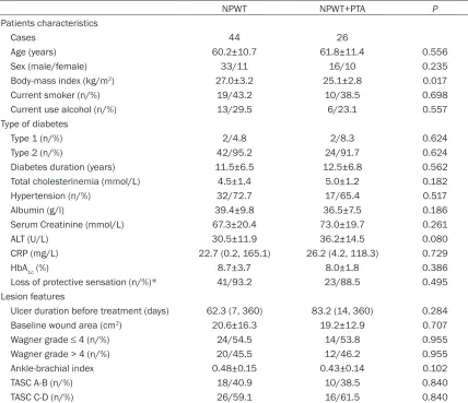
During the study, 70 patients were enrolled in our trial, and the Table 1 summarized their clini-cal characteristics at baseline and risk factors. Of the diabetic patients enrolled in this study, the patients in each group were predominantly male and the mean time of diabetes duration was more than 10 years. And the data showed
that no statistically significant demographic dif -ference existed between the treatment groups (P > 0.05).
Efficacy
An example of extreme distal PTA and NPWT was showed in Figure 2. All patients PTA-treated in NPWT-PTA group underwent
suc-cessfully revascularization and negative pres-sure therapy. Among the 26 procedures in NPWT-PTA group, 2 (7.7%) PTAs were exclusive-ly performed in the proximal segment, 10 (38.5%) exclusively in the infrapopliteal arter-ies, and 12 (53.8%) exclusively in the femoro-popliteal plus infrafemoro-popliteal arteries. The initial technical success of PTA was 92.3% in 24 patients (the patients underwent angioplasty were only one) in NPWT-PTA group, the other (2 patients) initially failed PTA procedures required bypass surgery because of suboptimal
dilata-tions in the context of heavy vessel calcifica -tions. The accumulative potencies at 8 and 20 weeks respectively were 96.2% and 84.6%. No patients died during hospitalization, and the procedure rule out any complications in the puncture site.
[image:4.612.92.520.95.464.2]A greater proportion of wound in NPWT-PTA group healed completely compared with NPWT Table 1. Clinical characteristics and risk factors of patients with diabetic foot ulcers with critical limb ischemia
NPWT NPWT+PTA P
Patients characteristics
Cases 44 26
Age (years) 60.2±10.7 61.8±11.4 0.556
Sex (male/female) 33/11 16/10 0.235
Body-mass index (kg/m2) 27.0±3.2 25.1±2.8 0.017
Current smoker (n/%) 19/43.2 10/38.5 0.698
Current use alcohol (n/%) 13/29.5 6/23.1 0.557
Type of diabetes
Type 1 (n/%) 2/4.8 2/8.3 0.624
Type 2 (n/%) 42/95.2 24/91.7 0.624
Diabetes duration (years) 11.5±6.5 12.5±6.8 0.562 Total cholesterinemia (mmol/L) 4.5±1.4 5.0±1.2 0.182
Hypertension (n/%) 32/72.7 17/65.4 0.517
Albumin (g/l) 39.4±9.8 36.5±7.5 0.186
Serum Creatinine (mmol/L) 67.3±20.4 73.0±19.7 0.261
ALT (U/L) 30.5±11.9 36.2±14.5 0.080
CRP (mg/L) 22.7 (0.2, 165.1) 26.2 (4.2, 118.3) 0.729
HbA1c (%) 8.7±3.7 8.0±1.8 0.386
Loss of protective sensation (n/%)* 41/93.2 23/88.5 0.495 Lesion features
Ulcer duration before treatment (days) 62.3 (7, 360) 83.2 (14, 360) 0.284 Baseline wound area (cm2) 20.6±16.3 19.2±12.9 0.707
Wagner grade ≤ 4 (n/%) 24/54.5 14/53.8 0.955
Wagner grade > 4 (n/%) 20/45.5 12/46.2 0.955
Ankle-brachial index 0.48±0.15 0.43±0.14 0.102
TASC A-B (n/%) 18/40.9 10/38.5 0.840
TASC C-D (n/%) 26/59.1 16/61.5 0.840
([61.5%] vs [40.9%], P=0.004) at 20 weeks. The time of wound healing, based on the com-plete ulcer closure (100% re-epithelization), was faster in NPWT-PTA group than NPWT group (48.3±32.8 vs 77.1±27.2, P=0.009) at 20 weeks. And the duration of therapy of NPWT in NPWT-PTA group was 33.12±15.86 days (means ± SD) versus 45.34±23.24 days in NPWT group (P=0.021).
ABI was assessed both before and after PTA. Figure 3A showed that a significant improve -ment of ABI in NPWT-PTA group were detected after the PTA, peaking at 8 weeks (0.83±0.19 vs 0.46±0.15, P=0.000), 20 weeks (0.72±0.17 vs 0.53±0.12, P=0.000) after the PTA, it still
was significantly higher than the baseline
(Figure 3A). There was no significant difference
was detected when it in NPWT group during the treatment (Figure 3B).
The healing time of complete ulcer closure in NPWT-PTA group was 83.5±10.0 days (95% CI 63.9-103.2). Healing time in NPWT group was 114.3±5.3 (95% CI 103.8-124.7) days; the Kaplan-Meier median time to complete ulcer closure was 70.0±30.0 (95% CI 11.3-128.8, P=0.023) days (Figure 3C). In separate multi-variate Cox proportional hazards regression models, the decrease of ABI (HR=15.000, 95% CI=2.243-100.333, P=0.005) and the increase of wound area (HR=0.926, 95%
CI=0.866-0.990, P=0.025) were significant risk increas -ing factors for days of wound heal-ing (Table 2). The survival function at mean of covariates was showed in Figure 3D.
Safety
[image:5.612.90.523.71.401.2]After adequate preparation of wound bed, 15.4% (4 of 26) NPWT-PTA-treated ulcers and
13.6% (6/44) NPWT-treated ulcers were
[image:6.612.92.517.75.368.2]surgi-cally closed after skin grafts transplantation. wound healing ratio, a shorter ulcer healing time, a higher ABI, and potential trend towards
Figure 3. ABI values in the two groups before and after therapy. A significant improvement of ABI in NPWT-PTA group were found after the PTA (A); There was no significant difference was found when it in NPWT group during the treat -ment (B). Kaplan-Meier estimates for time to complete ulcer closure (P=0.023). Continuous line: NPWT-PTA group; dash line: NPWT group (C). Survival Function at mean of covariates (D); *P < 0.05 when compared with the corre -sponding controls; #P < 0.05 when compared with the ABI at 8 week in NPWT-PTA group; &P < 0.05 when compared with the ABI at 0 week in NPWT-PTA group; $P < 0.05 compared with the ABI at 0 week in NPWT-PTA group.
Table 2. Hazard ratio of Patients with Diabetic Foot Ulcers with Critical Limb Ischemia
Hazard ratio (95% CI) P Baseline wound area (cm2) 0.926 (0.866-0.990) 0.025
Ankle-brachial index 15.000 (2.243-100.333) 0.005
Age (years) NS
Sex (male/female) NS
Diabetes duration (years) NS
Albumin (g/l) NS
Serum Creatinine (mmol/L) NS
ALT (U/L) NS
AST (U/L) NS
CRP (mg/L) NS
HbA1c (%) NS
Loss of protective sensation (n/%) NS Ulcer duration before treatment (days) NS
NS: No Significance; ALT: Alanine aminotransferase; AST: Aspartate aminotransfer-ase; CRP: C-Reactive Protein.
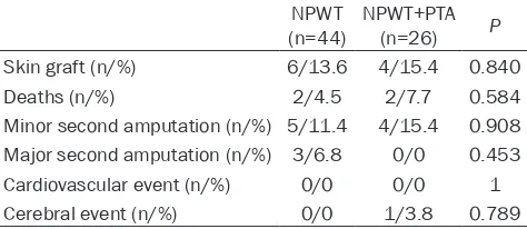
No major second amputation was observed in the NPWT-PTA group. Table 3 displays treat-ment-related rates for healing time, skin graft, deaths, minor second amputation, major sec-ond amputation, cardiovascu-lar event and cerebral event at 20 weeks. In all other
catego-ries, no significant statistically
differences were observed.
Discussion
This trial assessed the out-comes in diabetic foot ulcer with critical limb ischemia (CLI) through NPWT combined with
PTA, and our results firstly
[image:6.612.91.371.485.671.2]reducing the risk of major second amputation than NPWT in the patients with diabetic foot ulcerations. And it seemed to be a quite safe
therapy, with no significant differences
observed in proportion and distribution of adverse events compared with controlled group.
This study has unique characteristics that
dis-tinguish it from previous trials as followed: first -ly, we reported that the therapy NPWT com-bined with PTA has an obvious advantage to improve the diabetic wound healing and blood supply of foot; secondly, the ulcers enrolled into this trial were larger and more complex than previously reported. The ulcers in previous
study mainly focused on superficial neuropath -ic foot ulcers [20-22]. Thirdly, little was known that a prospective cohort trial included con-trolled group. And in this study, the negative controlled group (NPWT) had been set up to synthetically compare the therapeutic effect of NPWT-PTA.
The results indicated the difference in propor-tion of healing between treatment groups at the end of trial. The main reason attributed to the severity of these ulcers which posed a larg-er size, a deeplarg-er depth and accompanied with the presence of pre-existing infection. Such wounds needed debridement because they enable removal of devitalized and necrotic tis-sue. NPWT system in conjunction with debride-ment could contribute a lot advantages to the wound healing process [23]. And major clinical trials have observed that NPWT therapy
deliv-ered through the VAC system seems to effi -ciently stimulate a robust granulation tissue response compared with other therapies [24, 25]. As reported in this study, the wounds
treat-Thus the successful revascularization is far
more important to the final diabetic wound
healing. At present, although bypass surgery
still plays a significant role in the revasculariza -tion of CLI [12], increasing clinical experience over the past two decades shows that endovas-cular strategies (PTA) has low complication rates and high limb salvage rates comparable with bypass surgery [13-16]. In this study, we
have a low-risk and efficient procedure in PTA,
and we found that combination of NPWT and PTA had more advantage to improve wound healing compared with only NPWT treatment. This could explain the reason why the high level of blood supply of foot in NPWT-PTA group
which was achieved by opening specific path
-ways of flow. Meantime, depending soly on the
PTA was inadequated. Active infection may induce further hypoxia and tissue loss which means revascularization would be in vain. In this study, Cox proportional hazards regres-sion models showed that the decrease of ABI and the enlargement of wound area were
sig-nificant risk increasing factors. And our results
found that PTA treatment in NPWT-PTA group
significantly increased the ABI of wound tissue
compared with NPWT groupat 8 weeks, and also at 20 weeks, and in line with that described by Nylaende et al who claimed that the ABI at 3
and 12 months were highly significantly
improved in favor of PTA in the patients who diagnosed as peripheral arterial occlusive dis-ease without diabetes [27], the difference between our study was that their treatment didn’t include the NPWT. Also, these data were
confirmed by Aust et al who had found that a
[image:7.612.91.328.98.201.2]targeted peripheral vessel reopening before debridement helped to improve chronic lower extremity wounds [28]. However, the difference in our study is that the debridement in the latter Table 3. The Comparison of Adverse Events in the two
groups
NPWT
(n=44) NPWT+PTA (n=26) P Skin graft (n/%) 6/13.6 4/15.4 0.840 Deaths (n/%) 2/4.5 2/7.7 0.584 Minor second amputation (n/%) 5/11.4 4/15.4 0.908 Major second amputation (n/%) 3/6.8 0/0 0.453 Cardiovascular event (n/%) 0/0 0/0 1 Cerebral event (n/%) 0/0 1/3.8 0.789
Minor second amputation: amputation below foot; Major second amputation: amputation above foot.
ed with NPWT had achieved faster wound
closure. This result paralleled the findings
reported by Peter et al who determined
that NPWT was as safe as and more effi -cient than moist healing dressing for the treatment of diabetic foot ulcers [26]. The difference to ours was that their thera-pies were without PTA during the therapy of NPWT.
However, the majority of patients have a
observation was the treatment without nega-tive pressure wound therapy, and the applied wound was not diabetic foot.
A greater percentage of patients in NPWT group underwent a major second amputation (above foot) during the observing period. This different rate of second amputation probably resulted from the rapid and higher proportion wound healing. PTA could also contribute to the avoid-ance of the second major amputation, because
it can improve significantly blood supply of foot,
and it is a major independent prognostic factor for the major amputation [29]. Reiber et al believed that the major amputation (above-the-ankle) rate in patients who successfully under-went PTA is lower than the literature data [30], as it also showed in our study. We assumed that PTA has increased the likelihood of suc-cessfully conducting minor second amputa-tions, which would decrease the probability of major second amputation.
Limitations
This study has some potential disadvantages that are inherent to the procedure studied. The most important aspect is that this study was
confined by the small number of cases in the
two groups as well as by its retrospective nature; Secondly, although effective in improv-ing the early and medium period outcomes, PTA posed a poor long-dated patency rate, resulting from the evolution of the underlying pathology that causes recurrences very frequent in dia-betic patients; Thirdly, percutaneous oxygen pressure examination was not applied during the study.
In summary, the present study confirmed that NPWT-PTA combination was an efficient and
safe strategy for the diabetic foot ulcer accom-panied with critical limb ischemia. Treatment with NPWT-PTA System results in a higher pro-portion of wounds closure, shorter healing time, more adequate blood supply of foot, and a potential trend towards reduced risk for major second amputation than with sole NPWT thera-pies. Meantime it was a quite safe therapy. In the future, we will investigate the effect about
length of hospital stay, cost efficacy, and quali -ty of life in postoperative patients. We look for-ward to further understanding the NPWT-PTA Therapy System and improve the treatment for diabetic foot ulcer with critical limb ischemia in the near future.
Acknowledgements
The authors are thankful to Shanghai Key Laboratory of Diabetes for providing necessary help for carrying out the research work. The present study was supported by grants from Natural Science Foundation of China (8127- 0397 for Fang Liu) and Fund of Shanghai Health Bureau (No 201344197 for Jian Zhang). Disclosure of conflict of interest
None.
Address correspondence to: Jun Zhao, Department of Vascular Surgery, Shanghai Jiao-Tong University Affiliated Sixth People’s Hospital of Shanghai, Shanghai Clinical Medical Center of Diabetes, Multidisciplinary Collaboration Center of Diabetic Foot, 600 Yishan Road, Shanghai 200233, China. Fax : 86-21-64369586; E-mail: junnzhao@126.com; Fang Liu, Department of Endocrinology and Metabolism, Shanghai Key Laboratory of Diabetes, Shanghai Jiao-Tong University Affiliated Sixth People’s Hospital, Shanghai Clinical Medical Center of Diabetes, Shanghai Key Clinical Center of Metabolic Diseases, Multidisciplinary Collaboration Group of Diabetic Foot, Shanghai Institute for Diabetes, Shanghai 200233, China. Fax: 86-21-64368031; E-mail: f-liu@sjtu.edu.cn
References
[1] Singh N, Armstrong DG, Lipsky BA. Preventing foot ulcers in patients with diabetes. JAMA 2005; 293: 217-228.
[2] Lavery LA, Armstrong DG, Wunderlich RP, Tredwell J, Boulton AJ. Diabetic foot syndrome: evaluating the prevalence and incidence of foot pathology in Mexican Americans and non-Hispanic whites from a diabetes disease man-agement cohort. Diabetes Care 2003; 26: 1435-1438.
[3] Armstrong DG, Frykberg RG. Classifying dia-betic foot surgery: toward a rational definition. Diabet Med 2003; 20: 329-331.
[4] Wiwanitkit V. Vacuum-assisted closure and moist wound dressing in diabetic foot. J Cutan Aesthet Surg 2013; 6: 173.
[5] Ram M, Singh V, Kumawat S, Kumar D, Lingaraju MC, Uttam Singh T, Rahal A, Kumar Tandan S, Kumar D. Deferoxamine modulates cytokines and factors to accelerate cutaneous wound healing in diabetic rats. Eur J Pharmacol 2015; 764: 9-21.
re-sults of prospective randomized trial. Diabetes Care 2003; 26: 1701-1705.
[7] Li X, Liu J, Liu Y, Hu X, Dong M, Wang H, Hu D. Negative pressure wound therapy accelerates rats diabetic wound by promoting agenesis. Int J Clin Exp Med 2015; 8: 3506-3513.
[8] Schintler M. Negative pressure therapy: theory and practice. Diabetes Metab Res Rev 2012; 28: 72-77.
[9] Faglia E, Caravaggi C, Marchetti R, Mingardi R, Morabito A, Piaggesi A, Uccioli L, Ceriello A. Screening for peripheral arterial disease by means of the ankle-brachial index in newly di-agnosed type 2 diabetic patients. Diabet Med 2005; 22: 1310-1314.
[10] Prompers L, Huijberts M, Apelqvist J, Jude E, Piaggesi A, Bakker K, Edmonds M, Holstein P, Jirkovska A, Mauricio D, Ragnarson Tennvall G, Reike H, Spraul M, Uccioli L, Urbancic V, Van Acker K, Van Baal J, Van Merode F, Schaper N. High prevalence of ischemia, infection and se-rious comorbidity in patients with diabetic foot disease in European. Baseline results from the Eurodiale study. Diabetologia 2007; 50: 18-25.
[11] Alexandrescu V, Hubermont G, Philips Y, Guillaumie B, Ngongang Ch, Coessens V, Vandenbossche P, Coulon M, Ledent G, Donnay JC. Combined primary subintimal and endolu-minal angioplasty for ischemic inferior-limb ul-cers in diabetic patients: 5-year practice in a multidisciplinary ‘diabetic-foot’ service. Eur J Vas Endovasc Surg 2009; 37: 448-456. [12] Norgreen L, Hiatt WR, Dormandy JA, Nehler
MR, Harris KA, Fowkes FG; TASC II Working Group, Bell K, Caporusso J, Durand-Zaleski, Komori K, Lammer J, Liapis C, Novo S, Razavi M, Robbs J, Schaper N, Shigematsu H, Sapoval M, White C, White J, Clement D, Creager M, Jaff M, Mohler E, Rutherford RB, Sheehan P, Sillesen H, Rosenfield K. Inter-Society Consensus for the management of peripheral arterial disease (TASC II). Eur J Vasc Endovasc Surg 2007; 33 Suppl 1: S32-S55.
[13] Blevins WA, Schneider PA. Endovascular man-agement of critical limb ischemia. Eur J Vasc Endovasc Surg 2010; 39: 756-761.
[14] Conrad MF, Kang J, Cambria RP, Brewster DC, Watkins MT, Kwolek CJ, LaMuraglia GM. Infrapopliteal balloon angioplasty for the treat-ment of chronic occlusive disease. J Vasc Surg 2009; 50: 799-805.
[15] Romiti M, Albers M, Brochado-Neto FC, Durazzo AE, Pereira CA, De Luccia N. Meta-analysis of infrapopliteal angioplasty for chron-ic critchron-ical limb ischemia. J Vasc Surg 2008; 47: 975-981.
[16] Adam DJ, Beard JD. Cleveland T, Bell J, Bradbury AW, Forbes JF, Fowkes FG, Gillepsie I,
Ruckley CV, Raab G, Storkey H; BASIL trial par-ticipants. BASIL trial parpar-ticipants. Bypass ver-sus angioplasty in severe ischemia of the leg (BASIL): Multicentre, randomized controlled trial. Lancet 2005; 366: 1925-1934.
[17] Banwell PE. Topical negative pressure therapy in wound care. J Wound Care 1999; 8: 79-84. [18] Banwell PE, Teot L. Topical negative pressure
(TNP): the evolution of a novel wound therapy. J Wound Care 2003; 12: 22-28.
[19] Boulton AJ, Vinik AI, Arezzo JC, Bril V, Feldman EL, Freeman R, Malik RA, Maser RE, Sosenko JM, Ziegler D. Diabetic neuropathies: a state-ment by the American Diabetes Association. Diabetes Care 2005; 28: 956-962.
[20] Veves A, Sheehan P, Pham HT. A randomized, controlled trial of Prompgran (a collagen/ oxi-dized regenerated cellulose dressing) vs stan-dard treatment in the management of diabetic foot ulcers. Arch Surg 2002; 137: 822-827. [21] Veves A, Falanga V, Armstrong DG, Sabolinski
ML. Graftskin, a human skin equivalent, is ef-fective in the management of noninfected neu-ropathic diabetic foot ulcers: a prospective randomized multicenter clinical trial. Apligraf Diabetic Foot Ulcer Study. Diabetes Care 2001; 24: 290-295.
[22] Marston WA, Hanft J, Norwood P, Pollak R. The efficacy and safety of Dermagraft in improving the healing of chronic diabetic foot ulcers: re-sults of a prospective randomized trial. Diabetes Care 2003; 26: 1701-1705.
[23] Saxena V, Hwang CW, Huang S, Eichbaum Q, Ingber D, Orgill DP. Vacuum-assisted closure: microdeformations of wounds and cell prolif-eration. Plast Reconstr Surg 2004; 114: 1086-1096.
[24] Armstrong DG, Attinger CE, Boulton AJ, Fryberg RG, Kirsner RS, Lavery LA, Mills JL. Guidelines regarding negative pressure wound therapy in the diabetic foot: results of the Tucson expert consensus conference. Ostomy Wound Mana- ge 2004; 50 Suppl: 3S-27S.
[25] Armstrong DG, Lavery LA. Negative pressure wound therapy after partial diabetic foot am-putation: a multicentre, randomized controlled trial. Lancet 2005; 366: 1704-1710.
[26] Blume PA, Ayala J, Walters J, Ayala J, Lantis J. Comparison of negative pressure wound thera-py using vacuum-assisted closure with ad-vanced moist wound therapy in the treatment of diabetic foot ulcers. Diabetes Care 2008; 31: 631-636.
Angioplasty versus Conservative Treatment (OBACT) study. Vasc Med 2007; 12: 275-283. [28] Aust MC, Spies M, Guggenheim M, Gohritz A,
Kall S, Rosenthal H, Pichlmaier M, Oehlert G, Vogt PM. Lower limb revascularization preced-ing surgical wound coverage-an interdisciplin-ary algorithm for chronic wound closure. J Plast Reconstr Aesthet Surg 2008; 61: 925-933.
[29] Pecoraro RE, Ahroni JH, Boyko EJ, Stensel VL. Chronology and determinants of tissue repair in diabetic lower-extremity ulcers. Diabetes 1991; 40: 1305-1313.