Original Article
Neoadjuvant chemotherapy, wide resection and custom
prosthetic replacement for tumors of the
proximal femur
Ya-Nan Wu*, Dong-Yu Wang*, Wen-Zhi Bi, Gang Han, Jin-Peng Jia, Meng Xu
Department of Orthopedics and Rehabilitation, PLA General Hospital, Beijing 100853, PR, China. *Co-first au-thors.
Received December 6, 2015; Accepted March 19, 2016; Epub November 15, 2016; Published November 30, 2016
Abstract: Endoprosthetic reconstruction after bone tumor resection is accepted treatment for femoral tumors. The objective of this study is to evaluate the effectiveness and functional outcomes of custom prosthetic replacement of the proximal femur in patients undergoing oncologic resection. Data of 28 consecutive patients with proximal femur tumors receiving wide resection and prosthetic replacement (16 total hip replacements [THR], 12 bipolar half-hip replacements [HHR]) from January 2005-June 2014 were analyzed retrospectively. Postoperative lower limb func-tion was evaluated using American Musculoskeletal Tumor Society 1993 funcfunc-tion scores (MSTS 93). Mean follow-up was 28.6 months (range: 6-138 months). No patients had prosthesis-related complications or revision surgery, 3 had local recurrence of malignancy (8.3%), and 2 had venous thromboembolism resolved with medication. Mean MSTS 93 score was 84.8% (THR 85% vs. HHR 81%, P=0.044). Two patients (10.7%) suffered relapse and 9 (32.1%) died from lung metastasis. Survival rates were 91.7% at 1 year in both groups; 64.8% and 59.4% in THR and HHR groups after 30 months. In conclusions, Neoadjuvant chemotherapy, extended tumorectomy and special prosthetic replacement of proximal femur effectively treated femoral tumors, with minimal local recurrence and satisfactory functional outcomes.
Keywords: Femoral tumor, prosthesis, neoadjuvant chemotherapy, function reconstruction, half-hip replacement (HHR), total hip replacement (THR)
Introduction
The proximal femur is one of the most common locations for primary malignant bone tumors and metastatic bone tumors [1]. Approximately 16% of Ewing’s sarcomas, 13% of chondrosar-comas and 10% of osteosarchondrosar-comas develop at this location [2]. Amputation has generally been performed for patients with malignant tumors of the proximal femur, which may arrest the tumor but leads to patients’ physical dis-ability [3]. However, as a result of the growing awareness of bone tumors, along with improve-ments in chemotherapy, advances in surgical techniques, application of new reconstruction materials and improved imaging techniques, more and more patients with malignant tumors of the proximal femur can receive limb salvage surgery as a more acceptable alternative to amputation [3, 4].
noted for resulting in fewer complications than modular endoprosthesis, it is considered to be the most expensive option for replacement of the proximal femur [10]. On the other hand, modular endoprosthesis offers procedural and cost advantages for limb salvage surgeries, but may not produce functional outcomes equal to those of custom prosthesis. A recent study reported 99% limb salvage with no mechanical failure or periprosthetic fractures, concluding that modular endoprosthetic replacement was an exceptionally durable reconstructive option after resecting proximal femoral tumors [3]. Bernthal et al. [11] reported long-term survival of cemented bipolar proximal femoral replace-ment in 86 patients, concluding that endopros-thetic reconstruction represented a durable technique. In that study, survival was similar between modular prostheses and custom-designed prostheses.
We hypothesized that combined treatment of proximal tumors with effective pre- and postop-erative chemotherapy and custom-designed prosthesis would not only provide a better sur-gical outcome in terms of function, but would also reduce patients’ recurrence rate and increase their survival rate. Based on our prior experience with 104 cases of osteosarcoma near the knee, [12] preoperative MRI examina-tion would accurately determine the optimum tumor resection range and prosthesis size, helping to facilitate effective tumorectomy and preserve limb function. Therefore, the present study aimed to explore the effectiveness, key factors and functional outcomes of custom-designed prosthetic replacement of the proxi-mal femur in patients undergoing oncologic resection of femoral tumors and soft-tissue sarcomas.
Patients and methods
Study design and ethical considerations
Between January 2005 and June 2014, our surgical team performed extended tumorecto-my and special prosthetic replacement for 28 consecutive patients with tumors of the proxi-mal femur. All baseline, perioperative and post-operative data of these consecutive patients (19 males, 9 females) were analyzed retrospec-tively. The Internal Review Board of PLA General Hospital, Beijing, China, reviewed and approved
the study protocol. All patients provided signed informed consent for their data to be reviewed in this study.
Patient data
All lymphoma patients who underwent resec-tion of tumors of the proximal femur and received prosthetic replacement were required to meet the following conditions: have a solitary lesion; and have a pathological fracture or a high risk of pathological fracture (Mirels score > 9 points) [13]. All patients were diagnosed according to the results of preoperative biopsy and were staged according to Enneking bone tumor classification [14]. Based on hospital’s general rule, patients were provided for total hip replacement (THR). However, if surgeon decides and advises patients to receive half-hip replacement (HHR), patients will respect surgeon’ final decision. The economic burden among all patients was similar and it will not contribute any bias of current study. Current retrospective study included 2 groups: a total hip replacement group (THR group) with 16 patients, including 7 patients with osteosarco-ma, 4 with Ewing’s sarcoosteosarco-ma, 4 with mesenchy-mal chondrosarcoma and 1 with fibrosarcoma who all underwent total hip arthroplasty; and a half-hip replacement group (HHR group), with 12 patients, including 3 patients with osteosar-coma, 3 with malignant fibrous histiocytoma, 3 with chondrosarcoma, 2 with lymphoma and 1 with alveolar soft tissue sarcomas who all underwent hemiarthroplasty. All patients underwent bipolar prosthetic replacement.
Pre- and postoperative chemotherapy
Anaesthesia and intra- and postoperative medications
Intramuscular injection of atropine 0.5 mg was applied 30 min before anesthesia. Patients were monitored routinely for ECG, BP, SPO2, and BIS. After induction of anesthesia using intravenous infusion of midazolam 0.05 mg/ kg, etomidate 0.2 mg/kg, sufentanil 0.3 ug/kg, and rocuronium 0.6 mg/kg, patients received endotracheal intubation; mechanical ventila-tion VT 8 ml/kg, respiratory rate 10 to 14 beats/min, respiratory ratio of 1:2, to maintain the ETCO2 at 35~45 mmHg. Anesthesia was maintained with 1% sevoflurane, 3 mg·kg-1·h-1
propofol, 0.1~0.2 μg/(kg·min) remifentanil. Propofol and opioid administration were applied as needed based on BIS values, and rocuroni-um muscle relaxant was administered intraop-eratively if necessay. Antibiotics were adminis-tered from day of surgery using ceftriaxone (Roche Pharmaceuticals Ltd., Shanghai) 2 g intravenous infusion per day for 7 days.
Determining the extent of resection
During limb salvage surgery, the important goals are to prevent local recurrence, reduce the mortality rate and preserve the function of the affected limb. Preoperative MRI examina-tion was performed for all patients to evaluate the outcomes of neoadjuvant chemotherapy, and to accurately define the extent of resection required. Then the surgical procedure was determined based on that assessment. The tumor margins and the post-chemotherapy for-mation of the peritumoral fat boundary were detected according to the T1-weighted MRI images in combination with T2-weighted MRI images. Normal tissues at 3 cm beyond the tumor margins were excised. Pathological examination was performed for all patients after surgery and no tumor cells were observed in excised tissue outside the tumor margin. The pathway of preoperative biopsy was also com-pletely removed to help prevent tumor recur-rence. Overall, we found that precise preopera-tive design for each individual case improved the accuracy of resection and anatomic recon-struction. In our surgeries, the key to good post-operative functional outcomes was firm recon-struction of the gluteus medius muscle and focused and correct rehabilitation of femoral neck anteversion.
Surgical procedure
hip joint tension was detected and the hip joint stability was tested by 90° of hip flexion, 45° of internal flexion, overextension and external rotation. The leg length discrepancy was evalu-ated. The soft tissues around the acetabulum, such as the joint capsule, iliopsoas, etc., were tightened and sutured around the neck of the femur to build strength in the hip joint and pre-vent postoperative dislocation. The bone seg-ment preserved from osteotomy of the greater trochanter or the gluteus medius and minimus muscles were sutured to a ring-shaped struc-ture of the “greater trochanter” in the specially designed prosthesis to reconstruct the hip abductor muscles. The wound was closed after placing a drainage tube.
Postoperative management
The affected limb was placed in the position of abduction to neutral for 4-6 weeks after sur-gery. The drainage tube was removed 3-5 days after surgery. Intravenous injection of antibiot-ics was administered for about 7 days to pre-vent infection. Isometric exercise of quadriceps and calf muscles was started on the second postoperative day to enhance postoperative functional recovery and prevent thrombosis for-mation. The timing of postoperative ambulation was determined based on the status of soft tis-sue recovery. Weight-bearing was started 4-6 weeks after surgery (crutch walking).
Data on age and MSTS 93 are presented as mean ± SD (standard deviation). Other continu-ous data are presented as median (interquar-tile range) and categorical data are shown as count (%). To compare differences between two groups, the independent t-test and Mann-Whitney U test were implemented for continu-ous parameters and Fisher’s exact test was implemented for categorical variables. A Kaplan-Meier curve was generated to measure survival status of the study population and Gehan-Breslow-Wilcoxon test was implement-ed to examine the differences between two groups. A two-sided P < 0.05 was defined as statistical significance. Data analyses were performed using IBM SPSS Statistics for Windows (Version 22.0, IBM Corp., Armonk, NY, USA).
[image:4.612.91.391.84.268.2]Results
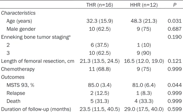
Table 1 summarizes the demographic and clini-cal characteristics and long-term outcomes of 28 patients. Patients in the HHR group were older than those in the THR group (48.3 years vs. 32.3 years, P=0.031). No statistically sig-nificant differences were found between the two groups in gender distribution, Enneking bone tumor staging, proportion of patients receiving chemotherapy and length of femoral resection. The mean length of femoral resec-tion for all 28 patients was 19.3 cm (SD=9.6, Table 1. Characteristics of 28 patients
THR (n=16) HHR (n=12) P Characteristics
Age (years) 32.3 (15.9) 48.3 (21.3) 0.031 Male gender 10 (62.5) 9 (75) 0.687 Enneking bone tumor staginga 0.190
2 6 (37.5) 1 (10) 3 10 (62.5) 9 (90)
Length of femoral resection, cm 21.3 (13.5, 24.5) 16.5 (12.0, 19.0) 0.121 Chemotherapy 11 (68.8) 9 (75) 0.999
Outcomes
MSTS 93, % 85.0 (3.4) 81.0 (6.4) 0.044 Relapse 2 (12.5) 1 (8.3) 0.999 Death 5 (31.3) 4 (33.3) 0.999 Duration of follow-up (months) 23.5 (11.5, 40.5) 29.0 (17.5, 40.0) 0.599
Data on age and MSTS 93 are shown as mean ± standard deviation, other continuous data are presented as median (interquartile range), and categorical data are shown as count (%). THR: total hip replacement; HHR: half-hip replacement; MSTS 96: Musculo-skeletal Tumor Society 1993. aTwo missing data were found.
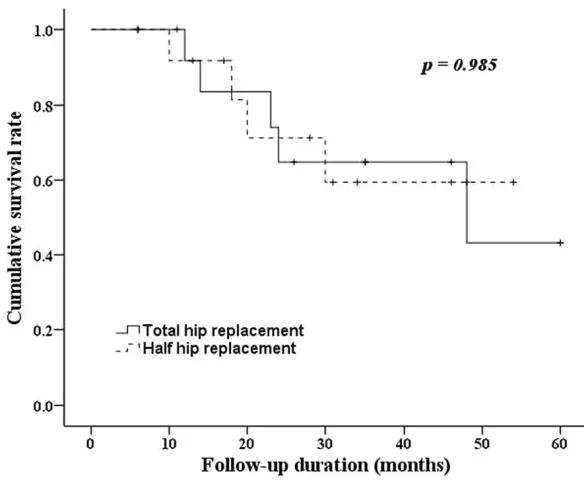
range: 10-60 cm). Most patients were males (67.9%) and 71.4% received chemotherapy. The THR group had higher MSTS 93 scores than the HHR group (85% vs. 81%, P=0.044). Local tumor recurrence was noted in 3 patients (10.7%). All 28 cases were not diagnosed as pulmonary metastasis in the beginning. However, there are 9 cases with late pulmonary metastasis and 4 of them got resection of lung tumor. All 9 cases (32.1%) were dead before end of study period. Mean follow-up of all patients was 30.5 months (SD=23.6, range: 6-108 months). The percentages of death and relapse were similar in both groups (Table 1). Figure 1 shows that the two groups of patients shared similar survival status. The survival rates for 1-year, 3-year and last visit were 91.7%, 64.8%, and 43.2%, respectively, in the THR group. In the HHR group, the survival rate was 91.7% at 1 year, which then dropped to and was maintained at 59.4% after 30 months (Figure 1).
Tumor recurrence was observed in 3 patients, including 2 patients with osteosarcomas and 1 with chondrosarcoma. Amputation was per-formed for these three patients. Prosthesis-related complications, including fracture, dislo-cation, implant breakage, modular failure, ac-
tumors of the proximal femur were treated using preoperative neoadjuvant chemotherapy, extended tumorectomy and custom prosthetic replacement. Results of this study revealed that local recurrence of femoral malignancies occurred in only 3 of the 36 patients receiving oncologic resection and customized bipolar prosthesis. The recurrence rate among the 36 patients was 8.3%. We believe that this low recurrence rate is related to the application of neoadjuvant chemotherapy and the perfor-mance of extended tumorectomy. In addition, evaluation of postoperative lower limb function using the American Musculoskeletal Tumor Society 1993 version (MSTS 93) demonstrated a mean functional score of 84.8%, represent-ing satisfactory functional outcomes of the lower extremities in the majority of patients. The percentages of death and relapse were similar between patients receiving THR and HHR, with 91.7% survival in both groups at one year, leveling off to 64.8% for the THR group and 59.4% for the HHR group after 30 months. Preoperative chemotherapy
Although the chemotherapy regimens for tumors of the proximal femur may be associat-ed with significant toxicity and long-term side effects [15], in our experience, administering Figure 1. Kaplan-Meier curve of 5-year overall survival in 28 patients. Data were
tested by Gehan-Breslow-Wilcoxon test.
etabular erosion in hemiar-throplasty, and migration, were not observed in any patient and no revision sur-gery was performed. Deep vein thrombosis was obs- erved in 2 patients, which resolved within two weeks after administering medic- ation.
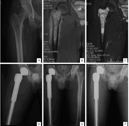
Pre- and post-operative co- nditions of the current spe-cial prosthetic replaceme- nt are demonstrated in Fi- gure 2, which depicts a 24- year-old male patient with Ewing’s sarcoma in the ri- ght proximal femur (Figure 2).
Discussion
effective chemotherapy is still the key to suc-cessful limb preserving surgery. We individual-ize the chemotherapy regimens, because it serves the Chinese population better. We con-sider tumor type and patient’s age. The appro-priate chemotherapy can improve the patient’s survival and produce a favorable local condi-tion for special prosthetic replacement. We carried out 3-4 cycles of standardized chemo-therapy for all malignant tumors before surgery. We combined three chemotherapy drugs dur- ing a 5-7-day cycle to control disease better.
[image:6.612.89.525.72.499.2]Application of neoadjuvant chemotherapy has helped to increase patients’ survival rates up to 80% [16]. Some investigators have noted that functional results and survival after proximal femur replacement were increased more for primary tumors than for metastatic disease [17], which also speaks to the value of chemo-therapy. Among our cases, we credit pre- and postoperative chemotherapy regimens, which differed depending on the tumor type, for the success of the replacement surgeries and the low recurrence rate.
Surgical options and functional recovery In order to achieve satisfactory outcomes for patients with tumors of the proximal femur who undergo prosthetic replacement, we consider that surgeons should pay close attention espe-cially to the following two aspects. First, 15°-20°of femoral neck anteversion should be maintained when placing the proximal femoral prosthesis. Second, the hip abduction should be reconstructed. For patients without tumor invasion to the greater trochanter, the superfi-cial bone block of the greater trochanter should be maintained. The bone block together with the gluteus medius and minimus should be fixed to the ring-shaped structure in the upper part of the prosthesis. For patients with tumor invasion to the greater trochanter, the gluteus medius and minimus should be detached from the greater trochanter and sutured to the ring-shaped structure of the prosthesis after oste-otomy. Reconstruction of the hip abduction can prevent hip dislocation. In the present study, the mean MSTS93 scores of the THR and HHR groups at the last follow-up were 85% and 81% respectively. Ogilvie et al. [7] reported a mean postoperative MSTS score of 67.7% for 34 patients who underwent prosthetic replace-ment of the proximal femur. Similarly, Finstein et al. [18] reported a postoperative MSTS score of 71% for 13 patients who underwent pros-thetic replacement of the proximal femur, and Zhang et al. [19] reported a mean MSTS score of 79% for 96 patients who underwent bipolar prosthetic replacement for proximal femoral tumors. In the present study, the THR group had higher MSTS scores than previous reports, while the mean score for HHR patients was similar to previous scores. This clearly suggests that the functional recovery of patients who undergo total hip arthroplasty is better than that of patients who undergo bipolar hip replacement. For patients with primary benign tumors and chemotherapy-sensitive malignant tumors of the proximal femur, we recommend total hip arthroplasty because the expected survival of patients with benign tumors or che-motherapy-sensitive malignant tumors is satis-factory after appropriate preoperative treat-ment, and the acceptable postoperative function can satisfy the physical and mental needs of patients. In the present study, although our overall recurrence rate was low (8.3%), we found no significant difference
between the THR and HHR groups in recur-rence. Other previous reports also found no dif-ference in the recurrence rate between patients receiving total hip arthroplasty and those receiving hemiarthroplasty [20].
Postoperative complications and functional recovery
reconstruction achieved good functional out-comes in patients who survived longer. We pre-fer to perform total hip arthroplasty for patients with primary benign tumors and chemotherapy-sensitive malignant tumors because it can give patients better function. During surgery, it is important for the femoral neck anteversion to be maintained and the hip abduction should be reconstructed to enhance postoperative func-tional recovery. After surgery, the patient’s sta-tus should be observed closely to prevent com-plications such as deep vein thrombosis. Limitations
This study has certain limitations, including pri-marily the small number of cases. Follow-up was also not long enough for about half of the patients. We did not report local control rates due to the effect of systemic therapy separat-ing the influence of wide margin and systemic therapy is not possible in a heterogeneous pop-ulation for sure. The MSTS 93 functional scores had shown statistically significant differences in current study. However, the significant clini-cal difference to explain why the THA patients did better cannot be fully explained only by MSTS 93 functional scores. We did not stratify the MSTS scores by degree of anteversion to show femoral anteversion and appropriate abductor repair/function improvements of patient outcome. We thought to maintain patients’ original anteversion angle is impor-tant and we did not perform additional studies, including restoring appropriate anteversion, offset, and abduction strength to explain hip biomechanics and function. We make the case that surgical techniques are keys to good out-come; however, it cannot be satisfactorily prov-en with retrospective evaluation and the long term result of THR with mega tumor prosthesis is not clear. Additional prospective study of functional outcomes in a larger patient popula-tion with bone tumors who require prosthetic replacement of the proximal femur are warrant-ed, with longer follow-up and detailed assess-ment of markers for recovery.
Conclusions
Neoadjuvant chemotherapy, extended tumor-ectomy and custom prosthetic replacement of the proximal femur were safe and effective in treating patients with femoral tumors. These measures resulted in minimal local recurrence
(8.3%) and satisfactory functional outcomes. The low recurrence rate is attributed to the effi-cacy of neoadjuvant chemotherapy. Our opera-tive results suggest that total hip arthroplasty should be applied for as many patients as pos-sible with primary benign tumors and chemo-therapy-sensitive malignant tumors because it produces better functional outcomes. The treatment protocol applied in this study may serve as a guide for making surgical decisions for patients requiring bone tumor resection. Acknowledgements
This study was supported by the National Natural Science Foundation of China (CN) (81472513).
Disclosure of conflict of interest None.
Address correspondence to: Dr. Wen-Zhi Bi, Depart- ment of Orthopaedics and Rehabilitation, PLA General Hospital, Fuxing Road 28, Beijing 100853, PR, China. Tel: +86-13601199993; E-mail: [email protected]
References
[1] Schneiderbauer MM, von Knoch M, Schleck CD, Harmsen WS, Sim FH and Scully SP. Pa-tient survival after hip arthroplasty for meta-static disease of the hip. J Bone Joint Surg Am 2004; 86: 1684-1689.
[2] Farid Y, Lin PP, Lewis VO and Yasko AW. Endo-prosthetic and allograft-Endo-prosthetic composite reconstruction of the proximal femur for bone neoplasms. Clin Orthop Relat Res 2006; 442: 223-229.
[3] Menendez LR, Ahlmann ER, Kermani C and Gotha H. Endoprosthetic reconstruction for the proximal femur. Clin Orthop Relat Res 2006; 450: 46-51.
[4] Bernthal NM, Greenberg M, Heberer K, Eck-ardt JJ and Fowler EG. What are the functional outcomes of endoprosthetic reconstructions after tumor resection? Clin Orthop Relat Res 2015; 473: 812-819.
[5] Meyers PA, Heller G and Vlamis V. Osteosarco-ma of the extremities: chemotherapy experi-ence at Memorial Sloah-Kettering. Cancer Treat Res 1993; 62: 309-322.
[7] Ogilvie CM, Wunder JS, Ferguson PC, Griffin AM
and Bell RS. Functional outcomes of endopros-thetic proximal femoral replacement. Clin Or-thop Rel Res 2004; 426: 44-48.
[8] Smith WS and Struhl S. Replantation of an au-toclaved autogenous segment of bone for treatment of chondrosarcoma. Long-term fol-low-up. J Bone Joint Surg Am 1988; 70: 70-75. [9] Benedetti MG, BOnatti E, Malfitano C and Do -nati D. Comparison of allograft-prosthetic com-posite reconstruction and modular prosthetic replacement in proximal femur bone tumors. Functional assessment by gait analysis in 20 patients. Acta Orthopaedica 2013; 84: 218-223.
[10] Mittermayer F, Krepler P, Dominkus M, Schwa-meis E, Sluga M, Heinzl H and Kotz R. Long-term follow-up of uncemented tumor endo-prostheses for the lower extremity. Clin Orthop Relat Res 2001; 388: 167-177.
[11] Bernthal NM, Schwartz AJ, Oakes DA, Kabo JM and Eckardt JJ. How long do endoprosthetic re-constructions for femoral tumors last? Clin Or-thop Relat Res 2010; 468: 2867-2874. [12] Bi W, Wang W, Han G, Jia J and Xu M.
Osteosar-coma around the knee treated With Neoadju-vant chemotherapy and a custom-designed prosthesis. Orthopedics 2013; 36: e444-450. [13] Mirels H. Metastatic disease in long bones. A
proposed scoring system for diagnosing im-pending pathologic fractures. Clin Orthop Relat Res 1989; 239: 256-264.
[14] Enneking WF, Spanier SS and Goodman MA. A system for the surgical staging of musculoskel-etal sarcoma. Clin Orthop Relat Res 1980; 153: 106-120.
[15] D’Adamo DR. Appraising the current role of chemotherapy for the treatment of sarcoma. Semin Oncol 2011; 38 Suppl 3: S19-S29.
[16] Glasser DB, Lane JM, Huvos AG, Marcove RC and Rosen G. Survival, prognosis,and thera-peutic response in osteogenic sarcoma. Can-cer 1992; 69: 698.
[17] Potter BK, Chow VE, Adams SC, Letson GD and Temple HT. Endoprosthetic proximal femur re-placement: Metastatic versus primary tumors. Surg Oncol 2009; 18: 343-349.
[18] Finstein JL, King JJ, Fox EJ, Ogilvie CM and Lackman RD. Bipolar proximal femoral re-placement prostheses for musculoskeletal neoplasms. Clin Orthop Relat Res 2007; 459: 66-75.
[19] Zang J, Guo W, Yang Y, Ji T and Zhang Y. [Bipo-lar prosthetic replacement for proximal femo-ral tumors]. Zhongguo Xiu Fu Chong Jian Wai Ke Za Zhi 2013; 41-44.
[20] Cannon CP, Mirza AN, Lin PP, Lewis VO and Yasko AW. Proximal femoral endoprosthesis for the treatment of metastatic. Orthopedics 2008; 31: 361-367.
[21] White RH and Henderson MC. Risk factors for venous thromboembolism after total hip and knee replacement surgery. Curr Opin Pulm Med 2002; 8: 365-371.
[22] Jawad MU and Brien EW. Proximal femoral re-construction using a constrained acetabulum in oncologic patients. Orthopedics 2013; 37: e187-e192.
[23] Chandrasekar CR, Grimer RJ, Carter SR, Till-man RM, Abudu A and Buckley L. Modular en-doprosthetic replacement for tumours of the proximal femur. J Bone Joint Surg 2009; 91B: 108-112.
Table 1. Osteosarcoma and mesenchymal chondrosarcoma
Chemotherapy drugs Dose Administration Time Ifosfamide 2 g/m2 Intravenous infusion D1-D5
Methotrexate 8-10 g/m2 Intravenous infusion D3
Doxorubicin 30-40 mg/m2 Intravenous infusion D5
[image:10.612.172.431.294.349.2]Cisplatin 120 mg/m2 Intravenous infusion D6 Notes: (a) in a course of treatment with methotrexate and cisplatin is not the same administration, for patients 30 years of age we use more Ifos-famide, methotrexate and doxorubicin this set of programs; for 30-year-old we use more than patients ifosfamide, doxorubicin and cisplatin this set of programs. (b) three cycles of preoperative chemotherapy, postoperative chemotherapy 6 cycles. (c) 3-4 weeks to repeat a course. (d) if found clear lung metastases, add vindesine 2 mg/m2 (D1, D8).
Table 2. Ewing’s sarcoma
Chemotherapy drugs Dose Administration Time Ifosfamide 2 g/m2 Intravenous infusion D1-D5
Vindesine 2 mg/m2 Intravenous infusion D1, D8
Doxorubicin 30-40 mg/m2 Intravenous infusion D5 Note: (a) per-op: three cycles of chemotherapy; post-op: six cycles of chemo-therapy. (b) 3-4 weeks to repeat a course.
Table 3. Malignant fibrous histiocytoma and alveolar soft tis-sue sarcoma
Chemotherapy drugs Dose Administration Time Ifosfamide 2 g/m2 Intravenous infusion D1-D5
Dacarbazine 200-400 mg/m2 Intravenous infusion D1-D5
Doxorubicin 30-40 mg/m2 Intravenous infusion D5 Note: (a) per-op: three cycles of chemotherapy; post-op: three cycles of che-motherapy. (b) 3-4 weeks to repeat a course.
[image:10.612.168.434.418.470.2]