Case Report
Endometriosis in the psoas major muscle: a case report
Fangxu Tao1, Jun Liu1, Zhifeng Kou2,3, Zhiyuan Wang4, Valerie Mika2,3, Yudong Xiao1, Yi Jiang5
Departments of 1Radiology, 5Pathology, The Second Xiangya Hospital, Central South University, Changsha
410011, Hunan Province, China; Departments of 2Biomedical Engineering, 3Radiology, School of Medicine, Wayne
State University, 818 W Hancock Street, Detroit 48201, USA; 4Department of Ultrasound, Hunan Cancer Hospital, The Affiliated Cancer Hospital of Xiangya School of Medicine, Central South University, Changsha 410013, Hunan
Province, China
Received March 27, 2016; Accepted September 11, 2016; Epub October 15, 2016; Published October 30, 2016
Abstract: Endometriosis occurs outside the pelvic cavity is uncommon. To our knowledge, until now there is no case like this has been reported. A 49-year-old Chinese woman only with CA125 abnormal was admitted to our clinic. After a series of examinations, a mass in the left psoas major muscle was discovered. Based on the clinical symp-toms, serologic markers and imaging features, we still could not rule out the possibility of malignant tumor. Finally,
a diagnosis of endometriosis was made based on intraoperative pathological findings. Then she received relevant
treatment. Three months later, both the lesion size and CA125 level decreased obviously. Retrospectively analysis
of the disease progression, we find that rare cases like this not only enrich the location spectrum of endometriosis,
but also help us gain more insight into the pathogenesis of it.
Keywords: Endometriosis, psoas major muscle, image
Introduction
Endometriosis is the presence of functional endometrial glands and stroma outside the uterine cavity; its incidence among childbear-ing women are estimated rangchildbear-ing from 3% to 10%, nearly 176 million in the world [1-3]. This disease damages female physical and psycho-logical health in terms of pelvic pain and/or infertility [4]. Compelling epidemiological evi-dence indicates that the implantation of endo-metrial tissues can occur in any organ, includ-ing brain, lung, bowel, abdominal wall, omen-tum, skin, and bladder [5, 6]. The pelvis, ova-ries, and Douglas pouch are the most com- monly involved targets [7, 8]. According to the onset location, the disease is generally divid- ed into two categories: the intra-pelvic and extra-pelvic.
Until now, the pathogenesis of endometriosis remains debatable. There are many proposed theories: 1) retrograde menstrual implantation, 2) vascular and lymphatic spread, iatrogenic implantation; 3) metaplastic of the pelvic peri-toneal cells; 4) immune system dysfunction
and autoantibody formation; and the theory that retrograde menstrual implantation is the most popular one [5, 9-12]. In clinic, the diverse locations, protean clinical manifestations and complicated pathogenesis hinder a clinician from making a correct diagnosis, especially for extra-pelvic cases. As estimated, the delay in diagnosis of endometriosis is about 6.7 years [13]. For the clinician, there is a long way before reaching the right diagnosis. Here, we present a case of endometriosis that occurred in the psoas major with atypical presentations.
Case report
Clinical data
uterine myoma seven years ago, but we did not get the details of the operation.
Routine examinations
To explore the etiology, the patient underwent a series of examinations and some valuable clues were discovered. The level of CA-199 (164.64 KU/L) and CA-242 (47.7 KU/L) also increased, the carcinoembryonic antigen (CEA) was normal. Then the Positron Emission To- mography-Computed Tomography (PET-CT) sh- owed that there was a soft-tissue-attenuation mass with increased glucose metabolism lo- cated in the left side of the psoas major: benign or low-grade malignant tumor was considered. The subsequent lumbar Magnetic Resonance
Imaging (MRI) confirmed the presence of the
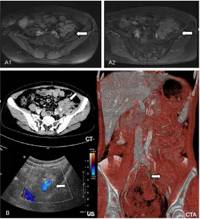
lesion, a miscellaneous intensity mass measur-ing 4.8×2.5 cm with heterogeneous enhance-ment in the same location (Figure 1A). There was no involvement of the abdominal cavity, no enlarged lymph nodes in the retroperitoneum. A presumed diagnosis that neoplasm originat-ed from the mesenchymal tissue was made. What a pity, the patient rejected further diag- nosis and treatment, and then she dischar- ged from hospital. After three months, the MRI revealed that the size of the mass increased to 7.0×4.2 cm (Figure 1A). In addition, the com-puted tomography angiography (CTA) display- ed that the neoplasm encasing the external iliac artery. The abdominal US demonstrated
a bright hetero-echoic irregular and ill-defined
mass located near the anterior-lateral of the left iliac vessels (Figure 1B). The serologic test showed that the level of CA125 was still ab- normal (237.40 KU/L, Table 1).
As was mentioned, it was a question about
how to interpret these findings: the mass was
benign or not? Is there relationship between
the lesion and history of pelvic surgery? After multidisciplinary consultations, the mass was tentatively interpreted as low-grade malignant tumor, with question about its origins: muscle, nerve tissue, or others. In this situation, did an exploratory laparotomy was indispensable. During the operation, we could see a mass measuring 10×10×8 cm in the left psoas major, with the peritoneum, iliac vessels and ureter adhesion. Considering the mass with rich blood supply and the adhesion of adjacent tissues, a decision that just did a biopsy rather than com-pletely removed it was made. Then the hema-toxylin and eosin (HE) staining showed endo-metriosis in the mass. While ectopic glands and stroma expressing cytokeratin (CK++) and cluster of Differentiation 10 (CD10+++) (Figure 2). The patient was diagnosed as endometrio-sis pathologically.
Treatment and follow up
We followed up the patient after she received GnRHa (Leuprorelin Acetate Microspheres) for more than three months. Effective treatment was indicated: the mass in size (4.8×2.1 cm) and the level of CA125 (84.31 KU/L, Table 1) decreased remarkably (Figure 3A). Moreover, we used Susceptibility Weighted Imaging (SWI)
and Diffusion Weighted Imaging (DWI) to find
some more valuable clues: multiple punctate signal voids within the mass on SWI; hyperin-tense on DWI, with no restriction of Brownian movement on corresponding Apparent Diffu- sion Coeffecient (ADC) map (Figure 3B). These
findings just reflected some features of
en-dometriosis. During our last two follow-ups, we saw that the level of CA125 have already reduced to normal (7.48 KU/L, Table 1). In the end, the treatment effects, laboratory tests, and imaging manifestations were in line with former pathological diagnosis. The patient was
finally diagnosed as endometriosis without any
doubt.
Discussion
As is known to all, endometriosis can occur in liver, cesarean section scar, rectus abdominis muscle, appendix, umbilical cord and so on [10, 14-16]. For this case, the onset location was very rare; the clinical symptoms were atypical and without classical cyclic pelvic pain; the radiology showed a solid mass mimicking a
Table 1. The value of CA125
Date CA125 (KU/L)
01/20/2015 489.58
04/30/2015 237.40
05/11/2015 214.35
05/29/2015 133.22
07/29/2015 84.31
08/26/2015 14.56
09/22/2015 7.48
neoplasm. All of these misled the doctor to make a false conclusion.
However, we still could find some subtle clues
that imply or support the truth: the abnormality of the CA125 and the history of operation. The current study reveals that CA125 has high sen-sitivity in the detection of endometriosis, espe-cially for late-stage ones [17, 18]. In clinic, the abnormality of CA125 in women can strongly suggest the possibility of endometriosis or epi-thelial ovarian cancer. If she just had a history of hysterectomy or intra-pelvic endometriosis,
we should firstly exclude endometriosis before
making any other diagnoses. For this case, we prefer to believe that the lesion was developed by iatrogenic cause, implanted the spread tis-sue during the surgery.
Besides what mentioned, we should admit that radiology especially MRI is useful in differenti-ating endometriosis from other diseases. The
and histology is still the gold standard [4]. So when we make a diagnosis about masses in women at the reproductive age, the endo- metriosis should be considered in the wide range of differential diagnoses, especially for patients with known endometriosis or those that have undergone pelvic surgery/cesarean before. Finally, rare cases like this not only enrich the location spectrum of endometriosis, but also help us gain more insight into the pathogenesis of it, and we will be more experi-enced when facing similar cases in the future.
Disclosure of conflict of interest
None.
[image:3.612.90.378.71.386.2]Address correspondence to: Dr. Jun Liu, Depart- ment of Radiology, The Second Xiangya Hospital of Central South University, No. 139 Middle Ren- min Road, Changsha, Hunan, China. Tel: +86 137- 87085002; E-mail: 2322349829@qq.com
Figure 1. Imaging manifestations. A1. Axial post-contrast T1-weighted im-age shows a heterogeneous enhancement mass in the left psoas major. A2. Three months later the mass increases in size. B. The mass encases the left external iliac artery on axial CT, US and coronal CTA. White arrow shows the lesion.
SWI confirmed the hemosid -erin and deoxy-hemoglobin deposited in the mass as signal voids. In addition, the appearances on DWI may be helpful to distinguish be- nign from malignant tumors [19]; additionally, adhesions are reported as an extremely common and important com-plication of the endometrio-sis, here the unclear fat spa- ce around the mass may just support the idea [5, 19]. Th- erefore, we could not deny the fact that MRI is a valu- able and reliable tool for as- sisting us in making a cor- rect diagnosis. We hope that in future there are more
spe-cific MRI signs of
References
[1] Giudice LC and Kao LC. Endometriosis. Lancet 2004; 364: 1789-1799.
[2] McLeod BS and Retzloff MG. Epidemiology of endometriosis: an assessment of risk factors.
[13] Nnoaham KE, Hummelshoj L, Webster P, d’Hooghe T, de Cicco Nardone F, de Cicco Nardone C, Jenkinson C, Kennedy SH, Zon- dervan KT; World Endometriosis Research Foundation Global Study of Women’s Health consortium. Impact of endometriosis on quali-Figure 2. Pathological examination results. A. Endometriosis in left psoas
[image:4.612.90.380.72.393.2]major on HE staining. B. Ectopic glands and stroma express CK and CD10.
Figure 3. MRI follow-up. A. The mass is iso-and low signal intensity on axial T1-weighted image; hyperintense on T2-weighted image; with heterogeneous enhancement after contrast administration. B. The SWI showes multiple punctate signal voids within the mass, and the contents of it are hyperin-tense on DWI and bright on ADC. White arrow shows the lesion.
[3] Taylor RN, Hummelshoj L, Stratton P and Vercellini P. Pain and endometriosis: Etiology, impact, and thera-peutics. Middle East Fertil Soc J 2012; 17: 221-225. [4] Dunselman GA, Vermeulen
N, Becker C, Calhaz-Jorge C, D’Hooghe T, De Bie B, Heikinheimo O, Horne AW, Kiesel L, Nap A, Prentice A, Saridogan E, Soriano D, Nelen W; European Socie- ty of Human R and Em- bryology. ESHRE guideline: management of women with endometriosis. Hum Reprod 2014; 29: 400-412.
[5] Woodward PJ, Sohaey R and Mezzetti TP. Endome- triosis: Radiologic-patholo- gic correlation. Radiogra- phics 2001; 21: 193-216. [6] Jubanyik KJ and Comite F.
Extrapelvic endometriosis. Obstet Gynecol Clin North Am 1997; 24: 411-440. [7] Fauconnier A and Chapron
C. Endometriosis and pel-vic pain: epidemiological evidence of the relation-ship and implications. Hum Reprod Update 2005; 11: 595-606.
[8] Farquhar C. Endometriosis. BMJ 2007; 334: 249-253. [9] Seydel AS, Sickel JZ, War-
ner ED and Sax HC. Ex- trapelvic endometriosis: di-agnosis and treatment. Am J Surg 1996; 171: 239. [10] Al-Jabri K. Endometriosis
at caesarian section scar. Oman Med J 2009; 24: 294-295
[11] Okeke TC, Ikeako LC and Ezenyeaku CC. Endome- triosis. Niger J Med 2011; 20: 191-199.
[image:4.612.91.377.442.580.2]study across ten countries. Fertil Steril 2011; 96: 366-373, e368.
[14] Huang WT, Chen WJ, Chen CL, Cheng YF, Wang JH and Eng HL. Endometrial cyst of the liver: a case report and review of the literature. J Clin Pathol 2002; 55: 715-717.
[15] Giannella L, La Marca A, Ternelli G and Menozzi G. Rectus abdominis muscle endo-metriosis: Case report and review of the litera-ture. J Obstet Gynaecol Res 2010; 36: 902-906.
[16] Paramythiotis D, Stavrou G, Panidis S, Pana- giotou D, Chatzopoulos K, Papadopoulos VN and Michalopoulos A. Concurrent appendi- ceal and umbilical endometriosis: a case re-port and review of the literature. J Med Case Rep 2014; 8: 258.
[17] Bedaiwy MA and Falcone T. Laboratory testing for endometriosis. Clin Chim Acta 2004; 340: 41-56.
[18] Kurdoglu Z, Gursoy R, Kurdoglu M, Erdem M, Erdem O and Erdem A. Comparison of the clinical value of CA 19-9 versus CA 125 for the diagnosis of endometriosis. Fertil Steril 2009; 92: 1761-1763.