Original Article
A simplified device for exposing rats to cigarette smoke
Xinnong Liu, Zhanqi Wang, Tianjia Li, Leng Ni, Rong Zeng, Changwei Liu
Department of Vascular Surgery, Peking Union Medical College Hospital, Peking Union Medical College & Chinese Academy of Medical Sciences, Beijing 100730, P. R. China
Received April 29, 2016; Accepted August 6, 2016; Epub October 15, 2016; Published October 30, 2016
Abstract: Objective: This study aims to explore a simplified device for exposing rats to cigarette smoke. Methods: Twenty-four rats were randomized into the following four groups: Group A, B, C, and D, in which rats were exposed to the cigarette smoke produced by 5, 3, or 1 cigarette, respectively. The smoke concentration inside the device was measured with portable detectors and the cotinine level in the serum was measured using an enzyme-linked immunosorbent assay (ELISA) kit. In addition, the WBCs and differential cell counts in BALF were performed with Wright-Giemsa staining, while NQO-1 and HO-1 in carotid arteries were detected by Western blot analysis. Results: The smoke concentration inside the device, as well as the cotinine in rats, significantly increased depending on the numbers of cigarettes between groups (P<0.01). The WBCs mainly for macrophages in BALF and oxidant stress index (NQO-1 and HO-1) increased with the increasing of cigarettes between groups (P<0.01). Conclusion: The pres -ent device could be used for exposing rats to cigarette smoke with a low cost and easy to perform. More importantly, it is easy to change the smoke concentration by only altering the numbers of cigarettes used at a time.
Keywords: Device, cigarette smoke, cotinine, rats, NQO-1, HO-1
Introduction
Faced with the enormous disease burden associated with smoking, related issues have
become a hot topic in research areas [1, 2]. To understand the various physiological and physicochemical processes associated with
tobacco consumption, a number of experimen -tal apparatuses aimed at exposing animals to
cigarette smoke have been designed [3]. As such, there is a diversity of “whole smoke exposure” system available, ranging from com
-mercial set-ups to bespoke machines [4, 5]. However, due to their high cost and technical
demands, many existing devices are
impracti-cal for widespread use in research.
While previous studies have devised a variety
of self-made apparatuses for exposing ani-mals to cigarette smoke [6-10], there is cur
-rently no standard model used for this purpose. Furthermore, there are advantages and disad
-vantages to each apparatus. For example, while the device reported by Brito et al. [6] can be
easily manufactured at a low cost, it was diffi
-cult to achieve homogeneous exposure of ani
-mals to cigarette smoke using this apparatus,
when exposed to the smoke. Meanwhile, the device reported by Bretthauer et al. [9] enabled homogenous exposure, but could only be used to expose one rat at a time. Moreover, most
studies related to smoking do not describe the methods used for exposure of animals to ciga
-rette smoke. Likewise, the smoke concentra -tion inside these devices is not always
record-ed, which makes it difficult to compare results
between studies [11-14].
The purpose of this study was to develop a novel low cost device for exposing rats to cigarette smoke. Here, we provide a detailed description of the components, assembly, and technical use of the device. In addition, we provide validation for the use of this device by
measuring the carbon monoxide (CO) and total particulate matter (TPM) concentration
inside the device as well as the levels of infl-ammatory and antioxidant biomarkers in rats. Methods
Animals and groups
-19214 Int J Clin Exp Med 2016;9(10):19213-19221 the Vital River Laboratory Animal Technology
Co. Ltd (Beijing, China). Rats were housed in a
standard laboratory environment with ambient
temperatures of 20-23°C, humidity of 43-45%, and a 12/12 h light/dark cycle. Animals had free access to standard solid claviform food
and autoclaved tap water.
Twenty-four rats were acclimated for one week to the aforementioned environment and then randomized into the following four groups (6 rats per group): Group A, B, and C, containing rats exposed to the cigarette smoke produced
by 5, 3, or 1 cigarettes at a time, respectively;
Group D, in which rats were not exposed to cigarette smoke. The protocols used in this study were performed according to approved guidelines from the Animal Care Committee of Peking Union Medical College.
Construction of the device used for cigarette smoke exposure
The present device was comprised of five
individual components, a glass chamber (len- gth × height × width=58 cm × 42 cm × 36 cm),
a metallic shelf (with a wire mesh on top of the shelf), a wire mesh fence, a cigarette holder,
and a glass cover (Figure 1), and was
assem-bled as follows: the metallic shelf (Figure 1B)
and wire mesh fence (Figure 1C) were sequen-tially placed in the glass chamber (Figure 1A), and the rats were placed within independent
grids on the metallic shelf. Subsequently, a cig -arette was placed on the cig-arette holder (Figure 1D) and lit, and the glass cover (Figure 1E) was placed on top of the wire mesh fence. Importantly, the bottom of the chamber was
designed to include two air inlets (red arrows shown in Figure 1A), and an air outlet was
enclosed with the cover and the top of the
chamber (red arrows shown in Figure 1F). In
this way, the smoke inside the device could rise vertically and accumulate up to peak concen -trations as the cigarettes burned out.
Cigarette smoke exposure
The rats in Groups A, B, and C were exposed to the cigarette smoke produced by 5, 3, or 1 ciga -rettes at a time, respectively, using the device described above. Rats were exposed to the
cigarette smoke for 1 h twice a day, for a total of seven days. When the first group of ciga -rettes burned out (approximately 10 min),
another group of cigarettes was lit to allow for continual smoke production. This process was repeated until the end of each exposure. Meanwhile, the rats in Group D were placed in a similar device, but lacking cigarette smoke, for 1 h twice a day, for seven days. The ciga
-rettes used in this study were Huangshan brand
(12 mg tar, 14 mg carbon monoxide (CO), and 1.2 mg nicotine per cigarette).
Measurement of TPM and CO levels
TPM is often used to reflect the smoke concen
-tration, especially for the concentration of the
particulate phase. In the present study, the
concentration of TPM inside the device was measured using an Aerosol Monitor (DUSTTRAK II-8530, TSI, USA). Meanwhile, the concentra
-tion of CO, one of the most important compo
-nents of the vapor phase of cigarette smoke, was measured using a KP-826 Gas Dete-ctor (Henan Zhongan Electronic Detection Technology Co., LTD, Henan, China). After light
-ing the cigarette(s), measurements were taken once per second for the first minute and then once per minute during the remainder of the
exposure.
Analyses of bronchoalveolar lavage fluid (BALF)
To examine the inflammatory response within the lungs of treated rats, total and differential
cell counts (macrophages, neutrophils, and
lymphocytes) in BALF were performed accord
-ing to previous studies [15]. Briefly, rats were anesthetized by intraperitoneal injection with 10% chloral hydrate at 3 ml/kg. Tracheas were
exposed, punctured with 14-gauge needles, clamped, and the lung was then lavaged 3 times each with 4 ml normal saline. The
recov-ery rate for each lavage was 90%-95%. The total and differential cell counts in BALF
were determined by globulimeter and Wright-
Giemsa staining, respectively. The relative pro
-portion of each cell type was determined mor -phologically by counting 300 cells/slide and
then was factored to the number (× 106/ml) of
total BALF cells collected in each group.
Measurement of cotinine in the serum
After seven days of exposure, all rats were euthanized and blood samples were harvested
by puncturing the abdominal aorta. Samples
10% glycerol). Protein samples (20 µg each) were then separated by SDS polyacrylamide gel electrophoresis (PAGE) and transferred to
nitrocellulose membranes. Membranes were
blocked in 5% skimmed milk in Tris-buffered saline + tween (TBST; 20 mM Tris-HCl [pH 7.6], 137 mM NaCl, 0.05% Tween-20) for 1 h at room temperature, washed three times with TBST, and incubated with primary antibodies specific to NAD(P)H quinone oxidoreductase 1 (NQO-1; 1:200; Santa Cruz Biotechnology, Inc., Dallas, TX, USA) and heme oxygenase-1 (HO-1) (1:500; BD Biosciences, Ltd., San Jose, CA, USA) over
-and centrifuged at 1,275 × g for 20 min. The serum was then collected and stored at -80°C prior to use. The concentration of cotinine in the serum was measured using an enzyme-linked immunosorbent assay (ELISA) kit (Lot COT4596; Calbiotech, Spring Valley, CA, USA), according to the manufacturer’s instruction.
Western’s blot analysis of NOQ-1 and HO-1 expression levels
[image:3.612.89.523.72.513.2]Total protein was extracted from the carotid arteries using lysis buffer (62.5 mM Tris-HCl [pH 6.8], 2% sodium dodecyl sulfate [SDS],
19216 Int J Clin Exp Med 2016;9(10):19213-19221
night at 4°C. An antibody specific to GAPDH
(glyceraldehyde 3-phosphate dehydrogenase;
1:5000; Proteintech, Chicago, IL, USA) was
used as a loading control within each indivi-
dual experiment. Following three washes, the
membranes were incubated with a horseradish peroxidase-conjugated goat
anti-mouse/anti-rabbit IgG (1:7000; Zhongshan Jinqiao Biote-chnology Co., Ltd, Beijing, China) for 1 h at
room temperature. Antigen-antibody complex-es were detected by enhanced chemilumi- nescence. Images were obtained, and the
gray values for each target protein were ana
-lyzed using an Alpha EaseFC system (Alpha Innotech Corporation, San Leandro, CA, USA).
Statistical analysis
Continuous variables were expressed as me-
[image:4.612.94.518.75.510.2]ans ± standard deviations (SD). Comparisons between groups were analyzed by one-way analysis of variance (ANOVA). All analysis was performed using SPSS 20.0 (SPSS Statistics Inc., Chicago, IL, USA). For all com -parisons, P<0.05 was considered statistically significant.
Results
Analysis of the concentrations of TPM and CO in each treatment group
As shown in Figure 2A, the peak concentra-tion of TPM increased depending on the numbers of cigarettes used with the values of TPM in group A, B, C, and D of 92.0 ± 8.2
mg/m3; 59.4 ± 7.1 mg/m3; 25.5 ± 4.6 mg/m3;
0.08 ± 0.01 mg/m3; respectively, (P<0.01).
Similarly, as shown in Figure 2B, the peak concentration of CO elevated with the num-bers of cigarettes used and its values in group A, B, C, and D was 233.1 ± 8.4 ppm; 138.0 ± 7.8 ppm; 89.6 ± 7.1 ppm; 0 ppm;
respectively, (P<0.01). Notably, peak TPM and
CO concentrations were achieved within only 12 s (Figure 2C and 2D), and the peak value
was stably maintained throughout the expo-sure process (Figure 2E and 2F).
Analysis of the serum cotinine concentrations in the rats in each treatment group
As shown in Figure 3, there was a significant increase in the serum concentration of cotinine in rats exposed to cigarette smoke compared
to that in the control group (P<0.01). Moreover, the serum concentration of cotinine increased depending on the number of cigarettes used (Group A=286.8 ± 36.9 ng/ml; Group B=233.3 ± 34.2 ng/ml; Group C=88.8 ± 9.3 ng/ml;
respectively, P<0.05).
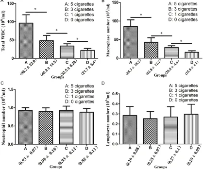
Total white blood cell (WBC) and differential cell count in the BALF of rats in each treat-ment group
There was a significant increase in the number of total WBCs (Figure 4A) and macrophages (Figure 4B) depending on the number of
cigarettes to which the rats were exposed (P<0.05). Furthermore, differential count
an-alyses indicated that macrophages were the
primary inflammatory cells recruited to the lungs of rats upon smoke exposure.
Mean-while, there were no changes in the neutrophil (Figure 4C) and lymphocyte (Figure 4D) counts between treatment groups (P>0.05).
Analysis of the expression levels of NOQ-1 and HO-1 in the carotid artery of rats in each treat-ment group
As shown in Figure 5, there were marked increases in the expression levels of the anti
-oxidant proteins HO-1 and NQO-1 in rats exposed to the smoke produced by one ciga -rette compared to that in the control group (P<0.01). In contrast, there was a significant decrease in the expression levels of these pro
-teins in the rats exposed to the smoke pro
-duced by three or five cigarettes compared
to the control group (P<0.01). However, no difference was detected in the expression levels of HO-1 and NQO-1 between Groups A and B (P>0.05).
Discussion
The development of a simplified, standard device for exposing rats to cigarette smoke
would not only contribute to a better
under-standing of smoke toxicity and the mechani-sms of smoke-related diseases, but would also facilitate comparisons of results bet-ween studies [16, 17]. The design of this smoke exposure device was inspired by the coal stoves that are often used to provide
warmth during the winters in china, and the underlying theory behind the device was based
on the “chimney effect” concept [18].
Notably, the function of the present device is
not dependent on any electric machine, which not only contributes to energy conservation, but also avoids any impact on the experim- ental results due to the noise associated with mechanical devices. Notably, only approxi-Figure 3. Graphic depiction of the serum concentra
19218 Int J Clin Exp Med 2016;9(10):19213-19221 Figure 4. Total and differential cell counts in bronchoalveolar lavage fluid (BALF) of rats exposed to cigarette smoke. *P<0.05. Data presented depict the average values from 6 biological replicates for each group.
mately 12 s were needed to reach the peak concentration within the device after lighting
the cigarettes. More importantly, however, the
peak smoke concentration was stably main -tained throughout the subsequent exposure process, even while changing cigarettes.
Because the cigarette holder can easily be
pulled out or pushed in through the air inlets
when changing cigarettes, the smoke concen -tration inside the device was not diminished.
To keep the rats separate, we included a wire mesh fence within the device. Each grid was
large enough to accommodate one rat, which ensured that all rats were homogeneously
exposed to the cigarette smoke by preventing clustering of the animals. To verify the stability of present device, we measured the smoke concentration and responses of rats when exposed to different numbers of cigarettes.
The concentrations of CO and TPM are the most frequently used index for reflecting the concentration of the vapor phase and particu
-late phase, respectively. Florek et al. [19] reported a new mechanical machine with
which they examined the severity of smoke
exposure by only modulating the CO concentra-tions within the device. Another machine
reported by Haussmann et al. [20] was used to
expose rats to different TPM concentrations. However, there are several disadvantages to
this machine, including a high cost and compli-cated procedures. Meanwhile, we observed
distinct CO (233.1 ± 8.4 ppm for 5 cigarettes, 138.0 ± 7.8 ppm for 3 cigarettes, and 89.6 ± 7.1 ppm for 1 cigarette) and TPM (92.0 ± 8.2
mg/m3 for 5 cigarettes, 59.4 ± 7.1 mg/m3 for 3
cigarettes, and 25.5 ± 4.6 mg/m3 for 1 ciga
the peak CO and TPM values among groups. These findings demonstrate that our device
would enable researchers to easily modulate
the exposure level of rats to cigarette smoke by altering the number of cigarettes used per treatment and would reflect the smoke concen
-tration by the index of CO and TPM at the same
time. In addition, to achieve distinct
concentra-tions of CO and TPM, researchers could likely adjust the size of the air inlets or outlets on the device; however, confirmation of this hypothe
-sis requires further study.
Due to its long half-life, cotinine, a metabolic product of nicotine in cigarette smoke, is one of the most commonly used biomarkers to reflect smoke exposure in animals and humans [21]. Based on the observed increase in the cotinine
concentration in rats exposed to smoke, com -pared to that in the control group, we conclud-ed that the rats had indeconclud-ed inhalconclud-ed the
ciga-rette smoke. Moreover, the cotinine concentra
-tion significantly increased depending on the number of cigarettes to which the rats were
exposed. While the cotinine concentrations
observed here were different from those
obtained in previous studies [8, 22], these
dis-crepancies could likely be explained by differ -ences in the method used to expose animals to
smoke or in the duration of smoke exposure.
Previous studies have shown that oxidative stress is the primary mechanism through
[image:7.612.99.510.74.477.2]which cigarette smoke exposure leads to the development of various diseases [23, 24]. In our present study, there was a significant
19220 Int J Clin Exp Med 2016;9(10):19213-19221
increase in expression levels of the antioxidant proteins NQO-1 and HO-1 within the carotid arteries of rats exposed to the smoke produced
by one cigarette compared to control group,
which was in keeping with the results obtained
in our previous study [25]. In contrast, there
was a significant decrease in the expression levels of NQO-1 and HO-1 in rats exposed to the smoke produced by three and five cigarettes
compared to control group, which was incon-
sistent with previous findings [26, 27]. The increased severity of smoke exposure in our present study may account for this difference; however, further studies are needed to confirm
this conclusion.
Regarding the inflammatory response within the lungs of rats exposed to cigarette smoke, the total WBC count in the BALF of these ani
-mals increased with numbers of cigarettes
used, and macrophages comprised the
pre-dominant inflammatory cell type. Furthermore, the magnitude of the increase of these par-ameters was smoke dose-dependent, which is consistent with the results of a previous study
[28]. In contrast, another previous report
indi-cated that neutrophils were the primary inflam
-matory cell type recruited to the lungs after smoke exposure [29]; however, we detected no significant differences in the number of neutro
-phils in the BALF samples harvested from dis -tinct treatment groups. In our present study,
the duration of smoke exposure was one week while the duration of exposure in previous stud
-ies ranged from 2 weeks to 36 weeks [28, 29]. As such, these differences in exposure lengths
could explain the distinct results obtained in
each study. However, further research is need
-ed to confirm this possibility.
In summary, there are many advantages to our
device. First, the severity of smoke exposure
can readily be adjusted by altering the num-
bers of cigarettes used. Second, because no
mechanical component was involved in the
device, it avoids negative effects due to noise.
More importantly, the present device could be
made up with a low cost and easy to perform. In addition, our device could be utilized to expose a variety of other small animals, such as mice, to cigarette smoke. There were also some limi
-tations to our study. First, a limited numbers of
rat were included in each treatment group. Second, the researchers need extra protection
from smoke exposure when using the present
device. Lastly, the cigarette smoke examined in the present study was sidestream smoke, not the mainstream smoke as humans.
Disclosure of conflict of interest
None.
Address correspondence to: Dr. Changwei Liu, Department of Vascular Surgery, Peking Union Medical College Hospital, Peking Union Medical College & Chinese Academy of Medical Sciences, No.1 Shuaifuyuan, Wangfujing Street, Beijing 100730, P. R. China. Tel: 86-010-69152501; Fax: 86-010-69152503; E-mail: liucw@vip.sina.cn
References
[1] Chen Z, Peto R, Zhou M, Iona A, Smith M, Yang L, Guo Y, Chen Y, Bian Z, Lancaster G, Sherliker P, Pang S, Wang H, Su H, Wu M, Wu X, Chen J, Collins R, Li L; China Kadoorie Biobank (CKB) collaborative group. Contrasting male and fe -male trends in tobacco-attributed mortality in China: evidence from successive nationwide prospective cohort studies. Lancet 2015; 386: 1447-56.
[2] Yang GH, Li YC, Wang ZQ, Liu B, Ye W, Ni L, Zeng R, Miao SY, Wang LF, Liu CW. Protective effect of melatonin on cigarette smoke-in -duced restenosis in rat carotid arteries after balloon injury. J Pineal Res 2014; 57: 451-8. [3] Thorne D, Adamson J. A review of in vitro ciga
-rette smoke exposure systems. Exp Toxicol Pathol 2013; 65: 1183-93.
[4] Okuwa K, Tanaka M, Fukano Y, Nara H, Nishiji -ma Y, Nishino T. In vitro micronucleus assay for cigarette smoke using a whole smoke expo -sure system: a comparison of smoking regi -mens. Exp Toxicol Pathol 2010; 62: 433-40. [5] Zhang W, Case S, Bowler RP, Martin RJ, Jiang
D, Chu HW. Cigarette smoke modulates PGE (2) and host defence against Moraxella ca -tarrhalis infection in human airway epithelial cells. Respirology 2011; 16: 508-16.
[6] Brito MV, Yasojima EY, Silveira EL, Yamaki VN, Teixeira RK, Feijó DH, Gonçalves TB. New ex -perimental model of exposure to environmen -tal tobacco smoke. Acta Cir Bras 2013; 28: 815-9.
[7] Zheng H, Liu Y, Huang T, Fang Z, Li G, He S. Development and characterization of a rat model of chronic obstructive pulmonary dis -ease (COPD) induced by sidestream cigarette smoke. Toxicol Lett 2009; 189: 225-34. [8] Jardim JR, Bizeto L, Fleig MA, Camelier A, Rosa
tobacco smoke toxicity in rodents]. Arch Bron -coneumol 2010; 46: 455-8.
[9] Bretthauer EW, Black SC, Satterwhite RL, Compton E, Moghissi AA. An inhalation device for exposing rats to cigarette smoke. Arch Envi -ron Health 1972; 25: 456-8.
[10] Castardeli E, Duarte DR, Minicucci MF, Azeve -do PS, Matsubara BB, Matsubara LS, Campa -na AO, Paiva SA, Zornoff LA. Exposure time and ventricular remodeling induced by tobac-co smoke exposure in rats. Med Sci Monit 2008; 14: BR62-66.
[11] Wang T, Chen X, Zhang W, Xiang X, Leng C, Jia Q. Roles of macrophage stimulating protein and tyrosine kinase receptor RON in smoke-induced airway inflammation of rats. Int J Clin Exp Pathol 2015; 8: 8797-808.
[12] Abobo CV, Ma J, Liang D. Effect of menthol on nicotine pharmacokinetics in rats after ciga -rette smoke inhalation. Nicotine Tob Res 2012; 14: 801-8.
[13] Bondy SC, Ali SF, Kleinman MT. Exposure of mice to tobacco smoke attenuates the toxic ef -fect of methamphetamine on dopamine sys -tems. Toxicol Lett 2000; 118: 43-6.
[14] Huang YC, Chin CC, Chen CS, Shindel AW, Ho DR, Lin CS, Shi CS. Chronic Cigarette Smoking Impairs Erectile Function through Increased Oxidative Stress and Apoptosis, Decreased nNOS, Endothelial and Smooth Muscle Con-tents in a Rat Model. PLoS One 2015; 10: e0140728.
[15] Qamar W, Sultana S. Farnesol ameliorates massive inflammation, oxidative stress and lung injury induced by intratracheal instillation of cigarette smoke extract in rats: an initial step in lung chemoprevention. Chem Biol Inter -act 2008; 176: 79-87.
[16] Winkler V, Mangolo NJ, Becher H. Lung cancer in South Africa: a forecast to 2025 based on smoking prevalence data. BMJ Open 2015; 5: e006993.
[17] Nabi H, Estaqiuo C, Auleley GR. Smoking and mortality--beyond established causes. N Engl J Med 2015; 372: 2169.
[18] Gamal EM, Szabo G, Nagy P, Bráth E, Peto K, Oláh A, Tamás R, Kovács A, Mikó I. [The role of pneumoperitoneum and the “chimney effect” on the development of port site metastasis. A new experimental animal model using Furka’s spleen tissue suspension]. Magy Seb 2005; 58: 89-92.
[19] Jardim JR, Bizeto L, Fleig MA, Camelier A, Rosa FW, Oliveira D, Azevedo D, Saldiva PH, Martins Mde A, Bonassa J, Nascimento OA. [An inhala-tion chamber model for controlled studies of tobacco smoke toxicity in rodents]. Arch Bron -coneumol 2010; 46: 455-8.
[20] Haussmann HJ, Anskeit E, Becker D, Kuhl P, Stinn W, Teredesai A, Voncken P, Walk RA. Comparison of fresh and room-aged cigarette sidestream smoke in a subchronic inhalation study on rats. Toxicol Sci 1998; 41: 100-16. [21] Nakajima M, Yamamoto T, Kuroiwa Y, Yokoi T.
Improved highly sensitive method for determi -nation of nicotine and cotinine in human plas -ma by high-perfor-mance liquid chro-matogra -phy. J Chromatogr B Biomed Sci Appl 2000; 742: 211-5.
[22] Florek E, Piekoszewski W, Wrzosek J. Relation -ship between the level and time of exposure to tobacco smoke and urine nicotine and cotinine concentration. Pol J Pharmacol 2003; 55: 97-102.
[23] Lambros ML, Plafker SM. Oxidative Stress and the Nrf2 Anti-Oxidant Transcription Factor in Age-Related Macular Degeneration. Adv Exp Med Biol 2016; 854: 67-72.
[24] Wang K, Jiang Y, Wang W, Ma J, Chen M. Escin activates AKT-Nrf2 signaling to protect retinal pigment epithelium cells from oxidative stress. Biochem Biophys Res Commun 2015; 468: 541-7.
[25] Yang G, Li Y, Wu W, Liu B, Ni L, Wang Z, Miao S, Wang L, Liu C. Anti-oxidant effect of heme oxy -genase-1 on cigarette smoke-induced vascular injury. Mol Med Rep 2015; 12: 2481-6. [26] Wei J, Fan G, Zhao H, Li J. Heme oxygenase-1
attenuates inflammation and oxidative dam -age in a rat model of smoke-induced emphy -sema. Int J Mol Med 2015; 36: 1384-92. [27] Ding S, Hou X, Yuan J, Tan X, Chen J, Yang N,
Luo Y, Jiang Z, Jin P, Dong Z, Feng L, Jia X. We -delolactone protects human bronchial epithe-lial cell injury against cigarette smoke extract-induced oxidant stress and inflammation responses through Nrf2 pathway. Int Immuno -pharmacol 2015; 29: 648-55.
[28] Beno M, Hurbankova M, Dusinska M, Volko -vová K, Staruchová M, Cerná S, Barancoková M, Kazimirová A, Kováciková Z, Bobek P, Ho -recký M, Mikulecký M, Kyrtopoulos SA. Some lung cellular parameters reflecting inflamma -tion after combined inhala-tion of amosite dust with cigarette smoke by rats. Cent Eur J Public Health 2004; 12 Suppl: S11-3.


![2 [(4 Benzhydrylpipérazin 1 yl)méthyl]acrylonitrile](data:image/gif;base64,R0lGODlhAQABAIAAAP///wAAACH5BAEAAAAALAAAAAABAAEAAAICRAEAOw==)