Original Article
Sunitinib treatment inhibited human breast cancer cell
migration through regulation furin interaction
with substrates
Shao-Ju Jin1*, Lei Ma2*, Qing Xu3*, Li-Ping Ren1, Yong-Chao Ma1,4
1The First Affiliated Hospital and Department of Pharmacology, Luohe Medical College, Luohe, Henan, China; 2Department of Emergency, General Hospital of Ningxia Medical University, Yinchuan, Ningxia, China; 3Depart -ment of Neurosurgery, The First People’s Hospital of Taicang, Taicang, China; 4Tumor Occurrence and Prevention Research Innovation Team of Luohe, Luohe, Henan, China. *Co-first authors.
Received October 27, 2015; Accepted January 9, 2016; Epub February 15, 2016; Published February 29, 2016
Abstract: It has been acknowledgement that human breast cancer (MCF-7) associated with high ability of migration and angiogenesis. In our present study, we aimed to explore the mechanism of sunitinib in the inhibition of MCF-7 cell migration and angiogenesis. MCF-7 cell line was used as breast cancer cell model for investigation the regula-tion of cell migraregula-tion upon sunitinib (0.5 µM and 2 µM) treatment. Western blot was used for detecregula-tion the expres-sions of MT1-MMP and VEGF-C. The activity of MMP2 and MMP9 was monitored with gelatin zymography assay. MCF-7 cell migration and invasion were detected by wound healing assay and transwell. Immunoprecipitation was used for detection the interaction among pro-MT1-MMP, Furin and VEGF-C. Results have shown that the expressions of MT1-MMP and VEGF-C were inhibited by sunitinib. Zymography assay demonstrated that the activity of MMP2
and MMP9 decreased significantly upon sunitinib treatment. The invasion and migration of MCF-7 were inhibited.
The interaction between Furin and its substrates pro-MT1-MMP, pro-VEGF-C decreased upon sunitinib treatment.
These findings indicated that sunitinib may be used for inhibiting the migration and invasion of MCF-7 through
down-regulated the interaction between Furin and its substrates (pro-MT1-MMP, pro-VEGF-C).
Keywords: MCF-7, sunitinib, furin, MT1-MMP, breast cancer, migration
Introduction
Breast cancer is the leading cause of the can-cer related death among woman in developed countries, accounting for 22% of new cases each year [1]. The occurrence of metastasis in tumorous progression is the most life-threaten-ing aspect of breast cancer. However, the underlying mechanisms of breast cancer initia-tion, progression and metastasis are still not fully understood [2]. Cell migration related pro-teins MT1-MMP, MMP2, VEGF must be cleaved by protein convertase, followed with maturation and activation.
Furin is the best-characterized representative of the mammalian subtilisin like family of pro-protein convertase. Many pro-protein precursors such as matrix metalloprotease, growth fac-tors, serum proteins, recepfac-tors, and adhesion molecules have been identified as the furin substrates [3-5]. MT1-MMP proenzyme
cleav-age by furin is considered to be a principal event in the activation of this substrate and it may be play an important role in MCF-7 cell migration and invasion [6].
Furin activation plays a vital role in tumorous process [7]. Furin inhibitor α1-PDX has been used to block furin activity and to prevent can-cer metastasis in cellular and animal studies [8]. Previous researches demonstrated that c-Src and furin together have been found to be upregulated in human cancers. As the most closely related members of the family of nonre-ceptor tyrosine kinases, up-regulation of c-Src correlates with a variety of human tumors, including breast cancer [9, 10]. Whether the ubiquitous c-Src participated in the interaction between furin and its substrates still remained unknown.
Furin play important roles in sunitinib inhibiting MCF-7 cell migration
se (RTK) inhibitor for the treatment of tumor angiogenesis, including vascular endothelial growth factor receptors and platelet-derived growth factor receptors [11]. Clinically, sunitinib antitumor activity was shown in renal carcino-ma, neuroendocrine and breast cancers [12, 13]. Both c-Src and AKT are down-stream of various tyrosine kinase receptor signaling path-ways. However, the antitumor effects of suni-tinib in inhibiting tumor migration and angio-genesis on breast cancer remained unknown. In the present study, we show that sunitinib induces tumor cell migration and invasion arrest in MCF-7 cells. The biological effects of sunitinib on tumor cell migration were associ-ated with inhibition of Furin and its substrates interaction. Our findings suggest the potential use of sunitinib for the treatment of breast can-cer cell metastasis.
Materials and methods
Cell culture and experimental reagents
MCF-7 cells were cultured in DMEM (Gibco) sup-plemented with 10% fetal bovine serum (FBS) and 100 units/mL penicillin and 100 µg/mL streptomycin. All the cells were cultured in a 5% CO2 humidified atmosphere at 37°C. Sunitinib (SU11248, Sigma PZ0012) was purchased from Sigma and dissolved in DMSO. In some experiments, logarithmic growth cells were incubated with sunitinib (0.5 or 2 µM) whenever necessary as indicated in the figure legend.
Primary antibodies against Furin, MT1-MMP, VEGF-C and β-actin were purchased from Santa Cruz Biotechnology (Santa Cruz, USA). Furin activity elisa kit (ABIN425690) was from Global Biotech (China). Gelatin zymography kit was from merck millipore (Millipore, USA), and MMP2/MMP9 elisa kits were purchased from Nanjing Jiancheng Bioengineering Institute (China).
Wound healing assay
MCF-7 cells were plated into 24-well plates and grown to confluence. The monolayer was artifi-cially wounded using the tip of a sterile 200 µL pipette. Cell debris was removed by washing with PBS. The cells were then incubated with sunitinib for appropriate time. The cells migrat-ed into the woundmigrat-ed areas were photographmigrat-ed.
Wound closure was photographed at the indi-cated times at the same spot with an inverted microscope equipped with a digital camera. The extent of healing was defined as the ratio of the difference between the original and the remaining wound areas compared with the orig-inal wound area.
Transwell invasion assay
Matrigel Invasion Chambers were hydrated for 4 h before starting the invasion assay. Log-phase cells (4×10⁴) were plated in 200 µL
DMEM containing 2% FBS in the upper cham-ber of the transwell and the lower chamcham-ber were filled with 500 µL DMEM containing 10% FBS. After incubation for 2 h, the cells were treated with sunitinib as previously description for 24 h. The cells were allowed to migrate for 10 h at 37°C and 5% CO2 followed by carrying out the invasion assay. The cells were fixed for 15 min at room temperature by replacing the culture medium in the bottom with 4% formal-dehyde dissolved in PBS. The cells that remained on the bottom of the chamber were stained with 0.1% crystal violet, the migrated clones were photographed under an optical microscopy. The cell number was counted at 12 different areas. Data were averaged from three parallel experiments, which were normalized to that of the control.
Elisa detection assay
MCF-7 cells were treated as previously descrip-tion. Total cellular proteins were used for detection the activity of Furin or the concentra-tions of MMP2/MMP9 according with kit procedures.
Gelatin zymography
Furin enzyme assay
The enzyme activity of Furin in vitro was per-formed at 37°C in 96-well flat-bottom black plates (Corning, USA). Assay was performed using Cbz-Arg-Ser-Lys-Arg-AMC as substrates. Whole cellular lysates of MCF-7 were incubated with Furin substrate in a final volume of 100 µL. The buffer consisted of 25 mM Mes, 25 mM Tris, and 2.5 mM CaCl2 (pH 7.4). The release of highly fluorescent 7-amino 4-methyl couma-rin (AMC) from Cbz-Arg-Ser-Lys-Arg-AMC was monitored by a Multi-Detection Microplate Readers (SpectraMax M5, USA) at excitation and emission wavelengths of 380 and 460 nm, respectively.
Western blot analysis
Cells were lysed in RIPA buffer [50 mM Tris (pH 7.4), 150 mM NaCl, 1% Triton X-100, 0.1% SDS, 1% sodium deoxycholate, 5 mM EDTA, 100 mM
NaF, and 1 mM Na3VO4] containing protease inhibitor cocktail for 30 min at 4°C. All cell lysates were centrifuged at 4°C at 16,000 g for 30 min. The protein concentration was deter-mined with the BCA method (Pierce, USA). Aliquots of cell lysates were fractionated by electrophoresis in SDS-PAGE, 8% for the analy-sis of Furin, 10% for the analyanaly-sis of MT1-MMP and VEGF-C. Total proteins were transferred to PVDF membrane using a wet trans blot system (Bio-Rad, Hercules, CA). Blots were then blocked for 1 h at room temperature with 10% BSA or 5% nonfat dry milk. Membranes were incubated overnight at 4°C with antibodies against Furin, MT1-MMP, VEGF-C and β-actin (1:1000). After subsequent washing, the mem-branes were incubated for 1 h with horseradish peroxidase conjugated goat rabbit or anti-mouse second antibody, diluted 1:5,000 in PBST. After washing, the membrane was pro-cessed using Super Signal West Pico
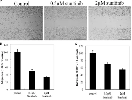
chemilu-Figure 1. Effects of sunitinib on the ability of MCF-7 cell migration and invasion. Confluent 90% MCF-7 cells were
wounded by 200 µl sterile pipette, then treated with varying concentrations of sunitinib for indicated time. The cells migrated into the wounded areas were photographed as A. The graph represents the mean ± S.E. of at least three independent experiments B. The ability of MCF-7 cell invasion was monitored by transwell assay C. *P<0.05
[image:3.629.99.533.84.413.2]Furin play important roles in sunitinib inhibiting MCF-7 cell migration
minescent substrate (Pierce, USA), followed by exposure to Fujifilm LAS3000 Imager (Fuji, Japan). Densitometric analysis was performed with Image J densitometer.
Co-immunoprecipitation
MCF-7 cells were washed twice with ice-cold PBS, lysed in 1 mL of RIPA buffer for 30 min on ice, clarified by centrifugation for 15 min at 10,000 g, and then the supernatant was sub-jected to immunoblot or immunoprecipitation. Cell lysates (500 µg) was incubated with 2 µg appropriate antibody (anti-Furin) overnight at 4°C. 50 µL of protein G was added and mixed at 4°C for 2 h with gentle agitation. The pellet was washed three times with RIPA buffer, boiled with 50 µL 2× loading buffer (Tris pH 6.8, 0.1% SDS, 10% glycerol, and 0.025% Bromophenol blue, 20 mM DTT) for 5 min prior to gel loading, and proteins were detected by western blot with anti-Furin, MT1-MMP and VEGF-C antibody. Some experiments substitut-ed the secondary antibody with Clean-Blot IP Detection Reagent for clear IP/Western blot results.
Statistical analysis
Western blots were quantified by measuring the relative density of protein bands recognized by a particular antibody using Image J software (NIH, USA). The results were expressed as mean ± standard deviation (SD). Statistical analysis was done with Student’s t-test for com-parison of two groups, differences with P<0.05 were considered statistically significant.
Results
Effects of sunitinib on the invasion and migra
-tion of MCF-7 cells
In order to explore whether the invasion and migration of MCF-7 cells were regulated by sunitinib treatment, we did wound healing assay and transwell assay. As our expected that sunitinib has obvious roles in modulation the invasion and migration of MCF-7 cells. The ability of invasion and migration of the cells that treated with sunitinib decreased signifi-cantly compared with control group (Figure 1).
Effects of sunitinib on the expressions of MMP2/9 in MCF-7 cell culture medium
MMP2 and MMP9 elisa kits were used for mon-itored the concentrations of MMP2/9 in the cell culture medium. The results have shown that the concentrations of MMP2/9 decreased sig-nificantly compare to the control group (Figure 2A and 2B). The results were expressed as mean ± SD from five independent experiments. We also detected the activity of MMP2/9 by gelatin zymography assay. As our expected, both the activity of MMP2 and MMP9 were all decreased (Figure 2C).
Effects of sunitinib on the expressions of furin and its substrates in MCF-7 cells
[image:4.629.103.529.81.245.2]We first found that the expression and activity of Furin have no obvious variation upon suni-tinib treatment (Figure 3A and 3B). However,
Figure 2. Effects of sunitinib on the expression and activity of MMP2 and MMP9 in MCF-7 cells. MCF-7 cells treated with sunitinib for indicated time, the concentrations of MMP2/MMP9 in cellular culture medium were detected by elisa assay (A and B). The results shown are representative of at least three independent experiments. At the same
time, the activity of MMP2 and MMP9 were detected with Zymography Assay (C). The statistically significant cutoff
the immunoblot indicated that a quantitative decrease in the intensities of MT1-MMP and VEGF-C bands were observed upon treatment with sunitinib (Figure 4A). These results indi-cated that some mechanisms maybe exit in regulation the maturation of MT1-MMP and VEGF-C.
The interaction between furin and its sub
-strates was inhibited by sunitinib in MCF-7
cells
It is unclear whether the binding between Furin and its substrates was affected upon sunitinib
treatment in MCF-7 cells. We then did co-immu-noprecipitation assay. Whole cell lysates were immunoprecipitated with anti-Furin antibody, and then detected the expressions of pro-MT1-MMP and pro-VEGF-C. As shown in Figure 4B, there was almost no band detection in sunitinib treated group. Similar results have been got for VEGF-C detection.
Discussion
[image:5.629.103.527.81.283.2]Clinical data has shown that sunitinib has been used in anticancer therapy through inhibiting cell proliferation or angiogenesis. However, its
Figure 3. Effects of sunitinib on the expression and activity of Furin in MCF-7 cells. The expression of Furin in MCF-7 cells upon sunitinib treatment was detected by western blot (A) and the activity of Furin was monitored by enzyme assay (B).
Figure 4. Effects of sunitinib on the interaction between Furin and pro-MT1-MMP, pro-VEGF-C in MCF-7 cells. MCF-7 cells were treated as previous description. Whole cell proteins were collected and 2 µg anti-Furin antibody was used for immunoprecipitation for overnight. The immunoprecipitates were used for detection the expression of
[image:5.629.100.529.345.491.2]Furin play important roles in sunitinib inhibiting MCF-7 cell migration
role in cancer cell metastasis remained unknown. The precursor proteins of cell metas-tasis association, such as MT1-MMP, MMP2, must be shorn by Furin in secretory pathway compartments [14]. Inhibition of Furin activity or interaction with its substrates may decrease substrate activation, proliferation rate and invasive potential of cancer cells. So, it is a potentially useful target for cancer therapeu-tics [15].
Our present study first found sunitib treatment may directly regulate the Capan-1 cell invasion and migration through modulation the matura-tion of MT1-MMP/VEGF-C. In our study, we first found that the ability of Capan-1 cells invasion and migration decreased upon sunitib treat-ment. To explore the mechanism, we then detected the effects of sunitib on the expres-sions of MT1-MMP or VEGF-C in Capan-1 cells. MT1-MMP and VEGF-C have played a vital role in regulation of cancer cell invasion and migra-tion. Upregulation of MT1-MMP can effectively cause elevated invasiveness in human cancer cell [16-18]. Results have shown that MT1-MMP or VEGF-C decreased significantly in accordance with c-Src activity, but the activity of furin had no obviously variation. The results indicated that regulation of MT1-MMP or VEGF-C not dependent on the down-regulation of furin, another mechanism may exist.
To be active, the zymogen of MT1-MMP or VEGF must be cleaved the pro-peptide by protein convertase. We then hypothesized that suni-tinib may directly inhibited the interaction between Furin and pro-MT1-MMP/pro-VEGF in vivo. So it is necessary to detect the effects of sunitinib on the interaction between Furin and its substrates. Our results showed that sunitib treatment decreased the formation of the com-plex between Furin and pro-MT1-MMP/pro-VEGF. Similar results got in interaction between Furin and VEGF-C.
In conclusion, we examined the role of sunitib in the process of Furin proteolysis its sub-strates. The potential inhibitor to block Furin and subsequent processing activity are more attractive therapeutic agents for MCF-7 cancer.
Disclosure of conflict of interest None.
Abbreviations
CHIP, Chromatin immunoprecipitation; MMPs, Matrix metalloproteinases.
Address correspondence to: Dr. Yong-Chao Ma, Luohe Medical College, 148 Daxue Road, Luohe
462002, Henan, China. Tel: +86-395-2985220; Fax: +86-395-2985220; E-mail: 14890698@ qq.com
References
[1] DeSantis C, Ma J, Bryan L, Jemal A. Breast can -cer statistics, 2013. CA Can-cer J Clin 2014; 64: 52-62.
[2] Schwertfeger KL, Cowman MK, Telmer PG,
Turley EA, McCarthy JB. Hyaluronan,
Inflam-mation, and Breast Cancer Progression. Front Immunol 2015; 6: 236.
[3] Fujisawa T, Kamimura H, Hosaka M. Functional localization of proprotein-convertase furin and its substrate TGFbeta in EGF receptor-express-ing gastric chief cells. Growth Factors 2004; 22: 51-59.
[4] Louagie E, Taylor NA, Flamez D. Role of furin in granular acidification in the endocrine pancre
-as: identification of the V-ATPase subunit Ac45
as a candidate substrate. Proc Natl Acad Sci U S A 2008; 105: 12319-12324.
[5] Yana I, Weiss SJ. Regulation of membrane type-1 matrix metalloproteinase activation by proprotein convertases. Mol Biol Cell 2000; 11: 2387-2401.
[6] Dangi-Garimella S, Krantz SB, Barron MR. Three-Dimensional Collagen I Promotes
Gem-citabine Resistance in Pancreatic Cancer through MT1-MMP-Mediated Expression of HMGA2. Cancer Res 2011; 71: 1019-1028. [7] Lopez de Cicco R, Bassi DE, Zucker S. Human
carcinoma cell growth and invasiveness is im-paired by the propeptide of the ubiquitous pro-protein convertase furin. Cancer Res 2005; 65: 4162-4171.
[8] Molloy SS, Thomas G. The Enzymes. Academic Press 2001; 199-235.
[9] Hilbig A. Src kinase and pancreatic cancer. Cancer Res 2008; 177: 179-185.
[10] Ischenko I, Guba M, Yezhelyev M. Effect of Src kinase inhibition on metastasis and tumor an-giogenesis in human pancreatic cancer. Angiogenesis 2007; 10: 167-182.
[12] Norton KA, Han Z, Popel AS, Pandey NB. Antiangiogenic cancer drug sunitinib exhibits unexpected proangiogenic effects on endothe-lial cells. Onco Targets Ther 2014; 7: 1571-1582.
[13] Valle JW, Faivre S, Hubner RA, Grande E, Raymond E. Practical management of sunitinib toxicities in the treatment of pancreatic neuro-endocrine tumors. Cancer Treat Rev 2014; 40: 1230-1238.
[14] Thomas G. Furin at the cutting edge: From
pro-tein traffic to embryogenesis and disease. Nat
Rev 2002; 3: 753-766.
[15] Bassi DE, Mahloogi H, Lopez De Cicco R.
Increased furin activity enhances the malig-nant phenotype of human head and neck can-cer cells. Am J Pathol 2003; 162: 439-447.
[16] Tolde O, Rosel D, Mierke CT. Neoplastic pro -gression of the human breast cancer cell line G3S1 is associated with elevation of cytoskel-etal dynamics and upregulation of MT1-MMP. Int J Oncol 2010; 36: 833-839.
[17] Seiki M. Membrane-type 1 matrix metallopro-teinase: a key enzyme for tumor invasion. Cancer Lett 2003; 194: 1-11.
[18] Zucker S, Pei D, Cao J, Lopez-Otin C.
Mem-brane type-matrix metalloproteinases