Original Article
Increased expression of LncRNA GAPLINC is associated
with tumor progression and predicts a poor prognosis
in hepatocellular carcinoma patients
Haikuan Wang
1, Shiqiang Shen
1, Yong Zhang
2, Xin Gu
2, Yongqiang Wang
21Department of Endoscopic Surgery, People’s Hospital of Wuhan University, Wuhan, Hubei Province, China; 2 De-partment of Gastrointestinal Surgery, Inner Mongolia People’s Hospital, Hohhot, China
Received December 26, 2016; Accepted March 16, 2017; Epub May 15, 2017; Published May 30, 2017
Abstract: Long non-coding RNAs (lncRNAs) have been investigated to be correlated with the behaviors and prog-nosis of hepatocellular carcinoma (HCC). However, the functional role of lncRNA GAPLINC in HCC has not been elucidated yet. The present study found that the expression of GAPLINC was up-regulated in HCC tissues and cell lines in comparison with tumor adjacent tissues and normal hepatocytes, respectively. Besides, high GAPLINC level was investigated to be correlated with tumor size (P < 0.001), number of tumors (P < 0.001) and TNM stage (P < 0.001) of HCC. Specially, patients with high GAPLINC expression displayed significantly lower overall survival rate and progression-free survival rate. Moreover, both univariate and multivariate COX regression analyses identified high GAPLINC expression as a risk factor of HCC poor prognosis. In addition, GAPLINC was verified to promote the
proliferation and metastasis of HCC cells in vitro assays. In conclusion, GAPLINC could promote HCC proliferation and metastasis and it may serve as a potentially prognostic marker and therapeutic target of HCC.
Keywords: Long non-coding RNA GAPLINC, hepatocellular carcinoma, prognosis, metastasis, proliferation
Introduction
Hepatocellular carcinoma (HCC) is one of the
most frequently happened cancers worldwide
and the second leading cause of cancer-relat-
ed death in men. Specially, China alone acc-
ounted for about 50% of the total number of
HCC cases and deaths worldwide [1]. Standard
treatment strategies, including surgical
resec-tion, liver transplantation and sorafenib could
only provide limited survival benefits due to
high postsurgical recurrence rates [2].
Besi-des, most HCC patients are usually diagnos-
ed at an advanced stage for lacking of typical
symptoms at early stage. The median survival
after the first treatment is only 23 months in
China [3]. Therefore, further investigations of
the molecular pathogenesis of HCC are
urgent-ly needed for identifying new diagnostic and
prognostic markers, which may shed light on
HCC early detection and promote the
develop-ment of novel therapeutic strategies, and thus
improve the overall prognosis of HCC patients.
Recently, long non-coding RNAs (lncRNAs), with
a length of 200 bp-100 kbp that lack
protein-coding potential, have been focused on by
many investigators for its potentially extensive
functions in almost every aspect of cell biology
from nuclear organization and epigenetic
regu-lation to post-transcriptional reguregu-lation and
splicing [4]. Although most of underlying
mech-anisms of lncRNAs functions remain obscure at
present, accumulating evidence has indicated
lncRNAs play essential roles in cancer
develop-ment and progression through various
regula-tory pathways, hierarchies and networks [4, 5].
LncRNA GAPLINC is a recently discovered
non-coding RNA, and has been reported to regulate
cell invasiveness of colorectal cancer, and
associates with the metastasis and poor
prog-nosis of gastric cancer [6, 7]. However, to the
best of our knowledge, the functional role of
GAPLINC in HCC has not been illustrated to
date.
was implicated to be associated with HCC
progression and poor prognosis. Furthermore,
multivariate Cox regression analysis revealed
that high GAPLINC expression was an
indepen-dent risk factor of HCC prognosis.
In vitro
assays, GAPLINC was showed to promote the
proliferation and metastasis of HCC cells.
These results indicated that lncRNA GAPLINC
may be considered as a potential prognostic
marker and therapeutic target of HCC patients.
Materials and methods
Cell lines and patient samples
The SUN449, HuH-6, GSG701, HCC LM3, HLE,
HuH-1, Hep3B, HuH-7, SMMC 7721, HepG2
and HK-HEP1 human HCC cell lines and the
LO2human immortalized normal hepatocytes
were obtained from the Cell Bank of Type
Culture Collection (Chinese Academy of Sci-
ences, Shanghai, China). Cells were cultured in
Dulbecco’s modified Eagle’s medium (DMEM,
Gibco, USA) or RPMI-1640 medium (Gibco,
USA) containing 10% fetal bovine serum (FBS,
Gibco, USA) and incubated at 37°C in an
atmo-sphere of 5% CO
2.
The latest follow-up was terminated on June
30, 2016. Overall survival (OS) was defined as
the interval between the date of surgery and
death or when censored at the latest date.
Progression-free survival (PFS) was defined as
the time from the date of surgery to the date of
disease relapse/progression or the date of
death or when censored at the latest date.
Patients died from causes other than HCC were
censored. This study was approved by the
Ethical Committee of the People’s Hospital of
Wuhan University and Inner Mongolia People’s
Hospital, and written informed consent was
obtained from each patient.
RNA extraction and real-time quantitative PCR
analysis (qRT-PCR)
[image:2.612.90.374.70.318.2]Total RNA was extracted from frozen tissues or
cultured cell lines using TRIzol Reagent (Takara,
Dalian, China). Then, cDNA was reversely
tran-scribed using the PrimeScript RT reagent kit
(Takara, Dalian, China) with 1 microgram of
total RNA. qRT-PCR was conducted to evaluate
the expression level of lncRNA GAPLINC using
SYBR Green PCR Master Mix (Takara, Dalian,
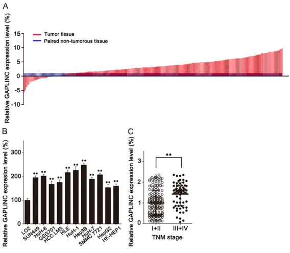
Figure 1. LncRNA GAPLINC is overexpressed in HCC. A. GAPLINC expressionwas analyzed in 274 HCC tissues and paired adjacent non-tumorous tissues by qRT-PCR assay. B. qRT-PCR assay was performed to detect the expression of GAPLINC in HCC cell lines and normal hepatocytes (LO2). C. The expres-sion levels of GAPLINC were compared on qRT-PCR assay for patients with TNM stage I+II and stage III+IV. **P < 0.01 based on the Student’s t-test. Data are represented as M ± SD.
China), which was performed on the ABI 7500
Fast Real Time PCR system (Applied Biosystems,
CA, USA). Comparative quantification was
determined with the 2
-ΔΔCtmethod. GAPDH used
as an internal control. The primer sequences
for GAPLINC were 5’-ACACACAGCAGCCTGGT-
TTC-3’ (sense) and 5’-ATGGCACAATCAGGGC-
TCTT-3’ (antisense); the primers for GAPDH
were 5’-GCACCGTCAAGGCTGAGAAC-3’ (sense)
and 5’-GGATCTCGCTCCTGGAAGATG-3’ (antise-
nse).
[image:3.612.92.342.96.585.2]All data was presented as M ± SD. For
statisti-cal comparisons, the χ
2test, the Fisher’s exact
test, the one-way analysis of variance and the
two-tailed Student’s
t
-test were performed
where appropriate. Kaplan-Meier method and
the log-rank test were conducted to evaluate
the difference of OS and PFS rates. Univariate
and multivariate Cox proportional hazards
mod-els were conducted to evaluate the survival
data.
P <
0.05 was considered to be
statisti-cally significant. All statistical analyses were
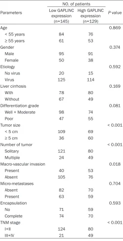
Table 1.
Correlation between GAPLINC expression and
clini-copathological characteristics of hepatocellular carcinoma
Parameters
NO. of patients
P value Low GAPLINC
expression (n=145)
High GAPLINC expression
(n=129)
Age 0.869
< 55 years 84 76
≥ 55 years 61 53
Gender 0.374
Male 95 91
Female 50 38
Etiology 0.592
No virus 20 15 Virus 125 114
Liver cirrhosis 0.169
With 78 80
Without 67 49
Differentiation grade 0.081 Well + Moderate 98 74
Poor 47 55
Tumor size < 0.001 < 5 cm 109 69
≥ 5 cm 36 60
Number of tumor < 0.001 Solitary 121 80
Multiple 24 49
Macro-vascular invasion 0.018 Present 40 53
Absent 105 76
Micro-metastases 0.704 Absent 82 70
Present 63 59
Encapsulation 0.593
No 71 59
Complete 74 70
TNM stage < 0.001
I+II 124 80 III+IV 21 49
Colony formation assay
The HCC cell lines were cultured in
6-well plates at a density of 100 cells
per well. Cells treated with different
strategies were incubated for 14
days. After being washed with PBS
and fixed with methanol, cell colonies
were stained with crystal violet.
Number of colonies containing more
than 50 cells was counted under a
microscope.
Cell migration and invasion assay
The migration and invasion abilities
of HCC cells were assessed by
Transwell assay and Matrigel assay
with boyden chambers (8 μm pore
size) (BD Biosciences, USA). For
Transwell assay, a volume of 200 μl
HCC cells (2.5 × 10
5/ml), suspended
in serum-free medium, was plated in
the upper chamber. The lower
cham-ber was filled with 600 μl medium
supplemented with 10% FBS. Then,
the cells were incubated in a
humidi-fied incubator supplemented with 5%
CO
2at 37°C for 24 hours. Subse-
quently, cells attached to the upper
side of the chamber were removed,
and cells migrated to the lower
sur-face of the membrane were fixed and
stained. For quantification, counts
were obtained from five random fields
at 100 × magnification. The Matrigel
assay was conducted similarly to the
Transwell assay except that the
boy-den chambers were precoated with
50 μl of 1 mg/ml Matrigel matrix (BD
Biosciences, USA) to form a matrix
barrier. Besides, the incubation time
was 48 hours for invasion assay.
carried out with the SPSS 19.0 software
pack-age (SPSS, Chicago, USA).
Results
LncRNA GAPLINC is overexpressed in HCC
tis-sues and cell lines
To investigate the functional role of lncRNA
GAPLINC in HCC, the expression level of
GAPLINC in HCC tissues was evaluated by
qRT-PCR assay. It was revealed that GAPLINC was
significantly overexpressed in HCC tissues rela
-tive to paired adjacent non-tumorous tissues
((1±0.813)%
vs.
(2.035±1.143)%,
P <
0.001).
(
Figure 1A
) Further detection of GAPLINC
expression in HCC cell lines showed that the
expression of GAPLINC in HCC cell lines was
much higher than in normal hepatocytes.
(
Figure 1B
) These results suggested that
lncRNA GAPLINC may play anoncogenetic role
in HCC.
LncRNA GAPLINC associates with HCC
pro-gression and poor prognosis
To further verify the function of GAPLINC in
HCC, the patients enrolled in this study were
the patients with high GAPLINC expression
(
Figure 2A
and
2B
). Additionally, univariate COX
regression analysis identified number of tumor,
macro-vascular invasion, TNM stage and high
GAPLINC expression as risk factors of HCC OS
and PFS (
Table 2
). By further analyzing these
factors with multivariate analysis, number of
tumor, macro-vascular invasion and high
GAPLINC expression were discovered to be
independent risk factors of HCC OS and PFS
(
Table 2
). Collectively, these results suggest
that GAPLINC may serve as a potential
recur-rence indicator and prognostic marker in HCC.
LncRNA GAPLINC could promote the
prolifera-tion and metastasis of HCC cells
To extend our investigation, the functional role
of GAPLINC in HCC was analyzed
in vitro
assay.
The expression of GAPLINC was knocked-down
with siRNA in Hep3B cells and overexpressed
in HepG2 cells (
Figure 2C
and
2D
). As a result,
GAPLINC interference dramatically suppressed
the proliferation of Hep3B cells on MTT assay
and colony formation assay (
Figure 3A
and
3B
).
In contrast, GAPLINC overexpression obviously
led to improved proliferation abilities of HepG3
Figure 2. LncRNA GAPLINC overexpression is correlated with HCC poor OSrate and PFS rate. (A and B) The median GAPLINC expression level was used as the cutoff and all the patients were divided into 2 groups based on the cutoff, including high GAPLINC expression group and low GAPLINC expres-sion group. Kaplan-Meier curves were used to estimate the overall survival rate (A) and progression free survival rate (B). (C and D) The expression of GAPLINC in HepG2 cells transfected with GAPLINC siRNAs (C) or cDNA (D) was investigated by qRT-PCR assay. **P < 0.01 based on the Student’s t-test or log-rank test. Data are represented as M ± SD.
divided into two groups, in-
cluding low GAPLINC
expres-sion group and high GAPLINC
expression group, with the
median GAPLINC expression
level working as the cutoff.
Correlation analysis between
GAPLINC expression and HCC
clinicopathological
character-istics discovered that high
GAPLINC expression was
cor-related with tumor size (
P <
0.001), number of tumors (
P
<
0.001) and TNM stage (
P <
0.001) of HCC (
Table 1
). Mo-
reover, patients with adv-
anced TNM stage displayed a
much higher GAPLINC exp-
ression level ((1±0.61)%
vs
.
(1.42±0.55)%,
P <
0.001)
(
Figure 1C
).
cells on MTT assay and colony formation ass-
ay (
Figure 3C
and
3D
). Besides, decreased
GAPLINC expression suppressed the migration
ability of Hep3B cells on Transwell assay (
Figure
3E
, up). Consistently, the invasion ability of
Hep3B cells was also reduced accompanied
with GAPLINC depletion. (
Figure 3E
, bottom)
Consistently, GAPLINC ectopic expression
sig-nificantly increased the migration and invasion
of HepG2 cells (
Figure 3F
). In conclusion, GAP-
LINC could promote the migration and invasion
of HCC cells.
Discussion
[image:5.612.100.523.96.573.2]HCC patients are usually asymptomatic at early
stage and have no access to surgical resection,
the only curative treatment strategy at present,
when diagnosed. Besides, adjuvant
therapeu-tic strategies are relatively rare for patients
Table 2.
Univariate analysis and multivariate analysis of clinicopathologic features for OS and PFS of
hepatocellular carcinoma patients
Parameters OS PFS
HR 95% CI P value HR 95% CI P value Univariate analysis
Age 1.287 0.959-1.725 0.092 1.250 0.934-1.672 0.133
≥ 55 years vs. < 55 years
Gender 1.066 0.782-1.453 0.688 1.083 .0798-1.471 0.608 Male vs. Female
HBs antigen 1.264 0.802-1.992 0.314 1.238 0.786-1.950 0.357 Negative vs. Positive
Liver cirrhosis 1.068 0.796-1.435 0.660 1.091 0.816-1.460 0.556 Without vs. With
Differentiation 1.037 0.767-1.401 0.814 0.991 0.734-1.337 0.952 Poor vs. Well + Moderate
Tumor size 1.178 0.868-1.599 0.293 1.141 0.842-1.546 0.394
≥ 5 cm vs. < 5 cm
Number of tumor 1.888 1.376-2.591 < 0.001 1.806 1.318-2.473 < 0.001 Multiple vs. Solitary
Macro-vascular invasion 1.599 1.188-2.151 0.002 1.645 1.225-2.208 0.001 Present vs. Absent
Micro-metastases 1.291 0.963-1.730 0.088 1.298 0.971-1.735 0.078 Absent vs. Present
Encapsulation 1.213 0.905-1.627 0.197 1.152 0.862-1.540 0.338 No vs. Complete
TNM stage 1.541 1.111-2.137 0.010 1.459 1.053-2.020 0.023 (III+IV) vs. (I+II)
GAPLINC 1.837 1.370-2.463 < 0.001 1.698 1.271-2.270 < 0.001 High vs. Low
Multivariate analysis
Number of tumor 1.925 1.208-3.067 0.006 1.899 1.195-3.017 0.007 Multiple vs. Solitary
Macro-vascular invasion 1.518 1.125-2.049 0.006 1.560 1.159-2.100 0.003 Present vs. Absent
TNM stage 0.845 0.517-1.382 0.502 0.814 0.499-1.326 0.408 (III+IV) vs. (I+II)
GAPLINC 1.660 1.222-2.255 0.001 1.527 1.126-2.069 0.006 High vs. Low
Figure 3. GAPLINC promotes proliferation and metastasis of HCC cells in vitro assay. Hep3B cells with GAPLINC silencing and overexpression were subjected to MTT (A and C), colony formation (B and D), Transwell (E and F, top) and Matrigel (E and F, bottom). (A) Cell proliferation of the siNC group, siGAPLINC.1 group and siGAPLINC.2 group was examined by MTT assay. (B) Colony formation assay was carried out to measure cell proliferation of the siNC group, siGAPLINC.1 group and siGAPLINC.2 group. Colonies containing more than 50 cells were counted and plot-ted. (C) Cell proliferation of the Vector group and GAPLINC group was examined by MTT assay. (D) Colony formation assay was carried out to measure cell proliferation of the Vector group and GAPLINC group. Colonies containing more than 50 cells were counted and plotted. (E) Representative images of the siNC group, siGAPLINC.1 group and
with advanced stage. The average 5-year
sur-vival rate is less than 12% [8], and only 3% in
advanced disease [9]. Therefore, early
diagno-sis is crucial for curative treatment. The
essen-tial roles of protein-coding genes in HCC
devel-opment and progression have been well
illuminated, but these genes only account for
1%-2% of transcribed RNAs [10-12].
Non-coding RNAs, including lncRNAs, are abundant
in human tissues and are investigated to be
crucial regulators of cellular transcription and
translation [10, 13].
Although only a small number of functional
lncRNAs have been well characterized till now,
they have been shown to control every level of
the multi-level regulated gene expression
path-ways, including chromatin remodeling, RNA
maturation (splicing, editing), transport and
protein synthesis [14]. Recent studies
implicat-ed that lncRNAs constitute an important
com-ponent of tumorbiology [15, 16]. In addition,
human plasma provides a convenient way for
the early detection of cancer. However, the
existing markers for HCC diagnosis are lack of
enough specificity and sensitivity. LncRNAs
could be measured in human plasma, thus it
may be a relatively non-invasive and potentially
effective tool for early diagnosis and prognosis
evaluation. Furthermore, aberrant expression
of lncRNAs in cancer marks the spectrum of
disease progression and may serve as an
inde-pendent predictor for patient outcomes [17,
18].
Numerous lncRNAs have been identified to be
involved in hepatocarcinogenesis, and mediate
HCC metastasis and proliferation [19-21].
Some lncRNAs have been reported to be
cor-related with HCC recurrence and prognosis
[22-31]. Therefore, lncRNAs are supposed to be
promising therapeutic targets of cancer [32,
33]. LncRNA GAPLINC is a 924-bp long lncRNA.
Hu et al. [6] reported that GAPLINC could
regu-late gastric cancer invasiveness via mediating
CD44 as a molecular decoy for miR211-3p, and
associated with gastric cancer prognosis. Peng
et al. [7] showed that GAPLINC could promote
colorectal cancer invasion by targeting snail
family zincfinger 2 (SNAI2) through binding
with PTB-associated splicing factor (PSF) and
non-POU-domain-containing octamer binding
(NONO) protein, and concluded that GAPLINC
may serve as a promising target for colorectal
cancer diagnosis and therapy. However, the
role of lncRNA GAPLINC in HCC has not been
elucidated.
The present study found that GAPLINC
expres-sion was up-regulated in HCC tissues and cell
lines in comparison with tumor adjacent
tis-sues and normal hepatocytes, which
suggest-ed that GAPLINC may also play an oncogenic
role in HCC. Indeed, correlation analysis
between GAPLINC expression and clinical
fea-tures showed that high GAPLINC level was
cor-related with tumor size (
P <
0.001), number of
tumors (
P <
0.001) and TNM stage (
P <
0.001)
of HCC. Kaplan-Meier method and the log-rank
test further detected that patients with high
GAPLINC expression displayed significantly
shorter OS rate and PFS rate. Moreover, high
GAPLINC expression was recognized as risk
factors of HCC poor prognosis on multivariate
COX regression analysis.
In vitro
assays,
GAPLINC was confirmed to promote the prolif
-eration and metastasis of HCC cells. Together,
these results indicated that lncRNA GAPLINC
could be considered as a predictor of HCC
recurrence and prognosis, and a potential
ther-apeutic target of HCC patients. However, there
are also some limitations of this study. The
detailed mechanisms of GAPLINC accelerating
HCC progression were not further explored, and
its therapeutic value needs more clinical
evidence.
In conclusion, our results confirmed that
lncRNA GAPLINC could promote HCC
prolifera-tion and metastasis and it may serve as a
potentially marker for HCC recurrence and
prognosis. In addition, it may be a promising
therapeutic target for individual treatment of
HCC patients.
Disclosure of conflict of interest
None.
the membrane and invaded cells through matrigel was shown relative to control. (F) Representative images of the
Vector group and GAPLINC group on Transwell assay and Matrigel assay. Corresponding quantification of migrated
Address correspondence to: Dr. Shiqiang Shen, De- partment of Endoscopic Surgery, People’s Hos- pital of Wuhan University, Wuhan 430060, Hubei Province, China. Tel: 88041911; Fax: 027-88041911-82011; E-mail: shensqwuhan@163.com
References
[1] Torre LA, Bray F, Siegel RL, Ferlay J, Lortet-Tieu-lent J and Jemal A. Global cancer statistics, 2012. CA Cancer J Clin 2015; 65: 87-108. [2] Gores GJ. Decade in review-hepatocellular
carcinoma: HCC-subtypes, stratification and
sorafenib. Nat Rev Gastroenterol Hepatol 2014; 11: 645-647.
[3] Choo SP, Tan WL, Goh BK, Tai WM and Zhu AX. Comparison of hepatocellular carcinoma in Eastern versus Western populations. Cancer 2016; [Epub ahead of print].
[4] Cheetham SW, Gruhl F, Mattick JS and Dinger ME. Long noncoding RNAs and the genetics of cancer. Br J Cancer 2013; 108: 2419-2425. [5] Yang G, Lu X and Yuan L. LncRNA: a link
be-tween RNA and cancer. Biochim Biophys Acta 2014; 1839: 1097-1109.
[6] Hu Y, Wang J, Qian J, Kong X, Tang J, Wang Y, Chen H, Hong J, Zou W, Chen Y, Xu J and Fang JY. Long noncoding RNA GAPLINC regulates CD44-dependent cell invasiveness and associ-ates with poor prognosis of gastric cancer. Cancer Res 2014; 74: 6890-6902.
[7] Yang P, Chen T, Xu Z, Zhu H, Wang J and He Z. Long noncoding RNA GAPLINC promotes inva-sion in colorectal cancer by targeting SNAI2 through binding with PSF and NONO. Oncotar-get 2016; 7: 42183-42194.
[8] El-Serag HB. Hepatocellular carcinoma. N Engl J Med 2011; 365: 1118-1127.
[9] Shen J, Siegel AB, Remotti H, Wang Q, Shen Y and Santella RM. Exploration of deregulated long non-coding RNAs in association with he-patocarcinogenesis and survival. Cancers (Ba-sel) 2015; 7: 1847-1862.
[10] Birney E, Stamatoyannopoulos JA, Dutta A, Guigo R, Gingeras TR, Margulies EH, Weng Z, Snyder M, Dermitzakis ET, Thurman RE, Kuehn MS, Taylor CM, Neph S, Koch CM, Asthana S, Malhotra A, Adzhubei I, Greenbaum JA, An-drews RM, Flicek P, Boyle PJ, Cao H, Carter NP, Clelland GK, Davis S, Day N, Dhami P, Dillon SC, Dorschner MO, Fiegler H, Giresi PG, Goldy J, Hawrylycz M, Haydock A, Humbert R, James KD, Johnson BE, Johnson EM, Frum TT, Rosen-zweig ER, Karnani N, Lee K, Lefebvre GC, Na-vas PA, Neri F, Parker SC, Sabo PJ, Sandstrom R, Shafer A, Vetrie D, Weaver M, Wilcox S, Yu M, Collins FS, Dekker J, Lieb JD, Tullius TD, Craw-ford GE, Sunyaev S, Noble WS, Dunham I, De-noeud F, Reymond A, Kapranov P, Rozowsky J,
Y, Zhu B and de Jong PJ. Identification and
analysis of functional elements in 1% of the human genome by the ENCODE pilot project. Nature 2007; 447: 799-816.
[11] Mattick JS. The genetic signatures of noncod-ing RNAs. PLoS Genet 2009; 5: e1000459. [12] Lander ES, Linton LM, Birren B, Nusbaum C,
Zody MC, Baldwin J, Devon K, Dewar K, Doyle M, FitzHugh W, Funke R, Gage D, Harris K, Hea-ford A, Howland J, Kann L, Lehoczky J, LeVine R, McEwan P, McKernan K, Meldrim J, Mesirov JP, Miranda C, Morris W, Naylor J, Raymond C, Rosetti M, Santos R, Sheridan A, Sougnez C, Stange-Thomann Y, Stojanovic N, Subramani-an A, WymSubramani-an D, Rogers J, Sulston J, Ainscough R, Beck S, Bentley D, Burton J, Clee C, Carter N, Coulson A, Deadman R, Deloukas P, Dun-ham A, DunDun-ham I, Durbin R, French L, GrafDun-ham D, Gregory S, Hubbard T, Humphray S, Hunt A, Jones M, Lloyd C, McMurray A, Matthews L, Mercer S, Milne S, Mullikin JC, Mungall A, Plumb R, Ross M, Shownkeen R, Sims S, Wa-terston RH, Wilson RK, Hillier LW, McPherson JD, Marra MA, Mardis ER, Fulton LA, Chinwalla AT, Pepin KH, Gish WR, Chissoe SL, Wendl MC, Delehaunty KD, Miner TL, Delehaunty A, Kram-er JB, Cook LL, Fulton RS, Johnson DL, Minx PJ, Clifton SW, Hawkins T, Branscomb E, Predki P, Richardson P, Wenning S, Slezak T, Doggett N, Cheng JF, Olsen A, Lucas S, Elkin C, Uberbach-er E, FraziUberbach-er M, Gibbs RA, Muzny DM, SchUberbach-erUberbach-er SE, Bouck JB, Sodergren EJ, Worley KC, Rives CM, Gorrell JH, Metzker ML, Naylor SL, Kucher-lapati RS, Nelson DL, Weinstock GM, Sakaki Y, Fujiyama A, Hattori M, Yada T, Toyoda A, Itoh T, Kawagoe C, Watanabe H, Totoki Y, Taylor T, Weissenbach J, Heilig R, Saurin W, Artiguenave F, Brottier P, Bruls T, Pelletier E, Robert C, Wincker P, Smith DR, Doucette-Stamm L,
Ru-benfield M, Weinstock K, Lee HM, Dubois J,
Rosenthal A, Platzer M, Nyakatura G, Taudien S, Rump A, Yang H, Yu J, Wang J, Huang G, Gu J, Hood L, Rowen L, Madan A, Qin S, Davis RW, Federspiel NA, Abola AP, Proctor MJ, Myers RM, Schmutz J, Dickson M, Grimwood J, Cox DR, Olson MV, Kaul R, Raymond C, Shimizu N, Kawasaki K, Minoshima S, Evans GA, Athana-siou M, Schultz R, Roe BA, Chen F, Pan H, Ramser J, Lehrach H, Reinhardt R, McCombie WR, de la Bastide M, Dedhia N, Blocker H, Hornischer K, Nordsiek G, Agarwala R, Aravind L, Bailey JA, Bateman A, Batzoglou S, Birney E, Bork P, Brown DG, Burge CB, Cerutti L, Chen HC, Church D, Clamp M, Copley RR, Doerks T, Eddy SR, Eichler EE, Furey TS, Galagan J, Gil-bert JG, Harmon C, Hayashizaki Y, Haussler D, Hermjakob H, Hokamp K, Jang W, Johnson LS, Jones TA, Kasif S, Kaspryzk A, Kennedy S, Kent WJ, Kitts P, Koonin EV, Korf I, Kulp D, Lancet D,
Lowe TM, McLysaght A, Mikkelsen T, Moran JV, Mulder N, Pollara VJ, Ponting CP, Schuler G, Schultz J, Slater G, Smit AF, Stupka E, Szustakowki J, Thierry-Mieg D, Thierry-Mieg J, Wagner L, Wallis J, Wheeler R, Williams A, Wolf YI, Wolfe KH, Yang SP, Yeh RF, Collins F, Guyer MS, Peterson J, Felsenfeld A, Wetterstrand KA, Patrinos A, Morgan MJ, de Jong P, Catanese JJ, Osoegawa K, Shizuya H, Choi S, Chen YJ and Szustakowki J. Initial sequencing and analysis of the human genome. Nature 2001; 409: 860-921.
[13] Mattick JS. The functional genomics of non-coding RNA. Science 2005; 309: 1527-1528. [14] Wapinski O and Chang HY. Long noncoding
RNAs and human disease. Trends Cell Biol 2011; 21: 354-361.
[15] Schmitt AM and Chang HY. Long noncoding RNAs in cancer pathways. Cancer Cell 2016; 29: 452-463.
[16] Prensner JR and Chinnaiyan AM. The emer-gence of lncRNAs in cancer biology. Cancer Discov 2011; 1: 391-407.
[17] Gupta RA, Shah N, Wang KC, Kim J, Horlings HM, Wong DJ, Tsai MC, Hung T, Argani P, Rinn JL, Wang Y, Brzoska P, Kong B, Li R, West RB, van de Vijver MJ, Sukumar S and Chang HY. Long non-coding RNA HOTAIR reprograms chromatin state to promote cancer metastasis. Nature 2010; 464: 1071-1076.
[18] Prensner JR, Iyer MK, Balbin OA, Dhanasek-aran SM, Cao Q, Brenner JC, Laxman B, Asan-gani IA, Grasso CS, Kominsky HD, Cao X, Jing X, Wang X, Siddiqui J, Wei JT, Robinson D, Iyer HK, Palanisamy N, Maher CA and Chinnaiyan AM. Transcriptome sequencing across a prostate
cancer cohort identifies PCAT-1, an unanno -tated lincRNA implicated in disease progres-sion. Nat Biotechnol 2011; 29: 742-749. [19] Yang X, Xie X, Xiao YF, Xie R, Hu CJ, Tang B, Li
BS and Yang SM. The emergence of long non-coding RNAs in the tumorigenesis of hepato-cellular carcinoma. Cancer Lett 2015; 360: 119-124.
[20] Yuan SX, Zhang J, Xu QG, Yang Y and Zhou WP. Long noncoding RNA, the methylation of genomic elements and their emerging cross-talk in hepatocellular carcinoma. Cancer Lett 2016; 379: 239-244.
[21] Sun J, Bie B, Zhang S, Yang J and Li Z. Long non-coding RNAs: critical players in hepatocel-lular carcinoma. Int J Mol Sci 2014; 15: 20434-20448.
[23] Peng W and Fan H. Long noncoding RNA CCHE1 indicates a poor prognosis of hepato-cellular carcinoma and promotes carcinogen-esis via activation of the ERK/MAPK pathway. Biomed Pharmacother 2016; 83: 450-455. [24] Lv J, Fan HX, Zhao XP, Lv P, Fan JY, Zhang Y, Liu
M and Tang H. Long non-coding RNA Unige-ne56159 promotes epithelial-mesenchymal transition by acting as a ceRNA of miR-140-5p in hepatocellular carcinoma cells. Cancer Lett 2016; 382: 166-175.
[25] Li J, Wang X, Tang J, Jiang R, Zhang W, Ji J and Sun B. HULC and Linc00152 act as novel bio-markers in predicting diagnosis of hepatocel-lular carcinoma. Cell Physiol Biochem 2015; 37: 687-696.
[26] Quagliata L, Matter MS, Piscuoglio S, Arabi L, Ruiz C, Procino A, Kovac M, Moretti F, Makows-ka Z, Boldanova T, Andersen JB, Hammerle M, Tornillo L, Heim MH, Diederichs S, Cillo C and Terracciano LM. Long noncoding RNA HOTTIP/ HOXA13 expression is associated with disease progression and predicts outcome in hepato-cellular carcinoma patients. Hepatology 2014; 59: 911-923.
[27] Zhang JY, Weng MZ, Song FB, Xu YG, Liu Q, Wu JY, Qin J, Jin T and Xu JM. Long noncoding RNA AFAP1-AS1 indicates a poor prognosis of hepa-tocellular carcinoma and promotes cell prolif-eration and invasion via upregulation of the RhoA/Rac2 signaling. Int J Oncol 2016; 48: 1590-1598.
[28] Chang L, Li C, Lan T, Wu L, Yuan Y, Liu Q and Liu Z. Decreased expression of long non-coding RNA GAS5 indicates a poor prognosis and promotes cell proliferation and invasion in he-patocellular carcinoma by regulating vimentin. Mol Med Rep 2016; 13: 1541-1550.
[29] Li T, Xie J, Shen C, Cheng D, Shi Y, Wu Z, Deng X, Chen H, Shen B, Peng C, Li H, Zhan Q and Zhu Z. Upregulation of long noncoding RNA ZEB1-AS1 promotes tumor metastasis and predicts poor prognosis in hepatocellular carci-noma. Oncogene 2016; 35: 1575-1584. [30] Calvisi D, Wang K, Guo WX, Li N, Gao CF, Shi
J, Tang YF, Shen F, Wu MC, Liu SR and Cheng SQ. Serum lncRNAs profiles serve as novel po -tential biomarkers for the diagnosis of HBV-positive hepatocellular carcinoma. PLoS One 2015; 10: e0144934.
[31] Zhou T and Gao Y. Increased expression of
ln-cRNA BANCR and its prognostic significance in
human hepatocellular carcinoma. World J Surg Oncol 2016; 14: 8.
[32] Takahashi K, Yan I, Haga H and Patel T. Long noncoding RNA in liver diseases. Hepatology 2014; 60: 744-753.