Original Article
Elevated plasma cathepsin B and cystatin C levels in
chronic obstructive pulmonary disease
Kok-Khun Yong1,2, Shih-Ming Tsao3,4, Thomas Chang-Yao Tsao4,5, Shun-Fa Yang1,6
1Institute of Medicine, Chung Shan Medical University, Taichung, Taiwan; 2Division of Pulmonary Medicine,
Puli Christian Hospital, Puli Township, Nantou County, Taiwan; 3Institute of Biochemistry, Microbiology and
Immunology, Chung Shan Medical University, Taichung, Taiwan; 4Division of Chest, Department of Internal
Medicine, Chung Shan Medical University Hospital, Taichung, Taiwan; 5School of Medicine, Chung Shan Medical
University, Taichung, Taiwan; 6Department of Medical Research, Chung Shan Medical University Hospital,
Taichung, Taiwan
Received February 4, 2016; Accepted June 4, 2016; Epub July 15, 2016; Published July 30, 2016
Abstract: Background: The aim of this study was to investigate differential changes in plasma levels of cathepsin B and its naturally inhibitory protein cystatin C in chronic obstructive pulmonary disease (COPD) patients during and 2 weeks as well as 8 weeks after acute exacerbation (AE). Materials and methods: Forty six COPD patients, including 44 male and 2 female, were included in this study. Plasma were collected in three different times, i.e., during, and 2 weeks as well as 8 weeks after AE. Plasma cathepsin B and cystatin C levels were measured in 46 adult patients with COPD and 18 healthy controls using a commercial enzyme-linked immunosorbent assay (ELISA). Results: The plasma levels of cathepsin B were significantly higher in COPD patients at 2 weeks and 8 weeks after AE when compared with those of healthy subjects. The plasma level of cystatin C showed significantly higher than the plasma levels of healthy subjects at time of AE, also 2 weeks and 8 weeks after AE. However, there was no significant dif-ference between the time of AE and 2 or 8 weeks after AE. Conclusions: The persistently significant higher plasma levels of cystatin C in COPD patients not only on AE but also at 2 and 8 weeks after AE than those in healthy sub-jects might represent a chronic inflammatory status in COPD. Moreover, plasma level of cathepsin B significantly increased at 2 weeks after AE and which returned to be non-significant at 8 weeks after AE in COPD patients. These findings might hint cathepsin B is one of the acute phase reactive protein in AE of COPD.
Keywords: Cathepsin B, cystatin C, chronic obstructive pulmonary disease
Introduction
Chronic obstructive pulmonary disease (COPD) is a disease of blocked airflow and no medica-tion is available that provides full recovery. COPD is caused by abnormal inflammation of the lungs due to harmful micro-particles or gases, and the deterioration is progressive [1]. Characteristics of COPD include chronic sys-temic inflammation at the airways, lung paren-chyma, and lung vessels. The number of macro-phages, T cells (mostly CD8+) and neutrophils increases in various parts of the lungs. Activated inflammatory cells would release many mediators, such as CRP [2-4], interleu-kin-6 (IL-6) [2, 4, 5], interleukin-8 (IL-8) [6, 7] and tumor necrosis factor (TNF) [8, 9], which can damage lung structure and prolong neutro-phil-caused inflammation.
Cathepsins are also called cysteine cathepsins because of a cysteine residual at the activation site [10]. Pathological studies have shown that cathepsin B is associated with rheumatoid arthritis [11] and cancers [12, 13]. Furthermore, cathepsin B can also be found in respiratory secretions [14]. Bronchial epithelial cells would secrete inactive cathepsin B, which is later acti-vated by neutrophil elastases [14]. Together with neutrophil elastase, activated cathepsin B can hydrolyze the extracellular matrix, causing damage to tissues [15].
[17] and apoptosis [18]. Therefore, one can evaluate the progress or occurrence of diseas-es by monitoring concentration changdiseas-es in cys-tatin C in the extracellular fluid or serum [19-22]. However, to the best of our knowledge, no study has investigated the prognostic value of cathepsin B and cystatin C in a cohort of patients with COPD. Therefore, we compared the plasma cathepsin B and cystatin C concen-trations of COPD patients before and after acute exacerbation.
Materials and methods
Subjects and specimen collection
The experiment specimens were collected from patients admitted to the Emergency Department of Puli Christian Hospital. The investigators first screened the patients; those COPD patients with cancers, inflamma-tory diseases (e.g., rheumatoid arthritis, liver inflammation, gingivitis and respiratory inflam-mation), severe heart failure (e.g., NYHA class IV), and asthma or with no smoking history (less than 10 packs per year) were excluded. In addition, we also recruited 18 healthy sub-jects (10 males and 8 females) to be the con-trol. After obtaining informed consent from all subjects, the investigators collected 3 mL lithi-um heparin-anticoagulant and 2 mL K2-EDTA-anticoagulant blood specimens from all of the 46 COPD subjects at AE, two weeks after AE, and eight weeks after AE for stability follow-up. But the blood samples from the eighteen healthy subjects were collected only once to be the control. The collected specimens were used
[image:2.612.90.368.97.267.2]for neutrophil count, the CRP test, and quantitative protein analysis for cathepsin B and cystatin C. After adding the blood, the tubes were slightly mixed to prevent coagulation and hemolysis. Tubes contain-ing lithium heparin were centri-fuged at 3500 rpm for ten min-utes. Plasma at the upper layer was then collected and used for CRP, cathepsin B and cystatin C analyses. Tubes with K2 EDTA were not centrifuged but used for neutrophil count (whole blood). Before commencement of this study, approval was obtained from the Institutional
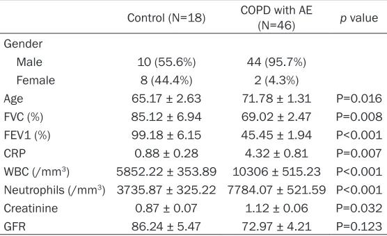
Table 1. Laboratory data of both controls and patients with chronic obstructive pulmonary disease (COPD)
Control (N=18) COPD with AE (N=46) p value Gender
Male 10 (55.6%) 44 (95.7%)
Female 8 (44.4%) 2 (4.3%)
Age 65.17 ± 2.63 71.78 ± 1.31 P=0.016
FVC (%) 85.12 ± 6.94 69.02 ± 2.47 P=0.008
FEV1 (%) 99.18 ± 6.15 45.45 ± 1.94 P<0.001
CRP 0.88 ± 0.28 4.32 ± 0.81 P=0.007
WBC (/mm3) 5852.22 ± 353.89 10306 ± 515.23 P<0.001
Neutrophils (/mm3) 3735.87 ± 325.22 7784.07 ± 521.59 P<0.001
Creatinine 0.87 ± 0.07 1.12 ± 0.06 P=0.032
GFR 86.24 ± 5.47 72.97 ± 4.21 P=0.123
Review Board of Chung Shan Medical University Hospital, and informed written consent to par-ticipate in the study was obtained from each person.
Measurements of plasma cathepsin B and cystatin C
The enzyme-linked immunosorbent assay (ELISA) was used to measure the plasma levels of cathepsin B and cystatin C in the blood sam-ples (R&D Systems, Abingdon, UK) [23]. For each plasma sample, 100 µL was directly transferred to the microtest strip wells of the ELISA plate and subsequently incubated for 2 hours at room temperature. After 3 washing steps, the detection antibody was added, and the reaction was incubated for 2 hours at room temperature. Antibody binding was detected with streptavidin-conjugated horseradish per-oxidase and developed with a substrate solu-tion. Next, the reaction was stopped, and opti-cal density was determined with a microplate reader set at 450 nm. Cathepsin B and cystatin C levels were quantitated with a calibration curve using human cathepsin B and cystatin C standards.
Statistical analysis
stability follow-up. The acquired data were expressed as mean ± SE. Spearman’s rank cor-relation was used for linear regression analysis to determine whether subjects’ cathepsin B, cystatin C, neutrophils and CRP are correlated with the progress of COPD.
Results
A summary of the demographic and clinical characteristics of the participants was present-ed in Table 1. The study included two different study groups, control subjects and COPD patients. The mean age of COPD patients in our data was 71.78 ± 1.31 and of healthy controls was 65.17 ± 2.63 (P=0.016). FVC (%) of the control group was 85.12 ± 6.94 and of the COPD group was 69.02 ± 2.47 (P=0.008). FEV1 (%) of the control group was 99.18 ± 6.15 and of the experimental group was 45.45 ± 1.94 (P<0.001). The blood cell counts of WBCs and neutrophils as well as CRP levels were signifi-cantly elevated in patients with COPD (WBCs, 10306/mm3; neutrophils, 7784/mm3 and CRP
level, 4.32) compared with those controls
(WBCs, 5852/mm3; neutrophils, 3736/mm3
and CRP level, 0.88) (P<0.001).
The blood cell counts of WBCs were significant-ly higher before treatment of COPD patients (10306 ± 515/mm3) compared with those
con-trol (5852 ± 353/mm3), two weeks after AE
(9029 ± 533/mm3), as well as eight weeks
after AE for stability follow-up (8935 ± 775/ mm3) (Figure 1A). The similar results were
found in the neutrophils counts as well as CRP levels (Figure 1B and 1C).
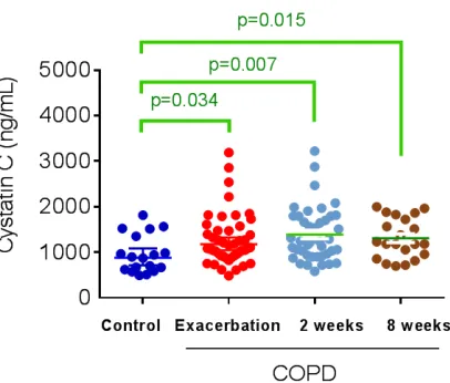
[image:3.612.93.300.72.397.2]Figure 2A presents the Cathepsin B levels in patients with COPD and the controls. The patients at 2 weeks and 8 weeks after AE exhib-ited significantly higher plasma Cathepsin B levels than the controls did (controls: 49.84 ± 4.75 ng/mL; patients at 2 weeks: 69.40 ± 6.06 ng/mL; P=0.048; patients at 8 weeks: 64.79 ± 4.81 ng/mL; P=0.034). Figure 2B presents the Cystatin C levels in patients with COPD and the controls. The COPD patients at AE, two weeks after AE, and eight weeks after AE follow-up exhibited significantly higher plasma Cystatin C
levels than the controls did (controls: 958 ± 98 ng/mL; AE: 1278 ± 84 ng/mL; P=0.034; patients at 2 weeks: 1390 ± 93 ng/mL; P=0.007; patients at 8 weeks: (1310 ± 96 ng/ mL; P=0.015). The results of cathepsin B/cys-tatin C ratio were presented in Figure 2C. In COPD patients, the ratio of cathepsin B/cys-tatin C was significantly different between the COPD patients at AE (0.045 ± 0.004) and two weeks after AE (0.056 ± 0.005) (P=0.025).
Discussion
Cathepsin B and cystatin C are both secreted proteins and clinically, they can be detected in the body fluids or blood specimens of humans. Some studies have shown that cathepsin B and cystatin C are closely associated with the occur-rence of some inflammatory diseases [17, 20, 24-27].
Interestingly, it can be found in Figure 2A that there was a trend of increase in cathepsin B from at AE to two weeks post AE. This may be because COPD is a slowly progressing
[image:4.612.316.519.82.255.2]inflam-matory response, and it is harder to see the response at the early onset of COPD. In con-trast, there was a reversed increase of cathep-sin B two weeks post AE (Figure 2A), which is the opposite of the finding that showed a drop in cathepsin B two weeks post AE in patients with pelvic inflammation [27]. In addition, in that pelvic inflammation study, cystatin C dropped before treatment but returned to normal after treatment. In this study, however, cystatin C in COPD patients was higher than in the control subjects (Figure 2B). The study of Chung et al., showed that cystatin C in patients with oral mucosal fibrosis was higher than in normal subjects [28]. In comparison, Chu et al. examined the association among cathepsin B, cystatin C and gout and showed an abnormal elevation of these two enzymes in chronic inflammation [26], suggesting that the higher concentration of cystatin C in COPD patients may be due to a compensatory mechanism in chronic inflammation, which explains why cystatin C in COPD patients is constantly higher than in the control subjects.
One limitation of this study is the lack of study subjects, and the fact that the expected num-ber of subjects was not reached. As a result, the present findings may not be adequate for representing the entire experiment. Secondly, there was a significant drop in the number of subjects for the second and the third blood sampling compared to first time, and therefore, the investigators have to be more careful with recruiting and tracking study subjects. Third, because COPD is a systemic inflammatory dis-ease, inflammatory symptoms at other parts of the body may also affect the results of the study. It is therefore necessary to be strict on the recruitment criteria. Lastly, it is important to recruit more people or to collect partial pul-monary specimens, such as tracheal fluid, to increase the accuracy and comprehensiveness of the results.
In conclusion, the persistently significant high-er plasma levels of cystatin C in COPD patients not only on AE but also at 2 and 8 weeks after AE than those in healthy subjects might repre-sent a chronic inflammatory status in COPD. Moreover, plasma level of cathepsin B signifi-cantly increased at 2 weeks after AE and returned to be non-significant at 8 weeks after AE in COPD patients. These findings might hint cathepsin B is one of the acute phase reactive protein in AE of COPD.
Acknowledgements
This study was supported by a grant (CSH-2009-D-002, CSH-2010-C-002) from Chung Shan Medical University Hospital, Taiwan.
Disclosure of conflict of interest
None.
Address correspondence to: Drs. Shun-Fa Yang and Thomas Chang-Yao Tsao, Institute of Medicine, Chung Shan Medical University, Taichung 402, Taiwan. Tel: +886-4-24739595 Ext. 34253; Fax: +886-4-24723229; E-mail: [email protected] (SFY); [email protected] (TCYT)
References
[1] Mannino DM and Buist AS. Global burden of COPD: risk factors, prevalence, and future trends. Lancet 2007; 370: 765-773.
[2] Broekhuizen R, Grimble RF, Howell WM, Shale DJ, Creutzberg EC, Wouters EF and Schols AM.
Pulmonary cachexia, systemic inflammatory profile, and the interleukin 1beta -511 single nucleotide polymorphism. Am J Clin Nutr 2005; 82: 1059-1064.
[3] Mannino DM, Ford ES and Redd SC. Obstructive and restrictive lung disease and markers of in-flammation: data from the Third National Health and Nutrition Examination. Am J Med 2003; 114: 758-762.
[4] Bolton CE, Ionescu AA, Shiels KM, Pettit RJ, Edwards PH, Stone MD, Nixon LS, Evans WD, Griffiths TL and Shale DJ. Associated loss of fat-free mass and bone mineral density in chronic obstructive pulmonary disease. Am J Respir Crit Care Med 2004; 170: 1286-1293. [5] Yende S, Waterer GW, Tolley EA, Newman AB,
Bauer DC, Taaffe DR, Jensen R, Crapo R, Rubin S, Nevitt M, Simonsick EM, Satterfield S, Harris T and Kritchevsky SB. Inflammatory markers are associated with ventilatory limitation and muscle dysfunction in obstructive lung disease in well functioning elderly subjects. Thorax 2006; 61: 10-16.
[6] Piehl-Aulin K, Jones I, Lindvall B, Magnuson A and Abdel-Halim SM. Increased serum inflam-matory markers in the absence of clinical and skeletal muscle inflammation in patients with chronic obstructive pulmonary disease. Respiration 2009; 78: 191-196.
[7] Schols AM, Buurman WA, Staal van den Brekel AJ, Dentener MA and Wouters EF. Evidence for a relation between metabolic derangements and increased levels of inflammatory media-tors in a subgroup of patients with chronic ob-structive pulmonary disease. Thorax 1996; 51: 819-824.
[8] Itoh T, Nagaya N, Yoshikawa M, Fukuoka A, Takenaka H, Shimizu Y, Haruta Y, Oya H, Yamagishi M, Hosoda H, Kangawa K and Kimura H. Elevated plasma ghrelin level in un-derweight patients with chronic obstructive pulmonary disease. Am J Respir Crit Care Med 2004; 170: 879-882.
[9] Takabatake N, Nakamura H, Abe S, Hino T, Saito H, Yuki H, Kato S and Tomoike H. Circulating leptin in patients with chronic ob-structive pulmonary disease. Am J Respir Crit Care Med 1999; 159: 1215-1219.
[10] Rossi A, Deveraux Q, Turk B and Sali A. Comprehensive search for cysteine cathepsins in the human genome. Biol Chem 2004; 385: 363-372.
[12] Loser R and Pietzsch J. Cysteine cathepsins: their role in tumor progression and recent trends in the development of imaging probes. Front Chem 2015; 3: 37.
[13] Olson OC and Joyce JA. Cysteine cathepsin pro-teases: regulators of cancer progression and therapeutic response. Nat Rev Cancer 2015; 15: 712-729.
[14] Guay C, Laviolette M and Tremblay GM. Targeting serine proteases in asthma. Curr Top Med Chem 2006; 6: 393-402.
[15] Jochum M, Gippner-Steppert C, Machleidt W and Fritz H. The role of phagocyte proteinases and proteinase inhibitors in multiple organ fail-ure. Am J Respir Crit Care Med 1994; 150: S123-130.
[16] Tidman M, Sjostrom P and Jones I. A Com- parison of GFR estimating formulae based upon s-cystatin C and s-creatinine and a com-bination of the two. Nephrol Dial Transplant 2008; 23: 154-160.
[17] Lah TT, Babnik J, Schiffmann E, Turk V and Skaleric U. Cysteine proteinases and inhibitors in inflammation: their role in periodontal dis-ease. J Periodontol 1993; 64: 485-491. [18] Nagai A, Ryu JK, Terashima M, Tanigawa Y,
Wakabayashi K, McLarnon JG, Kobayashi S, Masuda J and Kim SU. Neuronal cell death in-duced by cystatin C in vivo and in cultured hu-man CNS neurons is inhibited with cathepsin B. Brain Res 2005; 1066: 120-128.
[19] Nagai A, Terashima M, Harada T, Shimode K, Takeuchi H, Murakawa Y, Nagasaki M, Nakano A and Kobayashi S. Cathepsin B and H activi-ties and cystatin C concentrations in cerebro-spinal fluid from patients with leptomeningeal metastasis. Clin Chim Acta 2003; 329: 53-60. [20] Nagai A, Murakawa Y, Terashima M, Shimode
K, Umegae N, Takeuchi H and Kobayashi S. Cystatin C and cathepsin B in CSF from pa-tients with inflammatory neurologic diseases. Neurology 2000; 55: 1828-1832.
[21] Nakanishi T, Ozaki Y, Blomgren K, Tateyama H, Sugiura-Ogasawara M and Suzumori K. Role of cathepsins and cystatins in patients with re-current miscarriage. Mol Hum Reprod 2005; 11: 351-355.
[22] Chu SC, Wang CP, Chang YH, Hsieh YS, Yang SF, Su JM, Yang CC and Chiou HL. Increased cystatin C serum concentrations in patients with hepatic diseases of various severities. Clin Chim Acta 2004; 341: 133-138.
[23] Lee YT, Chen SC, Shyu LY, Lee MC, Wu TC, Tsao SM and Yang SF. Significant elevation of plas-ma cathepsin B and cystatin C in patients with community-acquired pneumonia. Clin Chim Acta 2012; 413: 630-635.
[24] Abboud RT and Vimalanathan S. Pathogenesis of COPD. Part I. The role of protease-antiprote-ase imbalance in emphysema. Int J Tuberc Lung Dis 2008; 12: 361-367.
[25] Angelidis C, Deftereos S, Giannopoulos G, Anatoliotakis N, Bouras G, Hatzis G, Panagopoulou V, Pyrgakis V and Cleman MW. Cystatin C: an emerging biomarker in cardio-vascular disease. Curr Top Med Chem 2013; 13: 164-179.
[26] Chu SC, Yang SF, Tzang BS, Hsieh YS, Lue KH and Lu KH. Cathepsin B and cystatin C play an inflammatory role in gouty arthritis of the knee. Clin Chim Acta 2010; 411: 1788-1792. [27] Tsai HT, Wang PH, Tee YT, Lin LY, Hsieh YS and
Yang SF. Imbalanced serum concentration be-tween cathepsin B and cystatin C in patients with pelvic inflammatory disease. Fertil Steril 2009; 91: 549-555.