Original Article
Mechanical stimulation promotes
osteogenic and chondrogenic differentiation
of synovial mesenchymal stem cells through BMP-2
Peiliang Fu, Song Chen*, Zheru Ding*, Ruijun Cong, Jiahua Shao, Lei Zhang, Qirong Qian
The Division of Adult Joint Reconstruction of Orthopedics Department, Changzheng Hospital affiliated to Second Military Medical University, No. 415 Fengyang Road, Huangpu District, Shanghai 200003, China. *Equal contribu-tors.
Received January 16, 2015; Accepted November 21, 2016; Epub February 15, 2017; Published February 28, 2017
Abstract: Mounting evidences have been shown that mesenchymal stem cells are present in various tissues includ-ing synovium. Given that environmental factors could have impacts on the differentiation of stem cells in vivo, we
elucidated that whether mechanical stimulation may influence the process. In this study, we investigated the effect
of cyclic mechanical stimulation on the osteogenic and chondrogenic differentiation of synovial mesenchymal stem cells (SMSCs) and its correlation with BMP-2. Cyclic mechanical stimulation was applied to SMSCs isolated from
bilateral hip, knee joints and unilateral ankle joint of rabbits. SMSCs were grouped into two classifications: treated
with mechanical stimulation (MS group) and without mechanical stimulation (no MS group). The mechanical stimu-lation was a cyclic tensile and compression: the amplitude and frequency of strain was 8%, 0.33 Hz and the peak of cyclic stress was 2.35 kilopascals (kPa) (1 kPa = 1.0 × 10³ N/m2). The osteogenic and chondrogenic
differentia-tion of SMSCs was determined by detecting expressions of osteogenesis markers (ALP and Cbfa1) and chondro-genesis markers (Sox9 and Col2a1). qRT-PCR and western blot showed that mechanical stimulation up-regulated expressions of osteogenesis markers (ALP and Cbfa1) and chondrogenesis markers (Sox9 and Col2a1) of SMSCs, respectively. In addition, BMP-2 was increased in SMSCs treated with mechanical stimulation during osteogenic and chondrogenic differentiation, and knockdown of BMP-2 leaded to the reversed result of the expression of
osteogen-esis markers and chondrogenosteogen-esis markers. To conclude, our study confirm that repetitive mechanical stimulation
increased the expression of BMP-2, and promoted osteogenic and chondrogenic differentiation of SMSCs.
Keywords: Mechanical stimulation, synovium mesenchymal stem cells, osteogenic differentiation, chondrogenic differentiation, BMP-2
Introduction
Articular cartilage is frequently damaged in dif-ferent pathological situations such as sports injuries, accidents, trauma and osteoarthritis [1, 2]. Although a number of approaches based on tissue and cellular therapies have been explored, more studies and mechanism need to be investigated in the process.
Mesenchymal stem cells (MSCs) are multi-potential non-hematopoietic progenitor cells that can differentiate into a variety of mesen-chymal lineages such as osteoblasts, chondro-cytes and adipochondro-cytes and have been found in many other adult tissues such as skeletal muscles, synovium, tendon and adipose
tis-sues [3-5]. Synovial mesenchymal stem cells (SMSCs) are attractive cell sources for bone/ cartilage regeneration because of their high expansion as well as osteogenic and chondro-genic potentials compared with other tissue-derived MSCs [6-8]. The clinical demands for bone-and cartilage-generating therapies incre- ase along with our population of aging people [9].
extracellular matrix (ECM) synthesis of the car-tilage explants and the cultured chondrocytes in vitro. Though the alteration of chondrocyte aggrecan and type-II collagen gene expression has been proved to be modulated by mechani-cal stimulation [9], and sulphate uptake shows the same correlation with mechanical stimula-tion [10], the exact mechanism still remains unknown for a decrease in biosynthesis sho wed inconsistence with the mRNA expression [9]. And in that case, the BMP-2 functioned as a morphogen may play an important role in this process since up-regulation of bone molec-ular markers, such as BMP-2, CoI I, ALP, has also been reported [14-16]. BMP-2 is a low-molecular-weight glycoprotein that functions as a morphogen and belongs to the transforming growth factor-β (TGF-β) super-family [17], and has been verified to be up-regulated in human MSCs (hMSCs) during osteogenic differentia-tion in response to chemical stimuladifferentia-tion [18, 19]. However, till present no study has focused on the effect of mechanical stimulation on the expression of BMP-2 in the differentiation process of SMSCs or the correlation between them.
In this study, we investigated the effect of cyclic mechanical stimulation on the osteogenic and chondrogenic differentiation of SMSCs and es- pecially revealed its correlation with BMP-2.
Materials and methods
Isolation and culture of SMSCs
SMSCs were generated from synovial mem-brane of bilateral hip, knee joints and unilateral ankle joint according to Suzuki’s method [6]. Cells were cultured in the Rabbit Mesenchymal
Stem Cell Medium (OriCell, USA) for 3 days. At the 3rd day, non-adherent cells were removed by PBS wash. Expansion of SMSCs lasted for 3 passages. The multi-differentiation potential of the cells were confirmed by osteogenic, adipo -genic, and chondrogenic differentiation assays. In vitro mechanical stimulation
SMSCs were 3D-embedded to small intestinal sub-mucosa scaffold (SIS) with the density of 1.0×10⁶ cells/ml respectively. To avoid complex
interaction between growth factors in serum and mechanical stimulation, SMSCs were cul-tured in serum-free medium at day 3 after 3D-embedding. Mechanical stimulation was applied using the Cell Stretcher System NS 500 (Scholar Tech, Osaka, Japan). The mechan-ical loading was repetitive. The amplitude and frequency of loading were set according to Ando, K.’s method and were adjusted for 8% uniaxial repeated tensile strain, 0.33 Hz; the 3D SIS was pressed with a cyclic pressure of 2.35 kilopascals (kPa) at its peak stress (1 kPa = 1.0×10³ N/m2) (21). The mechanical
stimulation lasted for 6 hours. Four hours after mechanical stimulation treatment, cells were harvested for further analysis.
Immuno blot
Cells were lysed with RIPA containing com- plete protease inhibitor cocktail (Roche, In- dianapolis, IN) and proteins were separated by SDS-PAGE electrophoresis. Proteins were then transferred onto a polyvinylidene fluoride membrane and then blocked with BSA. Primary antibodies against ALP, Cbfa1, Sox9, Col2a1 and BMP-2 were obtained from Cell Signal- ing Technology. Proteins were visualized using the SuperSignal West Pico Chemiluminescent Substrate (Pierce).
RNA isolation, reverse-transcribed PCR and quantitative real-time PCR
SMSCs were harvested and total RNA was extracted using TRIzol (Ambion, Life technolo-gies, U.S). Briefly, at least 106 cells were
[image:2.612.89.291.84.232.2]col-lected and washed by PBS, then added 1 mL TRIzol. The cell lysates and TRIzol were mixed thoroughly and left at room temperature for 5 min. 250 μL chloroform was added and shook vigorously for 15 sec. After 5 min, the mixture was centrifuged at 10000 rpm for 10 min. The
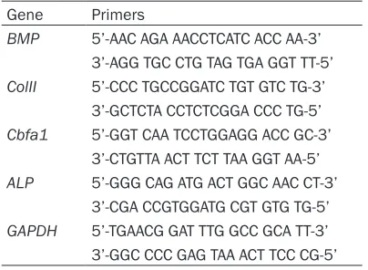
Table 1. The primers were used in study
Gene Primers
BMP 5’-AAC AGA AACCTCATC ACC AA-3’ 3’-AGG TGC CTG TAG TGA GGT TT-5’
ColII 5’-CCC TGCCGGATC TGT GTC TG-3’ 3’-GCTCTA CCTCTCGGA CCC TG-5’
Cbfa1 5’-GGT CAA TCCTGGAGG ACC GC-3’ 3’-CTGTTA ACT TCT TAA GGT AA-5’
ALP 5’-GGG CAG ATG ACT GGC AAC CT-3’ 3’-CGA CCGTGGATG CGT GTG TG-5’
Figure 1. Culture and identification of SMSCs. A. Morphology of SMSCs. Size scale: 1000 μm. B. SMSCs were analyzed for the indicated markers by flow cytometry.
aqueous phase was carefully removed and added with 550 μL isopropanol, then centri -fuged for 10 min at 12000 rpm. The pellet was air-dryed and dissolved in DEPC treated H2O after washing by 75% ethanol. RNA was reverse-transcribed using SuperScript III First-Strand Synthesis System Kit as the protocol instructed (Invitrogen). Quantitative real-time polymerase chain reaction (qRT-PCR) was performed using SYBR Green I (BioRad, Hercules, CA). The PCR parameters were 95°C for 15 sec, 60°C for 1 min for 40 cycles. A melting curve analysis was collected. Ct was determined in the expo-nential amplification phase, and the amplifica -tion plots were analyzed using SDS software (Applied Biosystems). The relative expression level (defined as fold change) of the target gene
described in Methods and Materials. SMSCs displayed spindle-like morphology (Figure 1A). Flow cytometric analysis demonstrated that SMSCs were CD90+CD73+CD105+CD44+CD34
-CD45- (Figure 1B). As expected, SMSCs were
multipotent as demonstrated by their ability to differentiate into adipocytes, osteoblasts and chondrocytes under appropriate conditions (Figure 1C). Together, the data showed that the SMSCs isolated in this study were MSCs. Mechanical stimulation promoted osteogenic and chondrogenic differentiation of SMSCs
[image:4.612.91.379.71.364.2]To investigate the effect of mechanical stimula-tion on the differentiastimula-tion of SMSCs, cells were cultured respectively and were treated with or Figure 2. Effect of mechanical stimulation on osteogenesis and
chongdrogen-esis of SMSCs. SMSCs cultured in osteo-inductive medium or chondro-induc-tive medium were treated with/without mechanical stimulation. At indicated time points, cells were harvested for detection of markers of osteogenesis and chondrogenesis. A. mRNA levels of ALP and Cbfa1 in cells treated with/ without mechanical stimulation under osteogenic condition were analyzed by qRT-PCR. B. Protein levels of ALP and Cbfa1 in cells treated with/without me-chanical stimulation were analyzed by western blot. C. mRNA levels of Sox9 and Col2a1 in cells treated with/without mechanical stimulation under chon-drogenic condition were analyzed by qRT-PCR. D. Protein levels of Sox9 and Col2a1 in cells treated with/without mechanical stimulation were analyzed by western blot. The data are represented as mean ± SD for triplicate samples.
*P < 0.05, **P < 0.01.
was measured by the follow-ing equation: 2-ΔΔCt (ΔCt =
ΔCttarget - ΔCtGAPDH; ΔΔCt =
ΔCtms - ΔCtno ms). The primers
were listed in Table 1. GAPDH was used as an internal control to norma- lize for differences in the amount of total RNA in each sample.
Statistical analysis
All quantitative assays were calculated from at least 3 replicate samples. Data were presented as mean ± SD. All the data analysis was done using SPSS 18.0 (SPSS Inc, Chicago, IL). The quantitative PCR results were determined by Stu- dent’s t-test (two-tailed). Di- fferences of multiple treat-ment groups were com- pared within individual ex- periments by one-way AN- OVA. *P < 0.05 was consid-ered significant. **P < 0.01 was considered remarkably different.
Results
Isolation and identification of SMSCs
without cyclic mechanical stimulation for 6 hours. At indicated time points, cells were harvested for detection of markers of osteo-genesis and chondroosteo-genesis. As detected by qRT-PCR, mRNA expression of ALP and Cbfa1, markers of osteogenesis, was significantly higher in cells treated with mechanical stimu- lation than that of without mechanical stimulation (Figure 2A). Consistently, the levels of ALP and Cbfa1 protein in cells treated with mechanical stimulation were significantly higher than those of without mechanical stimulation (Figure 2B).
Meanwhile, under chondrogenic conditions, mRNA expression of Sox9 and Col2a1, markers of chondrogenesis, was significantly higher in cells treated with mechanical stimulation than that of without mechanical stimulation (Figure
Knockdown of BMP-2 inhibited the osteogenic and chondrogenic differentiation of SMSCs promoted by mechanical stimulation treatment
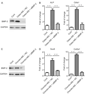
[image:5.612.92.379.73.357.2]To identify whether the osteogenic and chon-drogenic differentiation is mainly regulated by BMP-2, we utilized siRNA to inhibit the expres-sion of BMP-2 and further evaluated different markers (Figure 4A, 4B). We observed that the osteogenisis markers, ALP and Cbfa1 were down-regulated in SMSCs upon BMP-2 knockdown. The result of chondrogenisis markers was also proved (Figure 4C, 4D). The alteration of different markers suggested that BMP-2 played a crucial role in the regulation of osteogenic and chondrogenic differentiation. Moreover, the mechanical stimulation which promoted the oteogenisis and chondrogenisis Figure 3. Expression of BMP-2 by SMSCs with/without mechanical stimulation
treatment. During osteogenic or chondrogenic differentiation, SMSCs were treated with (MS group) or without mechanical stimulation (no MS group). And BMP-2 mRNA or protein in SMSCs were measured by qRT-PCR or western blot respectively. A. BMP-2 mRNA in SMSCs treated with/without mechani-cal stimulation during osteogenic differentiation. B. BMP-2 mRNA in SMSCs treated with/without mechanical stimulation during chondrogenic differentia-tion. C. BMP-2 protein in SMSCs treated with/without mechanical stimulation during osteogenic differentiation. D. BMP-2 protein in SMSCs treated with/ without mechanical stimulation during chondrogenic differentiation. The data are represented as mean ± SD for triplicate samples. *P < 0.05, **P < 0.01.
2C). The results were also confirmed by immuno blot (Figure 2D).
Mechanical stimulation pro-moted expression of BMP-2 in SMSCs during osteogenic and chondrogenic differen-tiation
differentiation was mainly through BMP-2 pathway.
Discussion
Our study demonstrated that repetitive mech- anical stimulation promoted the osteogenic and chondrogenic differentiation of SMSCs. Meanwhile, repetitive mechanical stimulation upregulated the expression of BMP-2 in SMSCs during osteogenic and chondrogenic differenti-ation. Differing from the previous single studies that only concentrated on the osteogenesis or chondrogenesis of SMSCs, our experiment indi-cated that both the osteogenesis and chondro-genesis capability of SMSCs could be
strength-the ostrength-ther durations of loading, while BMP-2 and bFGF also increased aggrecan and type II collagen mRNA expression when used sepa-rately. Similarly, Guilak et al [22] indicated that compression of the tissue to physiological strain magnitudes acted as a signal which regulated chondrocyte biosynthetic and cata-bolic responses by the thickness of cartilage, while enhanced compression under higher strains may take responsibility for tissue and cell damage.
[image:6.612.92.379.69.387.2]In the present study, the amplitude and fre-quency of strain were 8% and 0.33 Hz, res- pectively. The peak of cyclic stress was 2.35 kilopascals. It was reported that stretching Figure 4. Knockdown of BMP-2 inhibited the osteogenic and chondrogenic
dif-ferentiation of SMSCs promoted by mechanical stimulation treatment. siRNA was utilized to inhibit the expression of BMP-2 and further detected different markers. A. BMP-2 protein in SMSCs transfected with BMP-2 siRNA/negative control during osteogenic differentiation. B. mRNA levels of ALP and Cbfa1 in cells transfected with BMP-2 siRNA/negative control under osteogenic condi-tion were analyzed by qRT-PCR. C. BMP-2 protein in SMSCs transfected with BMP-2 siRNA/negative control during chondrogenic differentiation. D. mRNA levels of Sox9 and Col2a1 in cells transfected with BMP-2 siRNA/negative control under chondrogenic condition were analyzed by qRT-PCR. The data are represented as mean ± SD for triplicate samples. *P < 0.05, **P < 0.01.
ened by cyclic mechanical stimulation.
more than 10% and excessive time pressures were harmful to cells and tissues rather than beneficial [23-26]. However, it was sugge-sted that the expression of BMP-2 could be enhanced even if the stretching was over 12% [27]. In our preliminary experiment we found that the viability of SMSCs was decreased when treated with stretching more than 10% and pressure time over 3 h. Thus we adjusted strain at 8%, 0.33 Hz and the peak stress at 2.35 kilopascals.
The mechanisms of mechanical stimulation increasing the capability of osteogenesis and chondrogenesis remain diverse. Though it was reported that ECM reconstruction was involved in this progress [21, 28], we found that BMP-2 might be one of the essential factors mediating that mechanical stimulation promoted osteo-genesis and chondroosteo-genesis. In agreement with our findings, research on tendon-derived stem cells and bone marrow-derived stem cells demonstrated that mechanical loading might promote osteogenic differentiation by increas-ing BMP-2 expression [27, 29]. Altogether, our research demonstrates that BMP-2 might play a pivotal role during mechanical stimulation-mediated osteogenesis and chondrogenesis.
Conclusions
Cyclic mechanical stimulation promoted the osteogenic and chondrogenic differentiation of synovial mesenchymal stem cells (SMSCs). The expression of osteogenesis markers (ALP and Cbfa1) and chondrogenesis markers (Sox9 and Col2a1) were utilized to determine the osteogenic and chondrogenic differentiation of SMSCs. Mechanical stimulation up-regulated expressions of osteogenesis markers (ALP and Cbfa1) and chondrogenesis markers (Sox9 and Col2a1) of SMSCs, respectively. BMP-2 was increased in SMSCs treated with mechanical stimulation during osteogenic and chondro- genic differentiation, and knockdown of BMP-2 leaded to the reversed result of the expression of osteogenesis markers and chondrogenesis markers. To conclude, our study demontrated that repetitive mechanical stimulation increas- ed the expression of BMP-2, and promoted osteogenic and chondrogenic differentiation of SMSCs.
Acknowledgements
This work was sponsored by Natural Science Foundation of Shanghai (15ZR14140) and National Natural Science Foundation of China
(81000798) and National Natural Science Foundation of China (81601889).
Disclosure of conflict of interest
None.
Authors’ contribution
PF, SC, ZD participated in the design of the study, carried out the animal experiments, analyzed the results and drafted the script. RC participated in drafting the manu-script and revising the manumanu-script critically. JS and LZ participated in the evaluation of the results and data analysis. PF and QQ partici-pated in the design of the study. All authors read and approved the final manuscript.
Address correspondence to: Drs. Peiliang Fu and Qirong Qian, Joint Division of Orthopedics De- partment, Changzheng Hospital, No. 415 Fengyang Road, Huangpu District, Shanghai 200003, China. Tel: 0086-21-81885639; Fax: 0086-21-81885639; E-mail: fupeiliang@163.com (PLF); qianqr@163.com (QQ)
References
[1] Shintani N and Hunziker EB. Chondrogenic dif-ferentiation of bovine synovium: bone morpho-genetic proteins 2 and 7 and transforming growth factor beta1 induce the formation of different types of cartilaginous tissue. Arthritis Rheum 2007; 56: 1869-1879.
[2] Noel D, Gazit D, Bouquet C, Apparailly F, Bony C, Plence P, Millet V, Turgeman G, Perricaudet M, Sany J and Jorgensen C. Short-term BMP-2
expression is sufficient for in vivo osteochon -dral differentiation of mesenchymal stem cells. Stem Cells 2004; 22: 74-85.
[3] Pittenger MF, Mackay AM, Beck SC, Jaiswal RK, Douglas R, Mosca JD, Moorman MA, Sim-onetti DW, Craig S and Marshak DR. Multilin-eage potential of adult human mesenchymal stem cells. Science 1999; 284: 143-147. [4] Jo CH, Ahn HJ, Kim HJ, Seong SC and Lee MC.
Surface characterization and chondrogenic differentiation of mesenchymal stromal cells derived from synovium. Cytotherapy 2007; 9: 316-327.
[5] Jackson WM, Nesti LJ and Tuan RS. Potential therapeutic applications of muscle derived mesenchymal stem and progenitor cells. Ex-pert Opin Biol Ther 2010; 10: 505-517. [6] Suzuki S, Muneta T, Tsuji K, Ichinose S, Makino
[7] Sakaguchi Y, Sekiya I, Yagishita K and Muneta T. Comparison of human stem cells derived from various mesenchymal tissues: superiority of synovium as a cell source. Arthritis Rheum 2005; 52: 2521-2529.
[8] Liu J, Zhou L, Zhang L, Mao K, Xu J, Shi T, Su X, Wang X, Cui F and Liu Z. Bone morphogenetic protein-2 derived peptide loaded calcium sul-fate hemihydrate scaffold for enhanced bone tissue regeneration. Journal of Biomaterials and Tissue Engineering 2015; 5: 864-871. [9] Wozney JM and Seeherman HJ. Protein-based
tissue engineering in bone and cartilage re-pair. Curr Opin Biotechnol 2004; 15: 392-398. [10] Makris EA, Hadidi P and Athanasiou KA. The
knee meniscus: structure-function, pathophys-iology, current repair techniques, and pros-pects for regeneration. Biomaterials 2011; 32: 7411-7431.
[11] Zhang T, Wu X, Pu X, Huang H, Zhang Y, Li M and Xia H. Comparative study of different coat-ings on magnesium alloys: the effect on msc proliferation and osteogenic differentiation. Journal of Biomaterials and Tissue Engineer-ing 2015; 5: 579-585.
[12] Elder SH, Goldstein SA, Kimura JH, Soslowsky LJ and Spengler DM. Chondrocyte differentia-tion is modulated by frequency and duradifferentia-tion of cyclic compressive loading. Ann Biomed Eng 2001; 29: 476-482.
[13] Feng J, Ito M, Kureishi Y, Ichikawa K, Amano M, Isaka N, Okawa K, Iwamatsu A, Kaibuchi K, Hartshorne DJ and Nakano T. Rho-associated kinase of chicken gizzard smooth muscle. J Biol Chem 1999; 274: 3744-3752.
[14] Jaiswal N, Haynesworth SE, Caplan AI and Bruder SP. Osteogenic differentiation of
puri-fied, culture-expanded human mesenchymal
stem cellsin vitro. J Cell Biochem 1997; 64: 295.
[15] Halvorsen YD, Franklin D, Bond AL, Hitt DC, Auchter C, Boskey AL, Paschalis EP, Wilkison WO and Gimble JM. Extracellular matrix miner-alization and osteoblast gene expression by human adipose tissue-derived stromal cells. Tissue Eng 2001; 7: 729.
[16] Meng FG, He A, Zhang Z, Zhang Z, Yang Z, Hou C, Long Y, Kang Y, Chen W and Huang GX. Bone morphogenetic protein-7 induces chondrogen-ic differentiation in human mesenchymal stem cells cultured on tricalcium phosphate-colla-gen scaffolds. Journal of Biomaterials and Tis-sue Engineering 2015; 5: 711-721.
[17] Khan SN and Lane JM. Bone tissue engineer-ing: Basic science and clinical concept. Ortho-pedic Tissue Engineering 2004; 161.
[18] Oreffo RO, Kusec V, Romberg S and Triffitt JT.
Human bone marrow osteoprogenitors express estrogen receptor-alpha and bone morphoge-netic proteins 2 and 4 mRNA during os-teo-blastic differentiation. J Cell Biochem 1999;
[19] Frank O, Heim M, Jakob M, Barbero A, Schafer D, Bendik I, Dick W and Heberer M. Real-time quantitative RT-PCR analysis of human bone marrow stromal cells during osteogenic differ-entiation in vitro. J Cell Bio-chem 2002; 85: 737.
[20] Abe E, Yamamoto M, Taguchi Y, Lecka-Czernik B, O’Brien CA, Economides AN, Stahl N, Jilka RL and Manolagas SC. Essential requirement of BMPs-2/4 for both osteoblast and osteo-clast formation in murine bone marrow cul-tures from adult mice: antagonism by noggin. J Bone Miner Res 2000; 15: 663-673.
[21] Ando K, Imai S, Isoya E, Kubo M, Mimura T, Shioji S, Ueyama H and Matsusue Y. Effect of dynamic compressive loading and its combina-tion with a growth factor on the chondrocytic phenotype of 3-dimensional scaffold-embed-ded chondrocytes. Acta Orthop 2009; 80: 724-733.
[22] Guilak F, Meyer BC, Ratcliffe A and Mow VC. The effects of matrix compression on proteo-glycan metabolism in articular cartilage ex-plants. Osteoarthritis Cartilage 1994; 2: 91-101.
[23] Valhmu WB, Stazzone EJ, Bachrach NM, Saed-Nejad F, Fischer SG, Mow VC and Ratcliffe A. Load-controlled compression of articular carti-lage induces a transient stimulation of aggre-can gene expression. Arch Biochem Biophys 1998; 353: 29-36.
[24] Wong M, Siegrist M and Cao X. Cyclic compres-sion of articular cartilage explants is associat-ed with progressive consolidation and alterassociat-ed expression pattern of extracellular matrix pro-teins. Matrix Biol 1999; 18: 391-399.
[25] Loening AM, James IE, Levenston ME, Badger AM, Frank EH, Kurz B, Nuttall ME, Hung HH, Blake SM, Grodzinsky AJ and Lark MW. Injuri-ous mechanical compression of bovine articu-lar cartilage induces chondrocyte apoptosis. Arch Biochem Biophys 2000; 381: 205-212. [26] Mow VC, Wang CC and Hung CT. The
extracel-lular matrix, interstitial fluid and ions as a me -chanical signal transducer in articular carti-lage. Osteoarthritis Cartilage 1999; 7: 41-58. [27] Sumanasinghe RD, Bernacki SH and Loboa
EG. Osteogenic differentiation of human mes-enchymal stem cells in collagen matrices: ef-fect of uniaxial cyclic tensile strain on bone morphogenetic protein (BMP-2) mRNA expres-sion. Tissue Eng 2006; 12: 3459-3465. [28] Lui PP, Fu SC, Chan LS, Hung LK and Chan KM.
Chondrocyte phenotype and ectopic ossifica -tion in collagenase-induced tendon degenera-tion. J Histochem Cytochem 2009; 57: 91-100. [29] Rui YF, Lui PP, Ni M, Chan LS, Lee YW and Chan


