Original Article
Expression of tumor necrosis factor α induced protein-8
like-2 in allergic nasal mucosa of children and its
relationship with inflammatory cells infiltration
Ying Li, Yamin Shang, Qiwei Wang
Department of Pediatrics, Huaihe Hospital of Henan University, Kaifeng, China
Received December 9, 2015; Accepted February 18, 2016;Epub April 1, 2016; Published April 15, 2016
Abstract: To investigate the expression of tumor necrosis factor α induced protein-8 like-2 (TIPE2) mRNA and protein in nasal mucosa of allergic rhinitis children and its relationship with inflammatory cell infiltration. The nasal mucosal
tissue in 76 cases of allergic rhinitis children in our hospital were selected as the research object, and 30 cases of healthy children were selected as the control group. The expressions of TIPE2 mRNA and protein were detected
by real-time fluorescence quantitative PCR and immunohistochemistry; the inflammatory cells infiltration of nasal mucosa in two groups of children was analyzed using HE staining. And the relationship between TIPE2 and inflam
-matory cell infiltration was analyzed. Compared with the control group, the level of TIPE2 mRNA in nasal mucosa of children in the observation group was significantly decreased (P<0.05). The immunohistochemical staining showed that the expression of TIPE2 protein in nasal mucosa of children in the observation group was significantly lower than that of the control group (P<0.05). HE staining showed that there were a large number of eosinophils and mast cells infiltration in nasal mucosa of children in the observation group. The correlation analysis showed that the level of TIPE2 mRNA was negatively correlated with inflammatory cells infiltration. In conclusion, the TIPE2 protein was highly expressed in nasal mucosa of allergic rhinitis children, which was positively correlated with inflammatory cells infiltration.
Keywords: Tumor necrosis factor α induced protein-8 like-2 (TIPE2), allergic rhinitis, inflammatory cells
Introduction
Allergic rhinitis (AR) is a nasal mucosa allergic disease with the neurotransmitter release me- diated by IgE, the involvement of many im- munocompetent cells and cytokines after the atopic individual contacts allergen. Its patho-genesis may be related to genetics, immune, environment and other factors [1, 2]. Epide- miological studies have shown that the morbid-ity of allergic rhinitis is obviously increasing in children. It may lead to nasal obstruction, head-ache and other symptoms, which seriously af- fects the normal development, physical and psychological health in children [3, 4]. There- fore, to explore the pathogenesis of allergic rhi-nitis is important for the treatment of allergic rhinitis. Tumor necrosis factor α induced pro -tein-8 is a member in the tumor necrosis factor family, including TIPE1, TIPE2, TIPE3 and TIPE4, which plays an important role in the mainte-nance of cellular immunity and humoral
immu-nity [5-7]. TIPE2 may be a negative immuno-modulatory protein, which plays a negative regulatory role in innate immunity and acquired immunity [8]. The present study showed that TIPE2 protein was abnormally expressed in meningitis, atherosclerosis, primary liver can-cer and renal carcinoma [9-11]. In this study, the allergic rhinitis associated genes were sc- reened. The result showed that TIPE2 was ab normally expressed. Therefore, the expressions of TIPE2 mRNA and protein in allergic rhinitis mucosa as well as its relationship with inflam -mation were further analyzed. The purpose was to provide a certain basis for the pathogenesis of allergic rhinitis.
Subjects and methods
General data
object from June 2013 to July 2015. Inclusion criteria: ① All children conformed to AR diag- nosis and treatment criteria formulated in La- nzhou conference [12]; ② AR skin prink test and serum specific IgE detection were positive. ③ All the children had no other allergic disease such as asthma and dermatitis; ④ All family members signed the informed consents. There were 45 cases of male and 31 cases of female; aged 2-7 years old, the average age was 5.1±2 years; including 21 cases of dust mite allergy, 37 cases of pollen allergy and 18 cases of mixed allergy. At the same time, 30 cases of children receiving physical examination in our hospital were selected as the control group, including 19 cases of males and 11 cases of females, aged 2-7 years old; the average age was 4.9±1.9 years. There was no statistical significance in age, gender or other indicators between two groups of children (P<0.05). This study was conducted in accordance with the declaration of Helsinki. This study was conduct -ed with approval from the Ethics Committee of Huaihe Hospital of Henan University. Written informed consent was obtained from all partici-pants and (or) their parents.
Specimen collection and processing
In the observation group, the nasal mucosa tis-sues were obtained from the children when they received the medical examination. Some was for RNA extraction. The other was embed-ded with paraffin for immunohistochemical an-alysis and HE staining. In the control group, the nasal mucosa were obtained from the children when they received the medical examination. Some was for RNA extraction. The other was embedded with paraffin for immunohistochem -ical analysis and HE staining.
Real-time fluorescence quantitative PCR
The nasal mucosal tissue in the control group and observation group were extracted using the total RNA extraction kit (TaKaRa, Dalian, China), and reversely transcribed into cDNA for real-time fluorescent quantitative PCR tem -plate using the reverse transcription kit (Ta-KaRa, Dalian, China). RNA extraction and reve-rse transcription were in strict accordance with the kit instruction. The primers were designed according to TIPE2 mRNA sequence as follows: TIPE2-F: 5’-CCCATCATTGCAATAGCAGG-3’, TIPE- 2-R: 5’-GTTCAAACTTCTGCTCCTCA-3’. The β-ac-tin primer was designed for internal reference:
β-actin-F: 5’-GCGGGAAATCGTGCGTGAC-3’, β-ac-tin-R: CGTCATACTCCTGCTTGCTG-3’. After the primer was diluted, the specificity was opti -mized. The reaction mixture was prepared: 2*SYBR Green general qPCR Master Mix (TaKaRa, Dalian, China) 10 µl, upstream/down -stream primers (10 µmmol•L-1) 1 µl, cDNA 1 µl, the double distilled water was supplemented into 20 µl. The corresponding samples were prepared and added in the PCR board, 20 µl in each hole, centrifuged by 1500 rpm. The reac-tion mixture was thrown to the bottom of the tube. PCR was performed according to the fol-lowing reaction: predegeneration at 95°C for 30 s, degeneration at 95°C for 3 s, annealing and extension at 60°C for 30 s; The solubility curve was constructed. Finally, the data were read directly from the real-time fluorescence quantitative PCR instrument (Applied Biosyste-ms, Foster City, CA, USA).
Immunohistochemical analysis
The specimen tissue embedding with paraffin was cut into 5 μl slices, adhered on the slides, baked at 50°C oven for 1 h for dewaxing, hydrated with xylene and absolute ethyl alcohol with different concentrations successively, and then washed with the distilled water for 3 times. Then the slides were placed in the buffer solu-tion containing sodium citrate, heated for 8min for antigen retrieval, washed with PBS for 3 times; 3% hydrogen peroxide (75% methanol preparation) was dropped on the slides for 30 min to remove the endogenous catalase, wa- shed with PBS for 3 times, 5 min/each time. The mouse-human TIPE2 monoclonal anti-body (Santa-Cruz, CA, USA) was diluted accord-ing to 1:200, dropped on the slides, incubat- ed at 37°C for 2 h, washed with PBST for 3 times, 5 min/each time; 10% sheep-anti-mouse labelled second antibody (ZSGB-BIO, Beijing, China) diluted with goat serum was dropped on the slides, incubated at room temperature for 1 h, washed with PBST for 3 times, 5 min/each time. 50 μl DAB developing-coloring solution was dropped on the slides, flushed with tap water, dehydrated, dried, fixed with neutral re-sin and fixed under the microscope (Olympus, Tokyo, Japan).
HE staining
washed with ethyl alcohol with different con-centrations: xylene (I) 5 min→xylene (II) 5 min→100% ethyl alcohol→95% ethyl alco -hol→80% ethyl alcohol 1 min→75% ethyl alco -hol 1 min→washed with distilled water for 2 min, stained with hematoxylin for 5 min, flushed with tap water, differentiated with hydrochloric acid alcohol for 30 s. Immersed with tap water for 15 min or warm water (about 50°C) for 5 min. The eosin solution was placed. The routine dehydration, transparency, mounting and neu-tral resin sealing were performed.
Statistical analysis
All data were analyzed using SPSS 13.0 soft-ware (SPSS Inc, Chicago, IL, USA). The surement data were expressed by XS. The mea-surement data were compared with t test. The correlation was analyzed by Pearson. P<0.05 indicated that the difference was statistically significant.
Results
Comparison of TIPE2 mRNA levels in nasal mu-cosal tissue between two groups
In this study, the real-time fluorescence quanti -tative PCR system had higher specificity. The amplification curve repeatability was good wi-thout miscellaneous peak (Figure 1A). Quanti- tative PCR results showed that the level of TIPE2 mRNA in nasal mucosa tissue in the observation group was significantly decreased compared with the control group (Figure 1B).
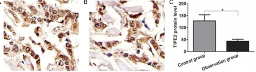
Comparison of TIPE2 protein levels in nasal mucosal tissue between two groups
As shown in Figure 2, the TIPE2 protein was mainly located in the cytoplasm membrane. The TIPE2 protein was moderately expressed in nasal mucosa tissue in the control group, while the TIPE protein was lowly expressed in the observation group. Gray degree quantita -Figure 1. Comparison of TIPE2 mRNA levels in nasal mucosal tissue between two groups. A. Amplification curve of
[image:3.629.103.532.80.222.2]TIPE2; B. TIPE2 mRNA level in nasal mucosal tissue of two groups.
[image:3.629.106.531.280.399.2]tive analysis showed that the level of TIPE2 pro-tein in nasal mucosa in the observation group was significantly lower than that of the control group, the difference was statistically signifi -cant (P<0.05).
Comparison of inflammatory cells infiltration in
nasal mucosal tissue between two groups
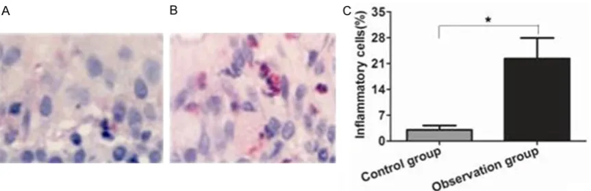
There was very rare inflammatory cells infiltra -tion in nasal mucosa in the control group, while there was a large number of inflammatory cells infiltrations in the observation group. Quanti-tative analysis showed that the proportion of inflammatory cells in nasal mucosa of the ob-servation group was significantly higher than that of the control group, the difference was statistically significant (P<0.05) (Figure 3).
Relationship between TIPE2 mRNA level and
inflammatory cells infiltration
The relationship between TIPE2 mRNA level and inflammatory cells infiltration showed that TIPE mRNA level was negatively correlated with inflammatory cells infiltration (P<0.05).
Discussion
Allergic rhinitis (AR) is a nasal mucosa allergic disease with the neurotransmitter release me- diated by IgE, the involvement of many im- munocompetent cells and cytokines after the atopic individual contacts allergen [13, 14]. Its essential conditions include [15]: ① specific antigen, namely the substance inducing the immunoreaction; ② atopic individual, namely individual difference and allergic constitution;
[image:4.629.100.532.79.219.2]③ encounter of specific antigen and atopic individual. The occurrence of allergic rhinitis has a great relationship with genetic, body weight and environment. In recent years, the incidence of allergic rhinitis shows an obvious rising trend in children has become a common disease in pediatric department and otorhino-laryngology department, which can not only affect the normal development of children, but also bring a lot of trouble for parents [16]. The pathogenesis of allergic rhinitis is still not completely clear. At present, many genes asso-ciated with allergic rhinitis have been found from the gene level, which provides a basis for clinical diagnosis and treatment. The earlier researches showed TIPE2 showed great differ-ence between the normal nasal mucosa and allergic rhinitis mucosa through transcriptome sequencing analysis. TIPE2 is tumor necrosis factor induced protein, which was discovered in meningitis mice model. It plays an important role in the regulation of inflammatory response and is considered as an immunoregulatory neg-ative protein [17, 18]. The recent studies sh- owed that the level of TIPE2 in atherosclerosis was significantly increased and was positively correlated with tumor necrosis factor [19, 20]. In addition, the levels of TIPE2 protein were not consistent in different tumor tissues. For exam-ple, its expression was decreased in primary hepatocellular carcinoma, but its expression was increased in renal cell carcinoma [21]. The above results indicated that TIPE2 protein played different roles in different diseases. So we analyzed the expressions of TIPE2 protein in nasal mucosa of allergic rhinitis children. Figure 3. Comparison of inflammatory cells infiltration in nasal mucosal tissue between two groups. Inflammatory cell level in nasal mucosal tissue of control group and observation group. A. HE staining showed inflammatory cells level in nasal mucosal tissue of control group; B. HE staining showed inflammatory cells level in nasal mucosal tissue of observation group; C. Quantitative analysis show inflammatory cells level in nasal mucosal tissue of two
The TIPE2 expression in normal nasal mucosa and allergic rhinitis mucosa was analyzed by real-time fluorescence quantitative PCR. The results showed that TIPE2 mRNA level was sig-nificantly lower than that of normal nasal muco -sa, indicating that TIPE expression was dec- reased in allergic rhinitis mucosa. The furth- er immunohistochemical analysis showed that the expression of TIPE protein in allergic rhinitis mucosa was significantly lower than that of the normal mucosa, which was consistent with the change of mRNA level. This result suggested that TIPE2 protein might play a negative regu-lating role in the occurrence of allergic rhinitis. An important pathological change of allergic rhinitis is inflammatory cells infiltration, espe -cially eosinophils and mast cells. Our study showed that the level of inflammatory cells infil -tration in allergic rhinitis mucosa was signifi -cantly increased. The correlation analysis be- tween the expression of TIPE2 mRNA and in- flammatory cells infiltration was analyzed. The results showed that they were negatively cor-related, suggesting that TIPE2 was an inflam -mation negative regulatory factor.
In conclusion, the expression of TIPE2 protein was lowest in allergic rhinitis mucosa, and it was negatively correlated with the degree of inflammatory cells infiltration.
Acknowledgements
We are grateful to all the participants in this study.
Disclosure of conflict of interest
None.
Address correspondence to: Dr. Ying Li, Department of Pediatrics, Huaihe Hospital of Henan University,
115 Ximen Street, Kaifeng 475000, Henan Province,
China. E-mail:yinglihn@163.com
References
[1] Seidman MD, Gurgel RK, Lin SY, Schwartz SR, Baroody FM, Bonner JR, Dawson DE, Dykewicz MS, Hackell JM, Han JK, Ishman SL, Krouse HJ, Malekzadeh S, Mims JW, Omole FS, Reddy
WD, Wallace DV, Walsh SA, Warren BE, Wilson
MN, Nnacheta LC; Guideline Otolaryngology
Development Group. AAO-HNSF. Clinical prac-tice guideline: Allergic rhinitis. Otolaryngol
Head Neck Surg 2015; 152: S1-43.
[2] Sastre J. Ebastine in allergic rhinitis and chron-ic idiopathchron-ic urtchron-icaria. Allergy 2008; 63 Suppl 89: 1-20.
[3] Phan H, Moeller ML, Nahata MC. Treatment of allergic rhinitis in infants and children: efficacy
and safety of second-generation
antihista-mines and the leukotriene receptor antagonist montelukast. Drugs 2009; 69: 2541-2576.
[4] Tao B, Ruan G, Wang D, Li Y, Wang Z, Yin G. Imbalance of Peripheral Th17 and Regulatory T Cells in Children with Allergic Rhinitis and Bronchial Asthma. Iran J Allergy Asthma Immu-nol 2015; 14: 273-279.
[5] Lou Y, Liu S. The TIPE (TNFAIP8) family in
in-flammation, immunity, and cancer. Mol Immu -nol 2011; 49: 4-7.
[6] Cui J, Hao C, Zhang W, Shao J, Zhang N, Zhang
G, Liu S. Identical expression profiling of hu
-man and murine TIPE3 protein reveals links to
its functions. J Histochem Cytochem 2015; 63: 206-216.
[7] Zhang Z, Liang X, Gao L, Ma H, Liu X, Pan Y, Yan W, Shan H, Wang Z, Chen YH, Ma C. TIPE1 in -duces apoptosis by negatively regulating Rac1 activation in hepatocellular carcinoma cells. Oncogene 2015; 34: 2566-2574.
[8] Zhang H, Zhu T, Liu W, Qu X, Chen Y, Ren P, Wang Z, Wei X, Zhang Y, Yi F. TIPE2 acts as a
negative regulator linking NOD2 and inflam -matory responses in myocardial
ischemia/re-perfusion injury. J Mol Med (Berl) 2015; 93:
1033-1043.
[9] Zhang YH, Yan HQ, Wang F, Wang YY, Jiang YN,
Wang YN, Gao FG. TIPE2 inhibits
TNF-α-in-duced hepatocellular carcinoma cell
metasta-sis via Erk1/2 downregulation and NF-κB acti -vation. Int J Oncol 2015; 46: 254-264. [10] Lou Y, Sun H, Morrissey S, Porturas T, Liu S,
Hua X, Chen YH. Critical roles of TIPE2 prote- in in murine experimental colitis. J Immunol 2014; 193: 1064-1070.
[11] Freundt EC, Bidere N, Lenardo MJ. A different
TIPE of immune homeostasis. Cell 2008; 133: 401-402.
[12] Zhang Y, Wang CS, Zhang L. Correlation study on the relationship between FOXP3 gene poly-morphism and allergic rhinitis. Chin J
Otorhino-laryngol Head Neck Surg 2010; 45: 397-400.
[13] Chen QY, Zhang H. Single nucleotide polymor-phisms and allergic rhinitis associated with antigen processing. Int J Otolaryngol Head
Neck Surg 2012; 36: 76-79.
[14] Bousquet J, Khaltaev N, Cruz AA, Denburg J, Fokkens WJ, Togias A, Zuberbier T, Baena-Cag -nani CE, Canonica GW, van Weel C, Agache I,
O, Kaliner MA, Kim YY, Kowalski ML, Kuna P, Le LT, Lemiere C, Li J, Lockey RF, Mavale-Manuel S, Meltzer EO, Mohammad Y, Mullol J, Naclerio R, O’Hehir RE, Ohta K, Ouedraogo S, Palkonen S, Papadopoulos N, Passalacqua G, Pawankar R, Popov TA, Rabe KF, Rosado-Pinto J, Scad
-ding GK, Simons FE, Toskala E, Valovirta E, van Cauwenberge P, Wang DY, Wickman M, Yawn BP, Yorgancioglu A, Yusuf OM, Zar H, Annesi-Maesano I, Bateman ED, Ben Kheder A, Boakye DA, Bouchard J, Burney P, Busse WW, Chan-Yeung M, Chavannes NH, Chuchalin A, Dolen WK, Emuzyte R, Grouse L, Humbert M, Jackson C, Johnston SL, Keith PK, Kemp JP, Klossek JM, Larenas-Linnemann D, Lipworth B, Malo JL, Marshall GD, Naspitz C, Nekam K, Niggemann B, Nizankowska-Mogilnicka E, Okamoto Y, Orru MP, Potter P, Price D, Stoloff
SW, Vandenplas O, Viegi G, Williams D; World Health Organization; GA(2)LEN; AllerGen. Aller-gic Rhinitis and its Impact on Asthma (ARIA) 2008 update (in collaboration with the World Health Organization, GA(2)LEN and AllerGen). Allergy 2008; 63 Suppl 86: 8-160.
[15] Agache I, Deleanu D, Khaltaev N, Bousquet J.
Allergic rhinitis and its impact upon asthma--update (ARIA 2008). Romanian perspective. Pneumologia 2009; 58: 255-258.
[16] Scaparrotta A, Attanasi M, Petrosino MI, Di
Filippo P, Di Pillo S, Chiarelli F. Critical apprais-al of Timothy grass pollen extract GRAZAX(®) in the management of allergic rhinitis. Drug Des Devel Ther 2015; 9: 5897-5909.
[17] Sun H, Gong S, Carmody RJ, Hilliard A, Li L, Sun
J, Kong L, Xu L, Hilliard B, Hu S, Shen H, Yang
X, Chen YH. TIPE2, a negative regulator of in-nate and adaptive immunity that maintains im-mune homeostasis. Cell 2008; 133: 415-426. [18] Wang Z, Fayngerts S, Wang P, Sun H, Johnson
DS, Ruan Q, Guo W, Chen YH. TIPE2 protein serves as a negative regulator of phagocytosis and oxidative burst during infection. Proc Natl Acad Sci U S A 2012; 109: 15413-15418. [19] Lou Y, Liu S, Zhang C, Zhang G, Li J, Ni M, An G,
Dong M, Liu X, Zhu F, Zhang W, Gao F, Chen YH,
Zhang Y. Enhanced atherosclerosis in
TIPE2-deficient mice is associated with increased
macrophage responses to oxidized low-density lipoprotein. J Immunol 2013; 191: 4849-4857. [20] Zhang G, Zhang W, Lou Y, Xi W, Cui J, Geng M,
Zhu F, Chen YH, Liu S. TIPE2 deficiency accel -erates neointima formation by downregulating smooth muscle cell differentiation. Cell Cycle 2013; 12: 501-510.
[21] Zhang Z, Qi H, Hou S, Jin X. TIPE2 mRNA