Int J Clin Exp Pathol 2016;9(7):6999-7009 www.ijcep.com /ISSN:1936-2625/IJCEP0026688
Original Article
Prognostic value of Thy1 in non-small cell
lung cancer: a RNA-Seq transcriptome analysis
Shuang Zhao1, Zhixin Qiu1, Yuting Jin1, Li Zhang2, Weimin Li1
1Department of Respiratory Medicine, West China Hospital, Sichuan University, Chengdu 610041, China; 2Key
Laboratory of Transplant Engineering and Immunology, Ministry of Health, West China Hospital, Chengdu 610041, China
Received February 26, 2016; Accepted May 21, 2016; Epub July 1, 2016; Published July 15, 2016
Abstract: Thy1 is a membrane glycoprotein, which can regulate cell-matrix and cell-cell interactions. Thy1 plays an important role in oncogenesis, tumor metastasis and survival. Thy1 has also been considered a major marker in many kinds of cancer stem cells. In order to investigate the expression and prognostic values of Thy1 in non small cell lung cancer (NSCLC), we cultured the stem cell-like sphere 95D cell lines in serum-free DMEM/F12. Colony for-mation assay, cell cycle and apoptosis were performed in vitro. Tumorigenicity was analyzed in vivo. The RNA expres-sion status of Thy1 in sphere 95D cell lines was investigated by RNA-Seq. Then we examined Thy1 expresexpres-sion via im-munohistochemistry (IHC) in 183 NSCLC samples and 96 normal lung samples. The stem cell-like sphere 95D cells
had a higher efficiency of cloning than adherence 95D in vitro, and a better tumorigenic potential in vivo. Sphere 95D cells were blocked in G0-G1 in Sphere 95D group significantly, but have no significant effect on the apoptosis compared with adherence 95D cells. The RNA expression of Thy1 in Sphere 95D cells was significantly up-regulated
compared with adherence 95D cells by RNA-seq. Of all NSCLC specimens, Thy1 was detected in the mesenchyme or nucleus by IHC, and the expression rates of Thy1 positive cells were much higher in NSCLC specimens than in normal lung samples. Mesenchymal Thy1 positive expression was an adverse predictor of prognosis in NSCLC
pa-tients, while nuclear Thy1 positive expression had no significant associations with overall survival. However, neither mesenchymal nor nuclear Thy1 positive expression was significantly related to clinicopathological characteristics in
NSCLC. Thus, Thy1 might serve as a vital therapeutic target for NSCLC.
Keywords: Thy1 (CD90), cancer stem cells, lung cancer, prognosis
Introduction
Lung cancer is the leading contributor to mor-bidity and mortality from cancer worldwide, with its 5-year overall survival (OS) being only about 17% [1]. Non small cell lung cancer (NSCLC) is the most common form and accounts for more than 85% of all lung cancer. Although there was great progress in the thera-py of NSCLC during the past years, many patients still died due to tumor metastases and resistant to conventional treatments. Recent studies reported that tumors had heteroge- neity, and only about 0.1%-2% cancer cells, named cancer stem cells (CSCs), were respon-sible for carcinogenesis [2]. The existence of CSCs is thought to contribute to cancer metas-tasis, recurrence and drug resistance. Thus, identifying lung CSCs may help to develop
novel, effective cancer diagnosis and treatment strategies.
found in human ovarian cancer cell lines, the growth rates and tumor sizes of the xenograft tumor with Thy1 transfectants were significant -ly reduced compared with their null counter-parts [6]. So far, the function of Thy1 still remains inconsistent. Further, there are limited studies about the role of Thy1 in patients with NSCLC.
In this study, the RNA expression status of Thy1 in stem cell-like sphere 95D cell line was inves-tigated by RNA-Seq. Then, relationships of Thy1 expression with clinicopathological features and prognostic value were analyzed in NSCLC patients.
Materials and methods
Patients and tissue specimens
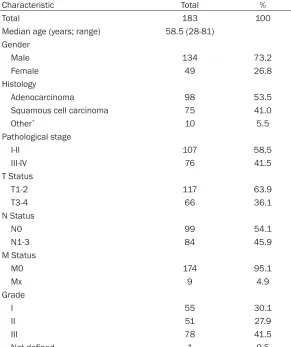
One hundred and ninety-five patients of West China Hospital during 2007 were subsequently recruited. All of these NSCLC patients received complete surgical resection for primary NSCLC without previous neoadjuvant therapy. Ninety-six normal lung tissues nearby the primary tumors were considered as controls. After sur-gical resection, patients underwent standard therapies according to the Clinical Oncology Information Network Guidelines on the nonsur-gical treatments for lung cancer [7]. Finally, One hundred and eighty-three patients with complete data were enrolled after a five-year follow-up. The stages of tumors were assessed on the basis of the American Joint Committee on Cancer (AJCC)’s tumor-node-metastasis (TNM) staging system [8], while their histologi-cal types and differentiation were based on the World Health Organization (WHO)’s classifica -tion for NSCLC [9]. This study was carried out by the permission of the Committee on Medical Ethics of West China Hospital, and all the enrolled patients signed a written informed consent.
Cell lines and animal models
Human lung cancer cell line 95D was obtained from the Laboratory of Stem Cell Biology, West China Hospital, Sichuan University. The adher-ence 95D cell lines were cultured in PRMI1640 supplemented with 10% fetal bovine serum (FBS) and 1% penicillin/streptomycin (Invitro- gen, Carsbald, CA, USA) at 37°C in a humidifi-ed atmosphere with 5% CO2 and 95% air. The
sphere 95D cells were cultured under the stem cell conditions: serum-free DMEM/F12 contain-ing 0.4% BSA (sigma), 20 ng/ml basic fibroblast growth factor (bFGF) (Peprotech Rocky Hill, NJ, USA), 20 ng/ml insulin growth factor (IGF) (Peprotech), 20 ng/ml epidermal growth factor (EGF) (Peprotech), and 1% N-2 Supplement (Invitrogen). And the sphere 95D cells were maintained on ultra low attachment plates (Corning, Corning, NY, USA). Four-six weeks old nude mice, male or female, were purchased from the Chengdu Experimental Animal Center of DaShuo (Chengdu, China). Each of them was injected with 1 × 10⁴ sphere 95D cells
subcuta-neously in the right flank and 1 × 10⁴
adher-ence 95D cells in the left flank. All the mice were housed under pathogen-free environ-ment, and all animal studies were performed on the basis of animal protocol approved by Animal Care and Use Committee of Sichuan University.
Colony formation assay
Approximately 500 cells were inoculated into a 6-well plate growing for 14 days, then the single cell containing wells were used for cell counting by using crystal violet exclusion. Every well was identified and analyzed for the ability of the cells to generate spheres.
Cell cycle analysis
About 1 × 10⁶ cells were gathered and washed
by phosphate-buffered saline (PBS), and then fixed with 70% ethanol overnight at 4°C. Then the cells were incubated in staining buffer (Sigma, USA) with propidium iodide (PI) (1 mg/ ml), R Nase A (10 mg/ml) and 0.1% (vol/vol) Triton X-100 in PBS for 30 min in the dark at room temperature. The cells’ percentages in different phases of cell cycles were determined via flow cytometry equipped with CXP Modifit software (BD, USA) [10].
Apoptosis analysis
Approximately 5 × 10⁵ cells were harvested
The prognostic value of Thy1 in non small cell lung cancer
RNA-Seq library preparation and sequencing
Briefly, total RNA samples were extracted from the sphere 95D cells and adherence 95D cells using TRIzol Reagent (Invitrogen, USA). Then the mRNA was enriched and purified from total RNA by the oligo (dT) magnetic beads. Cleaved mRNA fragments (about 200 bp) were synthe-sized the first strand of cDNA via using random hexamer-primer. Magnetic beads were then used to purify the double strand cDNA. Next, end reparation, 3’-end single nucleotide, adap-tor ligation, cDNA templates purification and enrichment were performed. Constructed lib- raries were sequenced by Illumina HiSeqTM 2000 according to the manufacturer’s instruc-tions. TopHat version 1.2.0 was used to aligne RNA-seq reads to genome reference (hg19) and identified splice variants of each sample. The genes were considered to be differentially expressed if their false discovery rates (FDRs) < 0.001 [11]. The gene expression levels were normalized to reads per kilowatt of exon model (RPKM) per million mapped reads in order to help for the comparison of transcripts between groups. Furthermore, genes were defined as upregulated if their mean log2 fold change ratio > 1.
Immunohistochemistry
The immunohistochemical staining of Thy1 were conducted with 4-μm-thick paraffin-em-bedded samples. Primary antibody was rabbit monoclonal antibody Thy1 (Abcan, USA) diluted 1:200. Secondary antibody was goat anti-rab-bit IgG (Dako, Shanghai, China). All samples were incubated at 4°C with the primary anti-body overnight. Immunohistochemistry was ful-filled according to the standard ChemMate™ Peroxidase/DAB Detection Kit techniques fol -lowing the manufacturer’s recommendations as reported before [12, 13]. We replaced the primary antibody by PBS as negative controls. The positive expression of Thy1 mesenchymal expression was visually evaluated and scored using a dual rate semi-quantitative method [14]. As for the nuclear Thy1 expression, posi-tive staining cells > 10% were considered as positive expression.
Statistical analysis
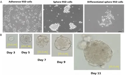
All data was analyzed by SPSS 19.0 Software (IBM SPSS Statistics, New York, NY, USA). Chi-squared test and variance analysis were used to determine the significant statistical differ -Figure 1. Phase-contrast microscopy representative images of 95D cell lines. A. Adherence 95D cells: were cultured by 10% fetal bovine serum. Sphere 95D cells: were cultured without fetal bovine serum. Differentiated sphere 95D
[image:3.612.93.522.73.330.2]ences among the groups. Kaplan-Meier was performed to survival evaluate curves, while log-rank tests were conducted to estimate their significance. Figures were completed by Graph-Pad Prism 6.0 (GraphGraph-Pad Software, Inc., San Diego, USA). All values were expressed as mean ± standard error of the mean (SEM), and levels of statistical significance of differences were set at P value < 0.05 (two-tailed).
Results
Stem cell-like spheres formation of 95D cells
When cultured in 10% fetal bovine serum, the 95D cells were adherent growth. Then, we cul-tured adherence 95D cells in a serum-free
medium (SFM) supplemented with growth fac-tors. After about 10-15 days, a small part of 95D cells formed spheres. Then, we incubated the spheres in 20% FBS containing medium for the differentiation, and the spheres regained to adherent growth (Figure 1A, 1B).
Identification of the function of stem cell-like sphere 95D cells
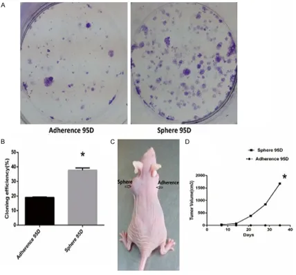
To further identify whether the stem cell-like sphere 95D cells owned the features of tumor initiating cells, we performed some experi-ments in both vitro and vivo. Approximately 500 cells were inoculated into a 6-well plate growing for 14 days. The cloning efficiency of sphere 95D cells was 37.67±1.609%, while the adher-Figure 2. The cloning efficiency and tumorigenic potential of sphere 95D cells and adherence 95D cells. A, B. Colony
[image:4.612.94.522.72.470.2]The prognostic value of Thy1 in non small cell lung cancer
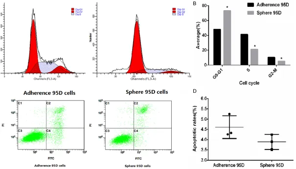
Figure 3. Cells cycle and apoptosis detection sell cycle detection. A, B. Approximately 1 × 10⁶ cells were collected and stained by PI. DNA content was detected
us-ing flow cycometry. Cluster of cells were gated out, and the profiles of cell cycles were investigated to quantitate cell cycle distribution. *P < 0.05. C, D. Cells were
ence 95D was 19.00±0.3819%. The results showed sphere 95D cells had a high efficiency of cloning than adherence 95D (P=0.001) (Figure 2A, 2B). To better understand the tumorigenic potential of tumor cells In vivo, xenograft models were established. After about 30 days, as few as 1 × 10⁴ sphere 95D cells
could generate xenografts and adherence 95D cells failed to do so (Figure 2C, 2D). In cell cycle analysis, approximately 1 × 10⁶ cells were
col-lected and stained by PI. DNA content was detected using flow cytometry. Cluster of cells were gated out, and the profiles of cell cycles were investigated to quantitate cell cycle distri-bution. Our results demonstrated that cells were blocked in G0-G1 in Sphere 95D group significantly (all P < 0.05) (Figure 3A, 3B). However, we did not observe the differences on the apoptosis between sphere 95D cells and adherence 95D cells (P=0.1315) (Figure 3C,
3D).
Aberrant mRNA expression in sphere 95D cells
Total numbers of RNA-seq reads obtained from the sphere 95D cells and adherence 95D cells were 11, 863, 292 (581, 301, 308 bp) and 11, 863, 292 (565, 244, 351 bp). Our results showed 637 genes were up-regulated. Through Gene ontology analysis, we found overrepre-sented GO terms in the stem cell like spheres
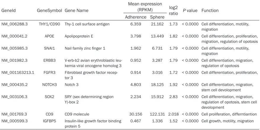
gene sets, were closely collated to cell prolifer-ation, motility and cell differentiation (All P < 0.05) (Table S1). Furthermore, we found some genes in sphere 95D cells were significantly up-regulated compared with adherence 95D cells (Table 1).
Thy1 expression and association with clinical characteristics
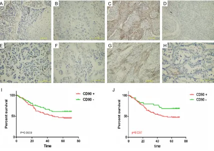
[image:6.612.87.526.97.317.2]To further verified the results of RNA-seq, we immunohistochemically analyzed the expres-sion of Thy1 in 183 NSCLC samples (Table S2) and 96 normal lung specimens nearby the tumors. Our results demonstrated that Thy1 was detected in the mesenchyme or nuclei (Figure 4). As showed in Table 2, the expression rates of Thy1 positive cells were much higher in NSCLC specimens than in normal lung samples (P < 0.001). Of all the NACLC specimens, there were 112 (61.2%) specimens with Thy1 nucle-us positive expression, and 148 (80.87%) spec-imens with Thy1 mesenchymal positive expres-sion. However, neither mesenchymal nor nucle-ar Thy1 positive expression was significant statistically associated with clinicopathological characteristics (All P > 0.5). Then, the Kaplan-Meier survival curves were used to access the correlations between the Thy1 expression lev-els in mesenchymal or nuclear and patients’ 5-year overall survivals (Figure 4I, 4J). The results suggested mesenchymal Thy1 positive
Table 1. List of some up-regulated genes in Sphere 95D cells as compared with adherence 95D cells by RNA sequencing
GeneId GeneSymbol Gene Name
Mean expression
(RPKM) log2
ratio P value Function Adherence Sphere
NM_006288.3 THY1/CD90 Thy-1 cell surface antigen 6.359 21.162 1.73 < 0.0000 Cell differentiation, motility, migration
NM_000041.2 APOE Apolipoprotein E 3.798 13.449 1.82 < 0.0000 Cell differentiation, proliferation, migration, regulation of opotosis NM_005985.3 SNAI1 Nail family zinc finger 1 1.962 6.731 1.79 < 0.0000 Cell differentiation, motility,
migration NM_001982.3 ERBB3 V-erb-b2 avian erythroblastic
leu-kemia viral oncogene homolog 3
0.952 3.287 1.79 < 0.0000 Cell differentiation, migration, regulation of opotosis NM_001163213.1 FGFR3 Fibroblast growth factor
recep-tor 3 0.914 3.016 1.72 < 0.0000 Cell differentiation, proliferation, NM_000435.2 NOTCH3 Notch 3 4.803 18.125 1.92 < 0.0000 Cell differentiation, migration,
stem cell devolopment NM_003106.3 SOX2 SRY (sex determining region
Y)-box 2
2.234 15.912 2.83 < 0.0000 Cell differentiation, migration, regulation of opotosis, stem cell devolopment
NM_001769.3 CD9 CD9 molecule 30.156 122.131 2.018 < 0.0000 Cell proliferation, differntiantion NM_000599.3 IGFBP5 Insulin-like growth factor binding
The prognostic value of Thy1 in non small cell lung cancer
expression was an adverse predictor of progno-sis in NSCLC patients (P=0.0397), while nucle-ar Thy1 positive expression had no significant associations with overall survival (P=0.0659).
Discussion
In the present study, we found the stem cell-like sphere 95D cells had a higher efficiency of cloning than adherence 95D in vitro and a bet-ter tumorigenic potential of tumor cells in vivo. On the other hand, sphere 95D cells were blocked in G0-G1 in Sphere 95D group signifi -cantly, but have no significant effect on the apoptosis compared with adherence 95D cells. Furthermore, we observed that Thy1 was up-regulated in stem cell like 95D cell lines and was overexpression in NSCLC patients. Mesenchymal Thy1 positive expression was a poor prognostic predictor for NSCLC patients.
[image:7.612.93.521.71.373.2]CSCs are a subpopulation of tumor cells that related to tumor proliferation and spreading. These cells have unlimited proliferation po- tential, capacity to self-renewal and ability to differentiate into a variety of specialized cells [15]. However, the methods of identifying CSCs are conflict nowadays and the amount of CSCs are too low for further analysis. Sphere forma-tion by a condiforma-tioned serum-free culture sys-tem was reported to be an effective method for enriching stem cell-like cells in numerous stud-ies. Sphere cells with the ability for tumorige-nicity and self-renewal are thought to be CSCs [16, 17]. Previous studies also reported they may be an important factor in tumor metasta-sis, resistance to current radiotherapy and che-motherapy [18, 19]. In our study, we compared the capacity of tumorigenicity and self-renewal in sphere 95D cell lines with adherence 95D cell lines. The results showed the sphere 95D Figure 4. Expression of Thy1 staining in NSCLC by immunohistochemistry and the correlations with overall surviv-als. A. Positive expression of nucleus CD90 in squamous cell carcinoma (SCC). B. Negative expression of nucleus CD90 in SCC. C. Positive expression of nucleus CD90 in adenocarcinoma (ADC). D. Negative expression of nucleus CD90 in ADC. E. Positive mesenchymal expression of nucleus CD90 in SCC. F. Negative mesenchymal expression of nucleus CD90 in SCC. G. Positive mesenchymal expression of nucleus CD90 in ADC. H. Negative mesenchymal
expression of nucleus CD90 in ADC. Original magnification, × 40. I. Survival of nucleus CD90 positive expression. J.
cells had a higher efficiency of cloning than adherence 95D in vitro and a better tumorigen-ic potential of tumor cells in vivo. We also observed cell cycle was arrested in G0/G1 in Sphere 95D group markedly. This result was consistent with another study in hepatocellular carcinoma (HCC) [20]; they found human poorly differentiated HCC cell lines can form spheres, while the well differentiated ones cannot. Fur- thermore, they also observed that cell cycle was blocked at G0-G1 in sphere cells compared with parental cells, and P21, which plays a cru-cial effect on the transition of the cell cycles from the G0/G1 to the S phase [21], was upreg-ulated in sphere cells.
By RNA-Seq, we found the expressions of many genes were different between sphere 95D and adherence 95D cells. These genes were closely collated to cell proliferation, motility and
[image:8.612.93.387.86.454.2]specimens, mesenchymal Thy1 positive expres-sion was an adverse predictor of prognosis in NSCLC patients, while nuclear Thy1 positive expression had no significant associations with overall survival. However, neither mesen-chymal nor nuclear Thy1 positive expression was significant statistically associated with clinicopathological characteristics in NSCLC. Kawamura et al. [30] conducted immunohisto -chemical analysis of clinical specimens (6 mesothelioma, 28 lung adenocarcinoma, 33 lung squamous cell carcinoma), and found the positive expression of Thy1 was higher in meso-thelioma than lung cancer. Study by Chen and his collaborators showed Thy1 positive expres-sion can serve as a prognostic maker for lung cancer [31]. However, Matsuwaki et al. have opposite opinions; their study suggested that Thy1 positive expression had no significant relationships with three-year recurrence-free
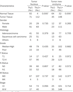
Table 2. Thy1 expression and association with clinical characteristics
Characteristics Tumor Nucleus P-value Tumor mes-enchyma P-value Thy1-
(No.) Thy1+ (No.) Thy1- (No.) Thy1+ (No.) Normal Tissue 92 4 0.000* 64 32 0.000*
Tumor Tissue 71 112 35 148
Gender
Female 20 29 0.735 12 37 0.265
Male 51 83 23 111
Histology
Adenocarcinoma 41 55 0.379 19 77 0.705 Squamous cell carcinoma 25 51 13 63
Others 5 6 3 8
Grade
Median-High 48 79 0.435 25 102 0.682
Poor 22 33 9 46
T Status
T1 14 17 0.427 6 25 0.972
T2-4 57 95 29 123
N Status
N0 39 60 0.857 17 82 0.572
N1 32 52 18 66
M Status
M0 67 107 0.737 32 142 0.377
MX 4 5 3 6
Stage
I-II 54 72 0.094 25 101 0.714
III-IV 17 40 35 148
No.: number. *P < 0.05.
The prognostic value of Thy1 in non small cell lung cancer
survival [32]. These inconsistencies might be explained by the limited patients’ number, the different cut-off scores and different primary antibody types or dilutions. Therefore, Thy1 might serve as a vital prognostic biomarker for NSCLC, and some large cohort studies with more suitable laboratory methodology are also needed to further demonstrate the relation-ships between Thy1 and the survival of NSCLC patients.
Thy1 operates as an important factor of regu-lating cell-matrix and cell-cell interactions, pro-liferation, adhesion, apoptosis, migration, me- tastasis, angiogenesis and fibrosis [33] in can -cers. Wang et al. found in lung squamous cell carcinoma, CD44 (+) CD90 (+) cells have a high-er ability to form sphhigh-eroids, and revealed mes-enchymal morphology [34]. In gastric cancer, Thy1 was overexpressed and could inhibit gas-tric cancer cell apoptosis through regulating the expression levels of SPARC proteins [35]. In melanoma, Thy1 could promote the adhe-sion and transmigration of melanoma cells to the activated endothelium via the interaction with the αvβ3-integrin [4, 36]. Furthermore, Sukowati et al. found in hepatocellular carcino-ma, expression of ABCG2 was upregrulated in CD90 (+) subpopulation after exposure to DOX, indicting CD90 involved in drug resistance [37]. Thus, Thy1 plays an important role in the prog-ress of the cancers, and might serve as a thera-peutic target for NSCLC.
In summary, our results have shown that Thy1 was up-regulated in stem cell like cells, and was overexpression in NSCLC patients. Al- though nuclear Thy1 positive expression was not associated with overall survival, mesenchy-mal Thy1 positive expression was an adverse predictor of prognosis for NSCLC patients. Thus, Thy1 might serve as a vital therapeutic target for NSCLC.
Acknowledgements
We thank Mrs. Fei Chen and Pro.Li Li for their help in the laboratory work. We are also Prof. Xianmin Mo for providing the lung cancer cell lines and laboratory. This work was supported by grants from the Nature Science Foundation of China (812410-68, 81372504).
Disclosure of conflict of interest
None.
Address correspondence to: Dr. Weimin Li, De- partment of Respiratory Medicine, West China Hospital, Sichuan University, Chengdu 610041, China. Tel: +86 (28) 85423998; E-mail: Weimi003@ yahoo.com
References
[1] Siegel R, Naishadham D and Jemal A. Cancer statistics, 2013. CA Cancer J Clin 2013; 63: 11-30.
[2] Reya T, Morrison SJ, Clarke MF and Weissman IL. Stem cells, cancer, and cancer stem cells. Nature 2001; 414: 105-111.
[3] Rege TA and Hagood JS. Thy-1, a versatile modulator of signaling affecting cellular adhe-sion, proliferation, survival, and cytokine/gro- wth factor responses. Biochim Biophys Acta 2006; 1763: 991-999.
[4] Saalbach A, Wetzel A, Haustein UF, Sticherling M, Simon JC and Anderegg U. Interaction of hu-man Thy-1 (CD 90) with the integrin alphavbe-ta3 (CD51/CD61): an important mechanism mediating melanoma cell adhesion to activat-ed endothelium. Oncogene 2005; 24: 4710-4120.
[5] Fiegel HC, Bruns H, Höper C, Lioznov MV and
Kluth D. Cell growth and differentiation of dif -ferent hepatic cells isolated from fetal rat liver in vitro. Tissue Eng 2006; 12: 123-130. [6] Abeysinghe HR, Pollock SJ, Guckert NL,
Veyberman Y, Keng P, Halterman M, Federoff
HJ, Rosenblatt JP and Wang N. The role of the THY1 gene in human ovarian cancer suppres-sion based on transfection studies. Cancer Genet Cytogenet 2004; 149: 1-10.
[7] The Royal College of Radiologists Clinical Oncology Information Network. Guidelines on the non-surgical management of lung cancer. Clin Oncol (R Coll Radiol) 1999; 11: S1-53. [8] Edge SB and Compton CC. The American Joint
Committee on Cancer: the 7th edition of the AJCC cancer staging manual and the future of TNM. Ann Surg Oncol 2010; 17: 1471-1474. [9] Travis WD, Brambilla E and Riely GJ. New
pathologic classification of lung cancer: rele -vance for clinical practice and clinical trials. J Clin Oncol 2013; 31: 992-1001.
[10] Jiang L, Man L, Dan L, Bojiang C, Wen Z, Lin M, Zeng J, Huang N, Huang Y, Mo XM and Li WM. BAD overexpression inhibits cell growth and induces apoptosis via mitochondrial-depen-dent pathway in non-small cell lung cancer. Cancer Cell Int 2013; 13: 53.
[11] Storey JD and Tibshirani R. Statistical signifi -cance for genomewide studies. Proc Natl Acad Sci U S A 2003; 100: 9440-9445.
[12] Qiu ZX, Wang L, Han J, Liu D, Huang W, Altaf K,
Prognostic impact of Raf-1 and p-Raf-1 expres-sions for poor survival rate in non-small cell lung cancer. Cancer Sci 2012; 103: 1774-1779.
[13] Huang Y, Liu D, Chen B, Zeng J, Wang L, Zhang S, Mo XM and Li WM. Loss of Bad expression confers poor prognosis in non-small cell lung cancer. Med Oncol 2012; 29: 1648-1655. [14] Zhao S, Qiu ZX, Zhang L and Li WM. Prognostic
values of ERK1/2 and p-ERK1/2 expressions
for poor survival in non-small cell lung cancer. Tumour Biol 2015; 36: 4143-4150.
[15] Baker DE, Harrison NJ, Maltby E, Smith K,
Moore HD, Shaw PJ, Health PR, Holden H and Andrews PW. Adaptation to culture of human embryonic stem cells and oncogenesis in vivo. Nat Biotechnol 2007; 25: 207-215.
[16] Yu SC, Ping YF, Yi L, Zhou ZH, Chen JH, Yao XH, Gao L, Wang JM and Bian XW. Isolation and characterization of cancer stem cells from a human glioblastoma cell line U87. Cancer Lett 2008; 265: 124-134.
[17] Cao L, Zhou Y, Zhai B, Liao J, Xu W, Zhang R, Li J, Zhang Y, Chen L, Qian H, Wu M and Yin Z. Sphere-forming cell subpopulations with can-cer stem cell properties in human hepatoma cell lines. BMC Gastroenterol 2011; 11: 71. [18] Sukowati CH, Rosso N, Crocè LS and Tiribelli C.
Hepatic cancer stem cells and drug resistance: Relevance in targeted therapies for hepatocel-lular carcinoma. World J Hepatol 2010; 2: 114-126.
[19] Diehn M, Cho RW, Lobo NA, Kalisky T, Dorie MJ, Kulp AN, Qian DL, Lam JS, Ailles LE, Wong M, Joshua B, Kaplan MJ, Wapnir I, Dirbas F, Somlo G, Garberoglio G, Paz B, Shen J, Lau SK,
Quake SR, Brown JM, Weissman IL and Clarke MF. Association of reactive oxygen species lev-els and radioresistance in cancer stem cells. Nature 2009; 458: 780-783.
[20] Hashimoto N, Tsunedomi R, Yoshimura K,
Watanabe Y, Hazama S and Oka M. Cancer stem-like sphere cells induced from de-differ-entiated hepatocellular carcinoma-derived cell lines possess the resistance to anti-cancer drugs. BMC Cancer 2014; 14: 722.
[21] Coqueret O. New roles for p21 and p27 cell-cycle inhibitors: a function for each cell com-partment? Trends Cell Biol 2003; 13: 65-70. [22] Woo SR, Oh YT, An JY, Kang BG, Nam DH and
Joo KM. Glioblastoma specific antigens, GD2
and CD90, are not involved in cancer stem-ness. Anat Cell Biol 2015; 48: 44-53.
[23] Knab LM, Ebine K, Chow CR, Raza SS, Sahai V, Patel AP, Kumar K, Bentrem DJ, Grippo PJ and Munshi HG. Snail cooperates with Kras G12D
in vivo to increase stem cell factor and
en-hance mast cell infiltration. Mol Cancer Res
2014; 12: 1440-1448.
[24] Yen WC, Fischer MM, Axelrod F, Bond C, Cain J, Cancilla B, Henner, WR, Meisner R, Sato A, Shah J, Tang T, Wallace B, Wang M, Zhang C,
Kapoun AM, Lewicki J, Gurney A and Hoey T.
Targeting Notch signaling with a Notch2/ Notch3 antagonist (tarextumab) inhibits tumor growth and decreases tumor-initiating cell fre-quency. Clin Cancer Res 2015; 21: 2084-2095.
[25] Karlsson G, Rörby E, Pina C, Soneji S, Reckzeh K, Miharada K, Karlsson C, Guo Y, Fugazza
C, Gupta R, Martens JH, Stunnenberg HG,
Karlsson S and Enver T. The tetraspanin CD9
affords high-purity capture of all murine hema-topoietic stem cells. Cell Rep 2013; 4: 642-648.
[26] Zhu J, Thakolwiboon S, Liu X, Zhang M and Lubman DM. Overexpression of CD90 (Thy-1) in pancreatic adenocarcinoma present in the tumor microenvironment. PLoS One 2014; 9: e115507.
[27] Tang KH, Dai YD, Tong M, Chan YP, Kwan PS,
Fu L, Qin YR, Tsao SW, Lung HL, Lung ML, Tong
DK; Law S, Chan KW, Ma S and Guan XY. A
CD90(+) tumor-initiating cell population with an aggressive signature and metastatic capac-ity in esophageal cancer. Cancer Res 2013; 73: 2322-2332.
[28] Jia Q, Zhang X, Deng T and Gao J. Positive cor-relation of Oct4 and ABCG2 to chemothera-peutic resistance in CD90(+)CD133(+) liver cancer stem cells. Cell Reprogram 2013; 15: 143-150.
[29] Scognamiglio G, D’Antonio A, Rossi G, Cavazza A, Camerlingo R, Pirozzi G, La Mantia E, Anniciello AM, Morabito A, Cantile M, Boscaino A, Sparano L, Botti G, Rocco G and Franco R. CD90 expression in atypical meningiomas and meningioma metastasis. Am J Clin Pathol 2014; 141: 841-849.
[30] Kawamura K, Hiroshima K, Suzuki T, Chai K,
Yamaguchi N, Shingyoji M, Yusa T, Tada Y,
Takiguchi Y, Tatsumi K, Shimada H and Tagawa
M. CD90 is a diagnostic marker to differentiate between malignant pleural mesothelioma and lung carcinoma with immunohistochemistry. Am J Clin Pathol 2013; 140: 544-549.
[31] Chen JF, Zhang LJ, Zhao AL, Wang Y, Wu N, Xiong HC, Liang Z, Li JY, Huang XF and Yang Y. Abnormal expression of Thy-1 as a novel tumor marker in lung cancer and its prognostic
sig-nificance. Zhonghua Yi Xue Za Zhi 2005; 85:
1921-1925.
[32] Matsuwaki R, Ishii G, Zenke Y, Neri S, Aokage
K, Hishida T, Yoshida J, Fujii S, Kondo H, Goya T, Nagai K and Ochiai A. Immunophenotypic
The prognostic value of Thy1 in non small cell lung cancer
[33] Rege TA and Hagood JS. Thy-1 as a regulator of cell-cell and cellmatrix interactions in axon regeneration, apoptosis, adhesion, migration,
cancer, and fibrosis. FASEB J 2006; 20:
1045-1054.
[34] Wang P, Gao Q, Suo Z, Munthe E, Solberg S,
Ma L, Wang M, Westerdaal NA, Kvalheim G and Gaudernack G. Identification and charac -terization of cells with cancer stem cell proper-ties in human primary lung cancer cell lines. PLoS One 2013; 8: e57020.
[35] Zhu GC, Gao L, He J, Long Y, Liao S, Wang H, Li X, Yi W, Pei Z, Wu M, Xiang J, Peng S, Ma J,
Zhou M, Zeng Z, Xiang B, Xiong W, Tang K, Cao
L, Li X, Li G and Zhou Y. CD90 is upregulated in gastric cancer tissues and inhibits gastric can-cer cell apoptosis by modulating the expres-sion level of SPARC protein. Oncol Rep 2015; 34: 2497-2506.
[36] Schubert K, Gutknecht D, Köberle M, Anderegg
U and Saalbach A. Melanoma cells use Thy-1 (CD90) on endothelial cells for metastasis for-mation. Am J Pathol 2013; 182: 266-276. [37] Sukowati CH, Anfuso B, Torre G, Francalanci P,
Table S1. Enrichment of genes involved in biological process in Sphere 95D cells
Gene Ontology
Number of genes
P value Up-regulated
genes Down-regulated genes
Cell proliferation 33 33 0.00132*
Cell motility 27 33 0.02861*
Cell migration 24 34 0.09278
Regulation of cell differentiation 26 34 0.04712*
Regulation of apoptotic process 32 29 0.38457
Regulation of cell adhesion 5 12 1
Stem cell development 5 1 1
Table S2. Patient characteristics
Characteristic Total %
Total 183 100
Median age (years; range) 58.5 (28-81) Gender
Male 134 73.2
Female 49 26.8
Histology
Adenocarcinoma 98 53.5
Squamous cell carcinoma 75 41.0
Other* 10 5.5
Pathological stage
I-II 107 58.5
III-IV 76 41.5
T Status
T1-2 117 63.9
T3-4 66 36.1
N Status
N0 99 54.1
N1-3 84 45.9
M Status
M0 174 95.1
Mx 9 4.9
Grade
I 55 30.1
II 51 27.9
III 78 41.5
Not defined 1 0.5
[image:12.612.161.452.266.613.2]