Original Article
MicroRNA-21 promotes endometrial carcinoma
proliferation and invasion by targeting PTEN
Wenrong Zhao1*, Peng Geng2*, Yunyun Li1, Xiaohe Wei1, Jingxin Cheng1
1Department of Obstetrics and Gynecology, 2Research Center for Translation Medicine, East Hospital, School of
Medicine, Tongji University, Shanghai, China. *Equal contributors.
Received June 21, 2017; Accepted August 2, 2017; Epub December 1, 2017; Published December 15, 2017
Abstract: Endometrial carcinoma is one of the most common gynecological malignant tumors. Recent evidence has demonstrated that miR-21 is involved in the proliferation and invasion of endometrial carcinoma. This study aims to explore the effect of biological behavior of miR-21 on endometrial carcinoma its relationship with PTEN. First, Collected endometrial carcinoma and adjacent non-tumor issues, the relative expression levels of miR-21 and PTEN mRNA were quantitated by real-time polymerase chain reaction (RT-PCR). Next, endometrial carcinoma cell line Ishikawa were transfected with miR-21 inhibitor. After transfection, real-time PCR was used to detect the expres-sion levels of miR-21. Then, the cells proliferation, the apoptotic rates and the invaexpres-sion rates were detected by MTT
method, flow cytometry, and Transwell assay. The expression levels of p-PTEN and PTEN proteins were detected by western blot. The results showed that miR-21 was significantly up-regulation and PTEN was down-regulation in
endometrial carcinoma tissues (P < 0.05). miR-21 inhibitor were successfully transfected into Ishikawa cell. The cell proliferation activity, and the number invasion cells in the 21 inhibitor group was obviously lower than the
miR-21 NC group and Normal group (P < 0.05). The apoptosis rate in the miR-miR-21 inhibitor group was significantly higher
than the miR-21 NC group and Normal group (P < 0.05). The expression levels of p-PTEN in miR-21 inhibitor groups
were significantly higher than miR-21 NC group and Normal group (P < 0.05). Therefore, we concluded that miR-21
could promote cell proliferation and invasion ability, and inhibite cell apoptosis in endometrial carcinoma partially by regulating its target gene PTEN on post-transcriptional level. In brief, miR-21 may be a new early diagnosis mark and therapy target in endometrial carcinoma.
Keywords: Endometrial carcinoma, miR-21, PTEN, apoptosis, proliferation, invasion
Introduction
Endometrial cancer is one of the most common tumors in female reproductive system, and the fourth common malignant cancer in developed countries. The morbidity of endometrial cancer also ranks the seventh place of female cancer in the developing countries [1]. In recent years, the morbidity has been constantly on the rise with marked youth-oriented tendency. The main reason of treatment failure is distant metastasis and local recurrence. Hence, the pathogenesis of endometrial cancer and eff- ective therapeutic target and early diagnosis mark has become the main subject in improv-ing the prognosis of endometrial cancer [2]. MicroRNAs (miRNAs) were a group of small and single strand noncoding RNA gene products about 21~23 nt long that were very
conserva-tive in the evolution. They were usually pro-cessed by Dicer from precursors RNA with a characteristic hairpin secondary structure. microRNA-21 (miR-21) is an important member of the non-coding small RNA molecules that acts as a oncogene in the 17q23.2. miR-21 has been proved to be highly expressed in almost all solid tumors including endometrial cancer. Thus, it is regarded as a carcinogenic miRNA. A great number of studies have confirmed that the 3’ non-coding region of inhibitor gene mRNA of multiple tumors contains a site for miR-21 specific recognition and binding [4].
analyze the miR-21 and PTEN protein level of endometrial cancer tissue and adjacent non-tumor tissue. The miR-21 inhibitor was trans-ferred into endometrial carcinoma cell and to observe the the effect of biological behavior of miR-21 on endometrial carcinoma.
Materials and methods
Clinical specimens, cell lines and cell cultures
18 fresh samples of endometrial carcinoma issues and adjacent non-tumor issues were col-lected from Shanghai dong-fang Hospital. None of the patients received radiochemotherapy before operating. All the specimens were con-firmed by clinical and pathological diagnosis and stored in liquid nitrogen. The endometrial carcinoma Ishikawa cell lines were purchased from the Institute of Cryobiology, Chinese Aca- demy of Sciences. The cells after resuscitation were cultured in high glucose DMEM medium, containing 10% fetal bovine serum and incu-bated at a constant incubator at 37°C, 5% CO2. The primers sequence of miR-21 was designed by PCR primer design software Primer Premier 5, and synthesized by Invitrogen company. has-mir-21: 5’-TCAACATCAGTCTG ATAAGCTA-3’, U6: 5’-TTCTCCGAACGTGTCACGT-3’.
Reagents and instruments
RPMU1640, fetal bovine serum and trypsase all were bought from Gibco (USA). Lipofecta- mineTM 2000 and Trizol were bought from TAKARA Company, U.S. SYBR PrimeScriptTM miRNA RT-PCR Kit was purchased from Pro- mega Company, U.S. The thiazolyl blue kit (MTT) was purchased from RiboBio Bio-chemistry Ltd. (Beijing). The propidiumiodide (PI) was bought from Sigma, U.S, as RNase A was from Fermen- tas (Canada). Lastly, the cell apoptosis kit was supplied by Hunan Clone times Biotechnology Co., Ltd. Rabbit anti-human p-PTEN, PTEN and
necrotic tissues should be avoided. Also, the distance of the adjacent tissues away from tumor tissues was 1 cm, and they ought to be cut into a size of 0.5 cm*0.5 cm*0.5 cm. The samples were then ground and the total RNA was extracted by Trizol. At the meantime, the quality and concentration of the total RNA extracted were analyzed as well as measured by Nanodrop 2000 Spectrophotometer. Follow- ing that, MMLV kit was utilized to reverse trans-fect such total RNA and synthesize cDNA. U6 was used as internal reference to undergo RT-PCR amplification. Three independent but repeated experiments were performed with 3 complex holes set for each time. The expres-sion abundance of target gene was relatively quantified by 2-ΔΔCt, and the average value of the complex holes were obtained for later sta-tistical analysis.
Expression of miR-21 in endometrial carci-noma cells after transfection with miR-21 inhibitor detected by RT-PCR
The mature miR-21 inhibitor and negative con-trol were synthesized by Invitrogen company. They were then transfected using Lipo-2000 transfection kit (Invitrogen, USA). The samples were divided into miR-21 inhibitor group, nega-tive control group, and normal group. After 48 hours of cell transfection, the RNA of each group was extracted utilizing TaqMan miRNA separation kit, while the changes in the expres-sion of miR-21 in the each group was detected by qRT-PCR, respectively.
Effect of mir-21 on cell proliferation were detected by MTT
adjust-ing cell density by microscope, the cells were placed into a 5% CO2 incubator at 37°C for a 5-day continuous culture. During each day, 20 μl of MTT with a concentration of 0.01 mol/L was added into the pores 4 hours prior to the end of daily culture. Next, 150 μl demithyl sulf-oxide (DMSO) dissolved Formazan granules were added in and shaken in oscillator for 5 to 10 minutes. Subsequently, microplate reader was utilized to measure its optical density (OD) at 490 nm of its wavelength, which is OD490. Lastly, the daily proliferation folds of cells in each groups compared to the first day (OD490/ fold) were calculated and a bar chart of cell pro-liferation was created using time as abscissa as well as OD490/fold as ordinates.
Effect of mir-21 on cell apoptosis were
detect-ed by flow cytometry
The cells of each groups in the logarithmic growth phase were digested and counted af- ter 48 h of the transfection. Subsequently, the cells were gathered in 5 ml centrifugal tubes with 3 holes per group. After centrifugal action, the supernatant was discarded, whereas the cell precipitation was washed with PBS once and centrifuged again before resuspended. Next, the cell suspension was supplemented with Annexin V-APC for staining and properly hid away from light at room temperature for 10 to 15 minutes. Lastly, flow Cytometry was used to detect the cell apoptosis in each group.
Effect of mir-21 on cell migration were detect-ed by transwell chamber invasion
The cells of each groups in the logarithmic gr- owth phase were digested and counted after
48 h of the transfection. On the upper Transwell chamber that was evenly paved with Matrigel, 200 µL of non-serum culture medium that con-tained 5×10⁴ cells was added. The Mixture was then incubated in a 5% CO2 incubator at 37°C for 24 hours. As the incubation finished, the cells were taken out and washed twice with PBS then fixed with 4% paraformaldehyde for 10 minutes. Afterwards, another 2 PBS washes were given before the cells were dyed with crys-tal violet for 15 minutes. Following another 2 PBS washes, the cells on the upper chamber were carefully wiped away with cotton bud. Eventually, pictures were taken in 8 random views under microscope for records.
Relative expression of p-PTEN, PTEN were detected by Western blot
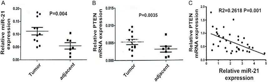
[image:3.612.92.522.73.205.2]The cells of each groups in the logarithmic growth phase were digested and counted after 48 h of the transfection. Afterwards, 150 μl of pre-cooling RIP A lysate that had been made into 1 mmol/L concentration by mixing with PMSF 1.5 μl was then supplemented, and the total protein was extracted. The process was performed on ice at 4°C centrifugal for 5 min at 10,000 r/min. After that, the supernatant was obtained to measure the protein concentration with BCA and denature at 99°C for 10 min. Subsequently, 50 μg protein was obtained for 10% SDSPAGE electrophoresis. The blots were electro transferred onto PVDF film and incubat-ed in sealincubat-ed suspension for 1 h. Following this, 1:100 diluted p-PTEN, PTEN primary antibody was added in to leave in overnight at 4°C. On the following day, the film was washed with TBST for 5 minutes and 3 times in a row. Lastly, Figure 1. Real-time PCR for the relative expression level of miR-21 (A) and PTEN mRNA (B) in endometrial carcinoma
tissues and the adjacent non-tumor tissues. There is a significant negative correlation between the two
1:5000 HRP-marked secondary antibody and GAPDH were added in to incubate at 37°C for 2 h.
Statistical analysis
All statistical analyses were performed with Student’s t-test and represented as mean ± standard deviation (SD) unless noted other-wise. No sample was excluded from the analy-sis. The p values were designated as *P < 0.05, **P < 0.01, and n.s.-non-significant (P > 0.05).
Results
Relative expression of miR-21 and PTEN mRNA in tumor and adjacent tissues
The RT-PCR results show that the miR-21 ex- pression in endometrial carcinoma was signifi-cantly higher than its corresponding adjacent tissue (Figure 1A). Whereas, the PTEN mRNA level in endometrial carcinoma tissue was sig-nificantly lower than that its corresponding adjacent tissue (Figure 1B). The expression lev-els of both endometrial carcinoma and
adja-of the miR-21 inhibitor group was obviously lower than the NC group and normal group (P < 0.05, Figure 2). The difference between the NC group and the mock group had no statistical significance (P > 0.05, Figure 2). In short, the miR-21 had successfully transfected into endo-metrial carcinoma Ishikawa cells.
miR-21 promote cell proliferation
The MTT assay result demonstrated that the proliferation activity of cells in the miR-21 inhib-itor transfection group was significantly lower than both the normal group (P < 0.05) and the NC group (P < 0.05, Figure 3). It was suggested the down-expression of miR-21 inhibited the proliferation ability of endometrial carcinoma Ishikawa cells.
miR-21 inhibite cell apoptosis
The flow cytometry results demonstrated that the apoptosis percentage of cells in the miR-21 inhibitor group was significantly higher than both the normal group (P < 0.05) and the NC group (P < 0.05, Figure 4). It was suggested that the down-expression of miR-21 promote cell apoptosis of endometrial carcinoma Ishi- kawa cells.
miR-21 promote cell migration and invasion
The Transwell chamber invasion results showed that the number of cells passed through Tr- answell chamber was significantly decreased in miR-21 inhibitor group (P < 0.05, Figure 5). It was suggested that the down-expression of miR-21 had inhibited endometrial carcinoma Ishikawa cell migration and invasion.
miR-21 downregulates the expressions of p-PTEN proteins
The western blot results showed that the expression of p-PTEN protein in the miR-21 fecting the endometrial carcinoma Ishikawa cells
with a miR-21 inhibitor. *P < 0.05 versus the normal or NC group.
inhibitor group was significantly higher than the normal group and NC group (P < 0.05, Figure 6). In addition, the PTEN protein expression had no significant difference (P > 0.05, Figure 6). It was suggested that the down-expression of miR-21 may upregulate the expressions of p-PTEN proteins directly or indirectly, but had no effect on the expression of PTEN proteins.
Discussion
microRNA (miRNA) combines with the 3’UTR region of target gene in the way of complemen-tary pairing to degrade target gene mRNA or repress the translation of target gene mRNA, and negatively regulate the expression of tar-get gene after transcription, thus affecting the proliferation, apoptosis, invasion and
[image:5.612.93.520.75.187.2]metasta-gus cancer, etc. A great number of studies have confirmed that the 3’ non-coding region of inhibitor gene mRNA of multiple tumors con-tains a site for miR-21 specific recognition and binding. Therefore, the inhibitor gene is the direct target molecule of miR-21. The high expression of miR-21 in tumor cells will mark-edly down-regulate the expression level of PTEN, TPM1, PDCD4 and other tumor suppres-sor gene, and this is probably because miR-21 is the molecular foundation of promoting tumor function [7-9]. A further study has shown that lowering the miR-21 level of tumor cell will up-regulate the expression of above-mentioned cells, and thus inhibit tumor cell proliferation, induce apoptosis and repress cell invasion and metastasis. Due to the difference of genetic background and tumor micro-environment in Figure 4. The effects of miR-21 on apoptosis of endometrial carcinoma Ishikawa cell lines. Using flow cytometry. *P
< 0.05 versus the normal or NC group.
Figure 5. The effects of miR-21 on endometrial carcinoma Ishikawa cell lines migration detected using Transwell invasion chambers. *P < 0.05 versus the normal or NC group.
sis of tumor. The researches in recent year show that miRNA is closely related to the development and process of endometrial cancer, and participates in regulating the molecular mechanism of endometrial cancer process and regulating the expression of progestational hormone [4, 6].
[image:5.612.90.369.236.449.2]esopha-different tumor cells, miR-21 may act on differ-ent target genes in differdiffer-ent tumors, affecting different cycles of tumor development and progress, and thus producing different effects. However, there are fewer reports on the expres-sion of miR-21 in endometrial cancer and its regulation on target gene [10].
PTEN, the upstream signal molecule of PI2K/ AKT pathway, is a cancer suppressor gene re- lated to the cryobiology, including cell growth, apoptosis, proliferation, and adhesion, and pl- ays a key role in the tumor development and process. Among all malignant tumors of gyne-cology, PTEN is closely related to endometrial carcinoma. The PTEN has a mutation rate of 23.5%~55%, and thus becomes a gene with the highest mutation rate in endometrial carci-noma, known as a house-keeping gene of endometrial cancer [11, 12]. The deficiency of PTEN will weaken its regulation on cell growth as well as control on apoptosis, adhesion and migration, and as a result facilitates the devel-opment and prognosis of carcinogenesis [13]. In terms of cell adhesion dependent signal, PTEN affects cell adhesion and metastasis, and inhibits the infiltration and metastasis of tumor cell by regulating FAK and SHC
phos-miR-21 and PTEN in endometrial cancer as well as adjacent tissues were detected via qRT-PCR. The results showed that the expression of miR-21 in endometrial cancer tissue was signifi-cantly higher than that of adjacent non-tumor tissue. miR-21 inhibitor was successfully trans-fected into cells. The cell proliferation and inva-sion activity in the miR-21 inhibitor group was obviously lower than the miR-21 NC group and Normal group. And the apoptosis rate in the miR-21 inhibitor group was significantly higher than the miR-21 NC group and Normal group (P < 0.05). The expression of p-PTEN protein in miR-21 inhibitor group were significantly lower than miR-21 NC group and Normal group. Above all, miR-21 is highly expressed in the endometrial cancer, and plays an oncogene effect in promoting the proliferation and inva-sion of endometrial and inhibiting apoptosis, and the partial mechanism may relate to the inhibition of PTEN on post-transcriptional level. In brief, miRNA-21 may become a new early diagnosis mark and therapy target in NSCLC. Acknowledgements
This work was supported by Shanghai Dongfang Hospital laboratory.
Disclosure of conflict of interest
None.
Address correspondence to: Dr. Jingxin Cheng, De- partment of Obstetrics and Gynecology, Shanghai Dongfang Hospital, 150 Jimo Road, Pudong New Area, Shanghai, China. E-mail: zhaowr321@163. com
References
[image:6.612.93.286.64.310.2][1] Torre LA, Bray F, Siegel RL, Ferlay J, Lortet-Tieu-lent J, Jemal A. Global cancer statistics, 2012. CA Can J Clin 2015; 65: 87-108.
[2] Li B, Lu W, Qu J, Zhang Y, Wan X. DICER1 re- gulates endometrial carcinoma invasion via histone acetylation and methylation. J Cancer 2017; 8: 933-939.
[3] Zuo K, Li M, Zhang X, Lu C, Wang S, Zhi K, He B. MiR-21 suppresses endothelial progenitor
cell proliferation by activating the TGFβ signal -ing pathway via downregulation of WWP1. Int J Clin Exp Pathol 2015; 8: 414-22.
[4] Abu Arab W. Video-assisted thoracoscopic sur-gery for non-small cell lung cancer. Minim Inva-sive Surg Oncol 2017; 1: 1-11.
[5] Bai J, Zhu X, Ma J, Wang W. miR-205 regulates A549 cells proliferation by targeting PTEN. Int J Clin Exp Pathol 2015; 8: 1175-83.
[6] Shang Y, Wang LQ, Guo QY, Shi TL. MicroRNA-196a overexpression promotes cell prolifera-tion and inhibits cell apoptosis through PTEN/ Akt/FOXO1 pathway. Int J Clin Exp Pathol 2015; 8: 2461-72.
[7] Mansoori B, Mohammadi A, Hashemzadeh S, Shirjang S, Baradaran A, Asadi M, Doustvandi MA, Baradaran B. Urtica dioica extract sup-presses miR-21 and metastasis-related genes in breast cancer. Biomed Pharmacother 2017; 16: 95-102.
[8] Shi L, He Y, Bai B, Chen M. Effects of microR-NA-21 inhibitor on apoptosis of type II alveolar epithelial cells in rats with hyperoxia-induced acute lung injury. Zhonghua Wei Zhong Bing Ji Jiu Yi Xue 2017; 29: 244-248.
[9] Ohkawa K, Asakura T, Tsukada Y, Matsuura T.
Antibody to human α-fetoprotein inhibits cell
growth of human hepatocellular carcinoma cells by resuscitating the PTEN molecule: in vi-tro experiments. Int J Oncol 2017; 50: 2180-2190.
[10] Kong Q, Wang W, Li P. Regulator role of HPV E7 protein on miR-21 expression in cervical carci-noma cells and its functional implication. Int J Clin Exp Pathol 2015; 8: 15808-13.
[11] Tsai CY, Wu JC, Fang C, Chang AY. PTEN, a negative regulator of PI3K/Akt signaling, sus-tains brain stem cardiovascular regulation dur-ing mevinphos intoxication. Neuropharmacol-ogy 2017; 123: 175-185.
[12] Moselhy J, Suman S, Alghamdi M, Chanda-rasekharan B, Das TP, Houda A, Ankem M, Damodaran C. Withaferin a inhibits prostate
carcinogenesis in a PTEN-deficient mouse
mo-del of prostate cancer. Neoplasia 2017; 19: 451-459.
[13] Shang Y, Wang LQ, Guo QY, Shi TL. MicroRNA-196a overexpression promotes cell prolifera-tion and inhibits cell apoptosis through PTEN/ Akt/FOXO1 pathway. Int J Clin Exp Pathol 2015; 8: 2461-72.
[14] Schultz KA, Rednam SP, Kamihara J, Doros L, Achatz MI, Wasserman JD, Diller LR, Brugières L, Druker H, Schneider KA, McGee RB, Foulkes WD. PTEN, DICER1, FH, and their associated tumor susceptibility syndromes: clinical fea-tures, genetics, and surveillance recommen-dations in childhood. Clin Cancer Res 2017; 23: e76-e82.
[15] Takahashi Y. Real-time intraoperative diagno-sis of lung adenocarcinoma high risk histologi-cal features: a necessity for minimally invasive sublobar resection. Minim Invasive Surg Oncol 2017; 1: 12-19.