Original Article
Roundabout1 distribution in neoplastic
and non-neoplastic diseases with a
focus on neoangiogenesis
Shuying Jiang1,2, Takao Hamakubo3, Kenichi Mitsui3, Ren Yagami4, Yukio Fujiyoshi5, Yoichi Ajioka2, Makoto Naito6,7
1Department of Orthoptist, Niigata College of Medical Technology, Kamishinnsakae-machi, Nishi-ku, Niigata, Niiga-ta-Pref, Japan; 2Division of Molecular and Diagnostic Pathology Graduate School of Medical and Dental Sciences, Niigata University, Asahimachi-Doori, Chuo-ku, Niigata, Niigata-Pref, Japan; 3Department of Quantitative Biology and Medicine, Research Center for Advanced Science and Technology, The University of Tokyo, Komaba, Meguro-ku, Tokyo, Japan; 4Aoyama Medical lmt. Sales Promotion Dept. Internal Affairs, Japan; 5Department of Anatomic Pathology and Molecular Diagnosis, Nagoya City University Graduate School of Medical Sciences and Medical School, Mizuho-cho, Mizuho-ku, Nagoya, Japan; 6Division of Pathology, Niigata Medical Center, Nishi-ku, Niigata, Niigata-Pref, Japan; 7Department of Cellular Function, Division of Cellular and Molecular, Pathology, Niigata Uni-versity Graduate School of Medical and Dental Sciences, Asahimachi-Dori, Chuo-ku, Niigata, Niigata-Pref, Japan
Received October 9, 2018; Accepted October 25, 2018; Epub December 1, 2018; Published December 15, 2018
Abstract: Slit and its receptor Roundabout (Robo) are important for neuronal development and neo-angiogenesis in various neoplastic and non-neoplastic diseases. Angiogenesis is a key factor for tumor growth and other
angio-genesis-dependent diseases including rheumatoid arthritis, and chronic inflammation Recently, over-expression of
Slit/Robo1 family proteins has been reported in several types of malignancy. We explored the expression of Robo1 in neoplastic and non-neoplastic diseases with a focus on newly formed blood vessels. Three hundred and thirty
four cases of malignancy and forty five cases of angiogenic diseases were recruited. Using the A7241A Robo1
monoclonal antibody, Robo1 expression was validated by immunohistochemistry. Among malignant cases, endo-thelial cells of newly formed blood vessels in 283 tumors (84.7%) exhibited positive staining with above antibody. In
non-neoplastic diseases, newly formed blood vessels were positive in 70.6% (12/17) cases of chronic
inflamma-tion, 100% (18/18) cases of pyogenic granuloma and 83.3% (5/6) cases of rheumatoid arthritis. Recently, newly anti-angiogenesis therapy is drawing attention as effective therapy for angiogenesis-dependent diseases without regard to their neoplastic or non-neoplastic nature. Our results showed a large number of neoplastic and non-neoplastic diseases showed positive staining for ROBO1 by immunohistochemistry. Thus, Robo1 targeted ther-apy may create new strategies for the treatment of angiogenic-dependent diseases through the suppression of an-giogenesis. Further, besides the majority of liver cell carcinomas (23/28, 82.1%), Robo1 was positive in 100% of the squamous cell carcinoma of the esophagus, uterine cervix, lung and skin. Thus, immunohistochemical evaluation of Robo1 may be useful as an additional diagnostic tool for liver cell carcinomas and squamous cell carcinomas.
Keywords: Angiogenesis, immunohistochemistry, inflammation, neoplasia, ROBO1
Introduction
The Slit family members and their receptor Roundabout (Robo) are important factors in neuronal development and leukocyte cell migration [1]. Recent work on mammals has indicated there are three Slit proteins and four forms of Robo (Robo1-4) [2, 3]. ROBO1 is the human homolog of the Drosophila roundabout gene, which defines a novel subfamily of the
Robo1 protein consists of five immunoglobulin-like (Ig) and three fibronectin type3-immunoglobulin-like (Fn3) domains in its extracellular region, and four cytoplasmic motifs termed CC0, CC1, CC2 and CC3 in its intracellular region [3].
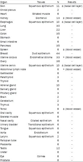
Table 1. Robo1 expression in human normal tissues
Organ Tssues N Results
Skin Squamous epithelium 11 ± (basal cell layer)
Chroid plexus 2
-Heart Striated muscle 6
-Kidney Glomeruli 13 ± (blood vessel) Esophagus Squamous epithelium 10 ± (basal cell layer)
Lung 30
-Liver 22
-Colon 35
-Stomach 6
-Small intestine 6
-Pancreas 35
-Spleen 6 ± (blood vessel)
Breast Duct epithelium 44
-Uterine corpus Endometrial stroma 33 + (blood vessel)
Ovary 36
-Uterine cervix Squamous epithelium 10 ± (basal cell layer) Abdominal lymph node 6 + (blood vessel)
Gallbladder 6
-Parathyroid 3
-Thyroid 6
-Adrenal gland 10
-Salivary gland 6
-Pituitary gland 3
-Brain 2
-Cerebellum 2
-Thymus 3
-Tonsil 6 ± (blood vessel)
Oral cavity Squamous epithelium 6
-Skeletal muscle 6
-Nasal cavity Ciliated epithelium 6
-Urinary bladder Transitional epithelium 6
-Tongue Squamous epithelium 6
-Aorta Endotheium 6
-Larynx Squamous epithelium 2
-Fallopian tube 6
-Plascenta 6
-Tastis 12
-Ureter 6
-Eye Cornea 6
-Prostate 6
-neoplastic diseases that may be classified to-gether as either angiogenesis-dependent [5] or angiogenic [7]. Angiogenesis-dependent dis-eases include diabetic retinopathy, rheumatoid arthritis, and chronic inflammation [5]. Various growth factors and receptors such as VEGF are
involved in angiogenesis [6], and the angiogenic properties of neuronal guidance factors, such as semaphorines, eph-rins, slits and neteph-rins, and their cognate receptors, have recent-ly attracted a great deal of attention [8, 9]. Involvement of most members of the Slit/Robo family has been reported, but their roles in the progression of angiogenesis-dependent disea- se including various cancers have not been clearly defined [10]. Robo1 is expressed in human primary lymphatic endo-thelial cells (hLECs) and Slit2/ Robo1 signaling promotes the lymphangiogenic activity of cul-tured hLECs [11]. In addition, Slit-Robo1 signaling pathways play a causal role in tumor angiogenesis in chemical-in- duced hamster squamous cell carcinogenesis [12]. MicroRNA- 218 inhibits the metastasis of gastric cancer by acting on the receptor Robo1 through angio-genesis, and this Slit-miR218-Robo1 pathway plays a pivotal role in gastric cancer metasta-sis [13]. Increased immunore-activity to Robo1 and Slit is an important factor for recurrence, clinical stage, and angiogenesis in endometrial cancer cases [14].
Slit/Robo family members may impact on the genesis of many solid tumors [15], and multiple Slit/Robo pathway genes are inactivated by hypermethyl-ation of their promotors in cervi-cal cancer progression [16]. Low Robo1 expression is asso-ciated with the development of cholangiocarcinomas [17], and homogenous deletions have been observed in lung and breast tumor cell lines [18]. Most mice with deletion of exon 2 of ROBO1 die because of delayed lung maturation, but survivors devel-op bronchial hyperplasia [19]. In mice, ROBO1
inactivation of both alleles for lung adenocarci-noma and lymphoma development [20]. Thus, in some tumors, it has been postulated as a tumor suppressor gene [21].
However, Ito et al showed contrasting overex-pression of Robo1 in hepatocellular carcino-mas [22]. Breast cancer cells and tissues derived from breast cancer patients express Robo1 and 2 receptors, and Slit treatment inhibits CXCR4-induced breast cancer cell che-motaxis, chemoinvasion and adhesion during metastasis [23]. Slit and Robo1 are both over-expressed in prostate tumors [24] while Robo1 and Robo4 were found to be overexpressed in colorectal cancer at the mRNA level and
immu-Materials and methods
Specimen characteristics
[image:3.612.89.526.85.178.2]This study was approved by the Ethics Com- mittees of Niigata Medical and Dental University and The University of Tokyo. The tissue speci -mens were collected at Niigata Medical and Dental University Hospital from 2003 to 2010, and consisted of 438 normal samples (heart, aorta, oral mucosa, salivary gland, breast, lar-ynx, lung, esophagus, stomach, small intestine, colon, liver, gallbladder, pancreas, spleen, kid-ney, ureter, urinary bladder, adrenal gland, tes-tis, ovary, uterus, Fallopian tube, placenta, prostate, thyroid, parathyroid, tonsil, thymus,
Table 2. ROBO1 expression in new blood vessel in angiogenesis-dependent diseases
New blood vessels Type N Immunostaining %
Malignant tumor 334 +++ 283/334 84.7
Chronic inflammation Granulation 17 +++ 12/17 70.6
Angiogenesis-dependent diseases
Granuloma pyogenicum 16 +++ 16/16 100 Rheumatoid arthritis 6 +++ 5/6 83.3
Bulla 6 -~± 1/6 16.7
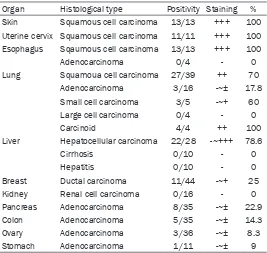
Table 3. Robo1 expression in human cancers and related non-neoplastic diseases
Organ Histological type Positivity Staining % Skin Squamous cell carcinoma 13/13 +++ 100
Uterine cervix Squamous cell carcinoma 11/11 +++ 100 Esophagus Sqaumous cell carcinoma 13/13 +++ 100
Adenocarcinoma 0/4 - 0
Lung Squamoua cell carcinoma 27/39 ++ 70 Adenocarcinoma 3/16 -~± 17.8 Small cell carcinoma 3/5 -~+ 60 Large cell carcinoma 0/4 - 0
Carcinoid 4/4 ++ 100
Liver Hepatocellular carcinoma 22/28 -~+++ 78.6
Cirrhosis 0/10 - 0
Hepatitis 0/10 - 0
Breast Ductal carcinoma 11/44 -~+ 25 Kidney Renal cell carcinoma 0/16 - 0 Pancreas Adenocarcinoma 8/35 -~± 22.9 Colon Adenocarcinoma 5/35 -~± 14.3 Ovary Adenocarcinoma 3/36 -~± 8.3 Stomach Adenocarcinoma 1/11 -~± 9
-: negative, ±: weakly positive, +: positive, ++: moderately positive, +++: strongly positive.
nohistochemistry demonstrated respective locations in tumor ce- lls, and endothelial cells of tumor vessels [25]. Thus, roles of the Slit/Robo pathway in human can-cers appear complex and tissue-specific expression patterns of Robo1 clearly require further in- vestigation.
[image:3.612.91.358.221.474.2]eye, brain, abdominal lymph node, skin, nasal cavity, tongue, pituitary gland, choroid plexus, and skeletal muscle (Table 1), 334 of various carcinomas and 45 angiogenic diseases (Table 2). The carcinomas comprised 28 cases of hepatocellular carcinoma, 13 of skin carcino-ma (squamous cell carcinocarcino-ma), 11 of cervical cancer (squamous cell carcinoma), 13 of eso- phageal squamous cell carcinoma, 4 cases of esophageal adenocarcinoma, 68 of lung can-cer (squamous cell carcinoma, adenocarcino-ma, small cell carcinoadenocarcino-ma, large cell carcinoadenocarcino-ma, and carcinoid tumor), 16 of renal cell carcino-ma, 35 of pancreas cancer (adenocarcinoma), 35 cases of colon cancer (adenocarcinoma),
HepG2 and human small cell lung carcinoma cell line NCI-H69 were obtained from American Type Culture Collection (ATCC). Aliquots of 1×10⁷ cells were homogenized with a radio immunoprecipitation assay (RIPA) buffer con-taining 50 mM Tris-HCl (pH 7.6), 150 mM NaCl, 1% Nonidet-P40, 0.5% Sodium Deoxycholate, 0.1% SDS and Protease Inhibitor Cocktail-Ethylenediaminetetraacetic acid free (Com- pleteTM, Roche Diagnostics KK), then subjected to SDS-PAGE followed by western blot analysis using the A7241A Robo1 monoclonal antibody at a final concentration of 1 ng/μl. The immuno-reacted band for Robo1 on membranes was detected by the ECL (Enhanced
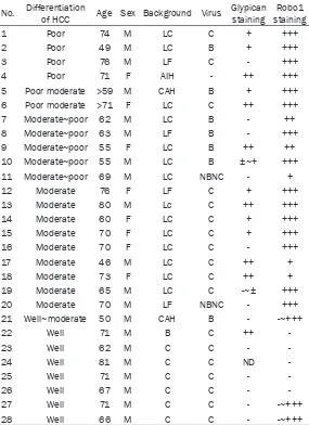
chemilumines-Table 4. Immunohistochemical expression of Robo1 and glypican by HCC
No. Differentiation of HCC Age Sex Background Virus Glypican staining stainingRobo1
1 Poor 74 M LC C + +++
2 Poor 49 M LC B + +++
3 Poor 76 M LF C - +++
4 Poor 71 F AIH - ++ +++
5 Poor moderate >59 M CAH B + +++ 6 Poor moderate >71 F LC C ++ +++
7 Moderate~poor 62 M LC B - ++
8 Moderate~poor 63 M LF B - +++
9 Moderate~poor 55 F LC B ++ ++
10 Moderate~poor 55 M LC B ±~+ +++
11 Moderate~poor 69 M LC NBNC - +
12 Moderate 76 F LF C + +++
13 Moderate 80 M Lc C ++ +++
14 Moderate 60 F LC C + +++
15 Moderate 70 F LC C + +++
16 Moderate 70 F LC C - +++
17 Moderate 46 M LC C ++ +
18 Moderate 73 F LC C ++ +
19 Moderate 65 M LC C -~± +++
20 Moderate 70 M LF NBNC - +++
21 Well~moderate 50 M CAH B - -~+++
22 Well 71 M B C ++
-23 Well 62 M C C -
-24 Well 81 M C C ND
-25 Well 71 M C C -
-26 Well 67 M C C -
-27 Well 71 M C C - -~+++
28 Well 66 M C C - -~+++
LC: Liver cirrhosis, LF: Liver fibrosis, CAH: Chronic active hepatitis, AIH: Autoimmune hepatitis, NBNC: non-B non-C, ND: not done, -: negative, ±: positive, +: positive, ++: moderately positive, +++: strongly positive.
36 of ovarian cancer (adeno-carcinoma) and 11 of gastric cancer (adenocarcinoma) (Ta- ble 3). As background hepato-cellular carcinoma-associated diseases, there were 18 cas- es of liver cirrhosis, two of pre-cirrhosis, 4 cases of liver fibro -sis, and 2 cases of chronic active hepatitis (Table 4). As angiogenic diseases 17 cases of chronic inflammation (gran -ulation), 16 of pyogenic granu-loma, 6 of rheumatoid arthri-tis and 6 of bulla were in- cluded (Table 2). All tissues were fixed in 10% formalde -hyde and embedded in pa- raffin.
Western blotting
[image:4.612.91.376.98.490.2]cence) method using SuperSignalTM West Dura Extended Duration Substrate (Life Technologies Japan) and an LAS-4000 Image analyzer (GE Healthcare Japan, Tokyo).
Immunohistochemistry
For detection of Robo1 protein in tissue sec-tions 3, 3’-diaminobenzidine (DAB) immunos-taining method was applied. The anti-Robo1 monoclonal antibody A7241A was generated by immunizing with an N-terminal peptide of Robo1, as described [22]. Simple stain multi PO (Nichirei, Tokyo, Japan) was used for sec-ondary antibody staining. The 1G12 glypican 3 antibody (Nichirei, Tokyo, Japan) specific for
hepatocellular carcinoma was applied for a comparison. Briefly, 3 μm sections were depar -affinized, rehydrated, and treated with 3% hydrogen peroxide for 15 minutes to inhibit endogenous peroxidase activity. Following anti-gen epitope retrieval by autoclaving in Tris-HCL buffer pH9.0, the slides were incubated with Robo1 antibody with a dilution of ×500 over-night at room temperature. Glypican 3 antibody was used without dilution. After incubation with a rabbit anti-mouse secondary antibody, detec-tion was with biotin-free horseradish peroxi-dase-labeled polymers obtained from DAKO Corporation (Tokyo, Japan). Staining was visual-ized using DAB substrate-chromogen solution with hematoxylin counterstaining.
Results
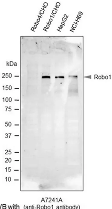
Reactivity of the A7241A anti-Robo1 antibody
To confirm the specific immunoreactivity of A7241A human Robo1 monoclonal anti-body, we performed western blot analysis. As shown in Figure 1, a band for Robo1 at 240 kDa was clearly detected in each lane for Robo1 transfected CHO cells (positive control), HepG2 cells and NCI-H69 cells, while no sig-nals were detected in the lane for Robo4 trans-fected CHO cells (negative control). No other indispensable bands were observed.
Robo1 expression in various normal tissues
The expression pattern of Robo1 in various nor-mal tissue is tabulated in Table 1. Robo1 was detected a membrane staining by immunohis-tochemistry. In normal tissues, weak staining was seen only in basal squamous cells in the skin, esophagus, tonsils, buccal mucosa, ton- gue, larynx, and uterine cervix. The other organs exhibited no staining for Robo1. Blood vessels of normal tissue showed weak staining in renal glomeruli, spleen, endometrium of uterine cor-pus, abdominal lymph nodes, and tonsil.
Robo1 expression in newly formed blood ves-sel in various cancer tissue
Results for expression patterns of Robo1 in newly formed blood vessels in various tumors and angiogenic diseases are summarized in
[image:5.612.101.286.71.417.2]Table 2. Among 334 malignancies, endothelial cells in 283 tumors (84.7%) exhibited positive staining with the Robo1 antibody, including Figure 1. Western blot analysis with the anti-Robo1
examples of squamous cell carcinoma of the cervix, squamous cell carcinoma of the skin, adenocarcinoma of the stomach, and squa-mous cell carcinoma and adenocarcinoma of the lung (Figure 2A-C). It was striking that posi-tive immunostaining for Robo1 was observed in vascular endothelial cells such as gastric ade-nocarcinoma and lung adeade-nocarcinoma which were Robo-1 negative in epithelial elements (Figure 2B, 2C).
Robo1 expression in newly formed blood ves-sels in non-cancerous tissues
Robo1 was also positive in newly organized vas-cular endothelial cells in various non-neoplastic angiogenic diseases (51/57, 89.5%) (Table 2). Newly formed blood vessels were positive for
cinoma) focal positive staining was observed. Adenocarcinomas tended to be less positive despite newly-formed blood vessels exhibiting strong expression (283 cases) (Figure 2C). Renal cell carcinomas exhibited no expression, along with large cell carcinomas of the lung.
Robo1 and glypican expression in hepatocel-lular carcinomas
Data for expression of Robo1 and glypican in individual hepatocellular carcinomas are tabu-lated in Table 4. In 28 cases, Robo1 was posi-tive in 23 (82.1%) (Figure 5A-C; Table 4). Metastases also displayed positive binding of the antibody and detection of minute HCC foci was possible (Figure 5B). Vascular invasion by HCC was also detected by
immunohistochemis-Figure 2. Robo1 expression in new-ly formed blood vessels of a ma-lignant tumor. Skin squamous cell carcinoma (A) gastric adenocar-cinoma (B), and lung adenocarci-noma (C). Robo1 immunostaining. H.E stain, (A, B) ×400; (C) ×1000.
Robo1 immunostaining in 70.6% (12/17) cases of chron-ic inflammation, 100% (18/18) cases of pyogenic granuloma (16/16) (Figure 3A) and 83.3% (5/6) cases of rheumatoid arthritis (Figure 3B). In con-trast, blood vessels of the bulla showed only weak stain-ing in only one case (1/6). Vascular endothelial cells in-volved in the granulation in pyogenic granuloma stained more intensely than those far from the granulation sites (Figure 3A).
Robo1 expression in epithe-lial cells of malignant tumors
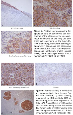
Data for Robo1 expression in a variety of malignancies (334 cases) are shown in Table 3. In addition to the majority of liver cell carcinomas, Robo1 was positive in 100% of the squa-mous cell carcinoma cases of the esophagus, uterine cervix, and skin (Figure 4A). In the lung, 70% of the squamous cell carcinoma cases and 60% of the small cell carcinoma and 100% of the carcinoid tumor cases displayed Robo1 expression (Figure 4B, 4C). In breast carcinoma (ductal
car-Figure 3. Robo1 expression in inflammatory diseases. Pyogenic granuloma
(A) and rheumatoid arthritis (B) and exhibit positive Robo1 staining in their newly formed blood vessels. In pyogenic granuloma, Robo1 staining in the newly formed blood vessels is stronger in the surface region than near the normal tissue (A). Robo1 immunostaining. Robo1 immunostaining, (A, B)
try (Figure 5C). In agreement with the results of Ito et al. [22], no Robo1 expression was seen in background non-neoplastic hepatic diseases such as cirrhosis and hepatitis (Figure 5B, 5C). Glypican 3 proved positive in the cytoplasm of 15 of 28 cases (53.8%).
Discussion
The term angiogenesis is generally applied to the growth of primitive microvessels that are the size of capillary blood vessels, a process that is orchestrated by a range of angiogenic factors, such as VEGF and angiogenesis inhibi
-cancers such as gastric and lung adenocarcino-mas (Figure 2B, 2C).
At present, a variety of anti-angiogenic drugs, such as Avastin and Endostatin, have been approved in many countries [31]. Anti-angio- genic therapy is widely applied [4, 6, 28, 29, 32]. For example, an anti VEGF agent was shown to be effective in certain cancers com-bined with chemotherapy [33]. MicroRNA218 inhibits the metastasis of gastric cancer by act-ing on the receptor Robo1, and this Slit-miR218-Robo1 pathway plays a pivotal role in gastric cancer metastasis [13]. Recently, it was
veri-Figure 4. Positive immunostaining for epithelial cells of squamous cell car-cinoma of the uterine cervix (A), squa-mous carcinoma of the lung (B), and small cell carcinoma of the lung (C). Note that strong membrane staining is apparent in squamous cell carcinoma of the uterus, but not in non-neoplastic squamous epithelium (right) except weakly in the basal layer. Robo1 immu-nostaining (A) ×100; (B, C) ×400.
Figure 5. Robo1 staining in neoplastic and non-neoplastic liver tissues. Nor-mal liver tissue (B, C) lacks staining, in contrast to HCC positivity. HCC ex-hibits strong membrane expression of Robo1 (A). A small focus of HCC can be seen surrounded by normal liver tissue (B). Tumor cells of HCC invading into vascular space are evident (C). Robo1 immunostaining (A, B) ×200; (C) ×40.
tors [6, 26, 27]. Folkman origi-nally suggested that tumor growth is dependent on angio-genesis [27]. In tumorigene-sis, tumor cell-induced angio-genesis and tumor metastasis both involve cell migration. The Slit2 protein attracts en- dothelial cells in newly formed blood vessels and promotes tube formation through the Slit/Robo signaling cascade [3, 28]. In this setting, Robo1, together with Robo4, may play important roles [3].
[image:7.612.86.379.72.535.2]fied in the mouse that inhibition of Robo1 by microRNA was effective for involution of gastric cancer through blocked angiogenesis [34]. These findings indicate that Slit/Robo signaling could be a novel therapeutic target of cancer therapy.
Previous studies on the expression of Robo1 in various tumors have generated contradictory results. In this study, squamous cell carcinoma of the skin, esophagus, and uterine cervix exhibited positive staining, whereas most ade-nocarcinomas of the esophagus, lung, and stomach did not. This result indicates a tissue specific expression pattern, with functions of Slit2/Robo1 signaling being cell-type depen-dent. In gastric cancer, activation of Slit/Robo1 signaling up-regulates the Wnt/β catenin path -way [35],and such activation of Wnt/β signal -ing also promotes genesis of colorectal cancer through Src signaling [36]. However, in our study, Robo1 was not found to be expressed in a large proportion of both gastric and colorec-tal cancers. The roles of Robo1 in carcinogen-esis and neovascularization of cancer tissue clearly must be considered separately.
In addition to the angiogenesis evident in tumors, our results showed this as a key com-ponent of some inflammatory diseases. Rheumatoid arthritis is an inflammatory angio -genic disease which is mediated by pro-inflam -matory and pro-angiogenic cytokines, charac-terized by VEGF decrease after the admini-stration of certain effective drugs [37]. In our study, Robo1 was positive in newly organized blood vessels, not only in cancerous tissues, but also in granulation, pyogenic granuloma and rheumatoid arthritis. These findings point to new strategies of neutralizing Robo1 activity for the inhibition of both tumor growth and also amelioration of non-neoplastic angiogenic diseases.
The findings in this study suggest that Robo1 might be a useful marker in some types of can-cer, especially in squamous cell carcinomas. This is the first study to specifically demon -strate utility of the Robo1 antibody for the diag-nosis of squamous cell carcinoma. Robo1 also appears useful as a marker of hepatocellular carcinoma (HCC), in line with data of an RNA experiment by Ito et al [22] which indicated 70% of cases to be positive. Glypican 3 is
well-known as the marker of hepatocellular carci-noma, but staining is focal [38]. Our study fur-ther showed that only 53.8% of the HCC cases were positive for Glypican 3 compared with 82.1% for Robo1, suggesting that the latter may be a more sensitive immunohistochemical marker. Furthermore, minute foci of HCC were detectable using Robo1.
Acknowledgements
This work was supported by the Program for Development of New Functional Antibody Technologies of the New Energy and Industrial Technology Development Organization (NEDO) of Japan, the Funding Program for World-Leading Innovative R&D on Science and Technology (FIRST program) of Japan Society for the Promotion of Science.
Disclosure of conflict of interest
None.
Address correspondence to: Dr. Yukio Fujiyoshi, Department of Anatomic Pathology and Molecular
Diagnosis, Nagoya City University Graduate School
of Medical Sciences and Medical School, 〒
467-8601 Mizuho-cho, Mizuho-ku, Nagoya, Japan. Tel: 052-853-8161; Fax: 052-851-4166; E-mail: yfuji@ med.nagoya-cu.ac.jp
References
[1] Rao Y, Wong K, Ward M, Jurgensen C and Wu JY. Neuronal migration and molecular conser-vation with leukocytechemotaxes. Genes Dev 2002; 16: 2973-2984.
[2] Kidd T, Brose K, Mitchell KJ, Fetter RD, Tessier-Levigne M, Goodman CS and Tear G. Round-about controls axon crossing of the CNS
mid-line and defines novel subfamily of evolutionary
conserved guidance receptors. Cell 1998; 92: 205-215.
[3] Legg JA, Herbert JM, Clissold P and Bicknell R. Slits and roundabouts in cancer, tumor angio-genesis, and endothelial cell migration. Angio-genesis 2008; 11: 13-21.
[4] Hanahan D and Weinberg R. The hallmarks of cancer. Cell 2000; 100: 57-70.
[5] Folkman J. Angiogenesis: an organizing princi-ple for drug discovery. Drug Discovery 2007; 6: 273-286.
[7] Carmeliet P and Jain RK. Principles and mech-anisms of vessel normalization for cancer and other angiogenic diseases. Nat Rev Drug Dis-cov2011; 10: 417-427.
[8] Klagsbrun M and Eichman A. A role for axon guidance receptors and ligands in blood vessel development and tumor angiogenesis. Cyto-kine Growth Factor Rev2005; 16: 535-548. [9] Shima D and Mailhos C. Vascular development
biology: getting nervous. Curr Opin Gen Devel-op2000; 10: 536-542.
[10] Fujiwara M, Ghazizadeh M and Kawanami O. Potential role of the Slit/Robo signal pathway
in angiogenesis. Vasc Med 2006; 11: 115-121.
[11] Yang XM, Han HX, Sui F, Dai YM, Chen M and Geng JG. Slit-Robo signaling mediates lym-phangiogenesis and promotes tumor lymphat-ic metastasis. Biochem Biophyslymphat-ic Res Com-mun2010; 396: 571-577.
[12] Wang LJ, Zhao Y, Han B, Ma YG, Zhang J, Yang DM, Mao JW, Tang FT, Li WD, Yang Y, Wang R and Geng JG. Targeting Slit-Roundabout signal-ing inhibits tumor angiogenesis in chemical-induced squamous cell carcinogenesis. Can-cer Sci2008; 99: 510-517.
[13] Tie J, Pan Y, Zhao L, Wu K, Liu J, Sun S, Guo XG, Wang B, Gang Y, Zhang YG, Qiao TD, Zhao QC, Nie YZ and Fan DM. MIR-218 inhibits invasion and metastasis of gastric cancer by targeting the Robo1 receptor. PLoS Gen 2010; 6: e1000879.
[14] Ma S, Liu X, Geng JG, Giuo SW. Increased SLIT immunoreactivity as a biomarker for recur-rence in endometrial carcinoma. Am J Obst Gynecol2010; 202: 68: e1-11.
[15] Mimmi S Ballard, Hink L. A roundabout way to cancer. Adv Cancer Res 2012; 114: 187-235. [16] Narayan G, Goparaju G, Arias-Pulido H, Kau-
fman AN, Schneider A, Dürst M, Mansukhani
M, Pothuri B and Murty VV. Promotor hyper -methylation-mediated inactivation of multiple Slit-Robo pathway genes in cervical cancer progression. Mol Cancer2006; 5: 1-10. [17] Mano Y, Aishima S, Fukuhara T, Tanaka Y, Kubo
Y, Motomura T, Toshima T, Iguchi T, Shirabe K, Maehara Y and Oda Y. Decreased roundabout 1 expression promotes development of intra-hepatic cholangiocarcinoma. Human Pathol 2013; 44: 2419-2426.
[18] Sundaresan V, Chung G, Heppell-Parton A,
Xiong J, Grundy Y, Roberts Y, James L, Cahn A, Bench A, Douglas J, Minna J, Sekido Y, Lerman M, Latif L, Bergh J, Li H, Lower N, Ogilvie D and Rabbitts P. Homozygoud deletion at 3p12 in breast and lung cancer. Oncogene 1998; 17: 1723-1729.
[19] Xian J, Clark C, Fordam R, Pannell R, Rabbitts TH and Rabbitts PH. Inadequate lung
develop-ment and bronchial hyperplasia in mice with a targeted deletion in the DUTT1/Robo1 gene. PNAS 2001; 98: 15062-15066.
[20] Xian J, Aitchison A, Bobrow L, Corbett G, Pan-nel R, Rabbitts T and Rabbitts P. Targeted
dis-ruption of the 3p12 gene, DUTT1/Robo1, pre -disposes mice to lung adenocarcinomas and lymphomas with methylation of the gene pro-motor. Cancer Res 2004; 64: 6432-6437. [21] Dallol A, Forgacs E, Martinez A, Sekido Y,
Walk-er R, Kishida T, Rabbitts P, MahWalk-er ER, Minna JD and Latif L. Tumour specific promotor region
methylation of the human homologue of the
Drosophila roundabout gene DUTT1 (ROBO1)
in human cancers. Oncogene2002; 21: 3020-3028.
[22] Ito H, Funahashi S, Yamauchi N, Shibahara J, Midorikawa Y, Kawai S, Kinoshita Y, Watanabe A, Hippo Y, Ohtomo T, Iwanari H, Nakajima A, Makuuchi M, Fukayama M, Hirata Y, Hamaku-bo T, Kodama T, tsuchiya M and Aburatani H.
Identification of ROBO1 as a novel hepatocel -lular carcinoma antigen and a potential thera-peutic and diagnostic target. Clin Cancer Res 2006; 12: 3257-3264.
[23] Prasad A, Fernandez AZ, Rao Y and Ganju RK. Mechanisms of signal transduction: Slit-pro-tein mediated inhibition of CXCR4-induced chemotactic and chemoinvasive signaling pa- thways in breast cancer cells. J Biol Chem 2004; 279: 9115-9124.
[24] Latil A, Chene L, Cochant-Priollet B, Mangin P, Fournier G, Berthon P and Cussenot O.
Quanti-fication of expression of netrins, Slits, and their
receptors in human prostate tumors.Int J Can-cer2003; 103: 306-315.
[25] Gröne J, Doebler O, Loddenkemper C, Hotz B, Buhr HJ and Bhargava S.Robo1/Robo4: differ-ential expression of angiogenic markers in colorectal cancer. Oncol Rep 2006; 15: 1437-1443.
[26] Folkman J. Angiogenesis. Ann Rev Med2006; 57: 1-18.
[27] Folkman J. Tumor angiogenesis: therapeutic implications. New Eng J Med 1971; 18: 1182-1186.
[28] Wang B, Xiao Y, Ding BB, Zhang N, Yuan XB, Gui L, Qian KX, Duan S, Chen ZJ, Rao Y and Geng JG. Induction of tumor angiogenesis by Slit-Robo signaling and inhibition of cancer growth by blocking Robo activity. Cancer Cell 2003; 4: 19-29.
[29] Hanahan D and Folkman J. Patterns and emerging mechanisms of the angiogenic switch during tumorigenesis. Cell 1996; 86: 353-364.
me-diated HUVEC migration by Robo4. Biochem
Biophys Res Comm2016; 469: 797-802. [31] Folkman J. Antiangiogenesis in cancer
thera-py-endostatin and its mechanisms of action. Experi Cell Res2006; 312: 594-607.
[32] Holash J, Thurston G, Rudge JS, Yancopoulos GD, Adjei AA, Bergers J, Pytowski B, Pegram S and Gordon MS. Inhibitors of growth factor re-ceptors, signaling pathways and angiogenesis as therapeutic molecular agents. Cancer Me-tastasis Rev2006; 25: 243-252.
[33] Jain RK, Duda DG, Clark JW and Loeffler JS.
Lessons for phase III clinical trials on
anti-VEGF therapy for cancer. Nature Clin Pract
2006; 3: 24-39.
[34] Zhang XY, Dong JQ, He Y, Zhao M, Liu Z, Wang N, Jiang MZ, Zhe Z, Gang L, Liu HM, Nie YZ, Fan DM and Tie J. miR-218 inhibited tumor an-giogenesis by targeting ROBO1 in gastric can-cer. Gene 2017; 615: 42-49.
[35] Shi R, Liu W, Liu B,Xu Z, Chen L and Zhang Z. Slit2 expression and its correlation with
sub-cellular localization of β-catenin in gastric can -cer. Oncol Rep 2013; 30: 1883-1889. [36] Zhang QQ, Zhou D, Lei Y, Zheng L, Chen SX,
Gou HJ, Gu QL, He XD, Lan T, Qi CL, Li JC, Ding YQ, Qiao L and Wang LJ. Slit2/Robo1 signaling promote intestinal tumorigenesis through
Src-mediated activation of the Wnt/β-catenin path -way. Oncotarget 2014; 6: 3123-3135. [37] Nagashima M, Asano G and Yoshino S.
Imbal-ance in production between vascular endothe-lial growth factor and endostatin in patients with rheumatoid arthritis. J Rheumatol 2000; 27: 2339-2342.
[38] Antelli F, Chuang ST, Yang XJ and Wang HL.