Original Article
Inhibition of γ‑secretase by retinoic acid chalcone (RAC)
induces G2/M arrest and triggers apoptosis
in renal cell carcinoma
Qing-Chun Li1, Hong-Jun Li2, Shi Liu3, Yun Liang2, Xue Wang2, Lei Cui4
1Department of Gastrointestinal Surgery, China-Japan Union Hospital of Jilin University, Changchun 130033, Jilin, China; 2Center of Physical Examination, China-Japan Union Hospital of Jilin University, Changchun 130033, Jilin, China; 3State Key Laboratory of Polymer Physics and Chemistry, Changchun Institute of Applied Chemistry, Chinese Academy of Sciences, Changchun 130022, Jilin, China; 4Jilin Institute for Drug Control, Changchun 130033, Jilin, China
Received January 11, 2014; Accepted February 28, 2015; Epub March 1, 2015; Published March 15, 2015
Abstract: The present study was devised to investigate the effect of RAC on inhibition of cell proliferation and
apoptosis of renal carcinoma cells. MTT assay and flow cytometry analysis were used to determine cell proliferation
and apoptosis along with cell cycle examination. Western blot analysis and immunohistochemistry were used for the detection of expression levels of Notch1 and Jagged1 in renal cell carcinoma (RCC) and normal kidney tissues.
The results revealed a significant inhibition of cell proliferation, G2/M phase cell cycle arrest and cell apoptosis at 30 μM concentration of RAC after 72 h. In ACHN and 769-P cells, the population in G2/M phase was increased to 45.27, and 54.23% respectively on treatment with 30 μM RAC for 72 h. In 769-P and ACHN renal carcinoma cells treatment with 30 μM RAC caused 69.71 and 59.27% of the cells to undergo apoptosis compared to 5.23
and 4.93% respectively in control cells. The positive staining rates of Notch1 and Jagged1 in renal carcinoma tissues were 95.3 and 93.0% compared to normal kidney tissues 36.4 and 42.4% respectively. Treatment of renal
carcinoma tissues caused a significant decrease in staining rates of Notch1 and Jagged1 after 96 h. Thus RAC can
be a potent agent in the treatment of renal cell carcinoma.
Keywords: Renal cell carcinoma, apoptosis, Notch1, Jagged1
Introduction
Among all the adult malignant neoplasms, renal cell carcinoma (RCC) alone accounts for 2-3%.
There are around one lakh fifty thousand cases
detected and seventy eight thousand deaths globally caused by renal cell carcinoma every year [1]. Of the patients diagnosed with renal cell carcinoma, 25-30% is detected at meta-
static stage which makes the treatment difficult
[2]. It is reported that multikinase inhibitors increase progression-free survival rates, how- ever, the effective therapy for patients with metastatic advanced-stage RCC remains limited [3, 4]. Renal cell carcinoma requires additional tumorigenic events, suggesting the importance of these pathways in the deve- lopment of novel agents [5].
It is reported that in renal carcinoma cells the Jagged1 is overexpressed which determines
poor outcome in RCC patients [6]. Latter it was shown that Notch plays a role in the tumorigenesis of renal carcinoma cells and
γ-secretase inhibitor inhibits cell proliferation
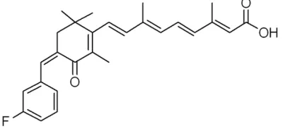
morphological differentiation, proliferation, and gene expression of neuroblastoma [14] and astrocytoma cells [15]. Recurrent malignant cerebral gliomas have been treated with ATRA [16, 17] and 13-cis RA [18]. Retinoids are known to have anti-proliferation, anti-migration, and anti-invasive activity against human malignant gliomas [19, 20], suggesting that retinoids are suitable anticancer agents to inhibit progression of tumors. In the present study the effect of retinoic acid amide (Figure
1) having more bioavailability compared to the parent compound on human liver cancer apoptosis was investigated.
Materials and methods
Reagents and antibodies
RAC, antibodies used against Notch1 (polycl- onal rabbit anti-human Notch1; ab27526) and Jagged1 (polyclonal goat anti-human Jagged1; sc-6011) were purchased from Sigma-Aldrich (St. Louis, MO, USA). The peroxidase-conjugated
mouse anti-goat IgG antibody and glyceralde-hydes-3-phosphate dehydrogenase (GAPDH)
was purchased from Santa Cruz Biotechnology,
Inc. and the γ-secretase inhibitor,
N-[N-(3,5-difluorophenacetyl)-l-alanyl]-S-phenylglycine
t-butyl ester (DAPT), was purchased from Merck Biosciences (Darmstadt, Germany). The Anne-xin V-fluorescein isothiocyanate (FITC)
apopto-sis detection kit was purchased from Beckman Coulter (Fullerton, CA, USA).
Cell culture
Human renal carcinoma cell lines, 786-O, 769-P, ACHN, Caki-1, HEK293, OS-RC-2, and TUHR- 14TKB were obtained from The Cell Bank of Type Culture Collection of Chinese Academy of Sciences, Shanghai Institute of Cell Biology (Shanghai, China). The cells were cultured in
DMEM containing penicillin 100 units/ml,
streptomycin 100 mg/ml and 10% FBS (PAA). The cells were maintained in an incubator at 37°C with 5% CO2 atmospheric condition. MTT assay
Cell proliferation was assessed at various time points by 3-(4,5-dimethylthiazol-2-yl)-2,5-diph-
enyltetrazoliumbromide (MTT) assay. Briefly, 1
× 105 cells were seeded in each well of a
96-well plate (BD Falcon, Franklin, NJ) and
allowed to adhere for 8 h. Then, 5 mg/mL MTT
(Sigma, Germen) was added to each well and
incubated for 4 h. The cells were lysed by
adding 150 μL/well of dimethyl sulfoxide and
read at 490 nm absorbance wavelength in microplate reader (MPR-A4i; Tosoh Corporation, Tokyo, Japan). The experiments were repeated at least three times independently.
Flow cytometry apoptosis assay
Analysis of apoptosis was accessed by an AnnexinV-FITC/propidium iodide double stai-
ning kit (Genmed Bioscience, China) following the manufacturer’s protocols. Briefly, the cells
were plated in six wells. Cells were continuously cultured for 48 and 72 h, and then to be
harvested. Before reading on the flow
cytometer, cell suspensions were washed in PBS, resuspended with a 1 × binding buffer
and exposed to 5 μL of Annexin V-FITC (20 μg/ mL) and 10 μL of propidium iodide (PI; 50 μg/
mL). After incubation of 20 min in the dark, the
samples were subjected to a FACScan flow
cytometer [equipped with CellQuest and ModFITLT for Mac V1.01 software (Becton
Dickinson, San Jose, CA)]. The experiments
were repeated at least three times indepen- dently.
Cell cycle analysis
The cells were treated with various concen-
trations of RAC (10, 20, 30 and 50 μM) for 72 h
at 37°C. After 72 h RAC treatment cells were
harvested, washed in PBS and then fixed in
cold 70% ethanol. The cells were then stained with propidium iodide while treating with RNase and analysed with Beckman Coulter EPICS XL-MCL cytometer (Beckman Coulter).
Multi-Cycle DNA Content and Cell Multi-Cycle Analysis
[image:2.612.90.290.76.169.2]Ethical statement
The present study was approved by the ethical committee of China-Japan Union Hospital of Jilin University (no. 2010-98; Changchun, Chi- na). Each patient was involved after providing informed written consent.
Tissue samples
For western blot analysis, fresh tumor tissues
(later verified as clear cell RCC) and normal
(non-tumor) kidney tissues were obtained intraoperatively from eight patients who underwent radical nephrectomy at the
Department of Urology, China-Japan Union
Hospital of Jilin University. The tissue samples were then snap-frozen in liquid nitrogen and stored at -80°C prior to analysis. For immunos-taining, a total of 129 patients with
pathologically verified clear cell RCC were
enrolled consecutively. All patients underwent nephrectomy (partial or radical) performed at
the Department of Urology, China-Japan Union
Hospital of Jilin University, between 2010 and 2014.
Western blot analyses
The renal carcinoma cells were washed with ice-cold phosphate-buffered saline and then lysed in lysis buffer containing 10 mM Tris (pH 7.5), 150 mM NaCl, 10 mM ethylenediamine-
tetraacetic acid (EDTA), 1% sodium dodecyl sulfate (SDS), 1 mM sodium orthovanadate,
and a mixture of protease inhibitors (1 mM
phenylmethylsulfonyl fluoride, 1 μg/mL pep-statin A, 2 μg/ mL aprotinin). The lysates were
sonicated for 10 s, centrifuged for 20 min at 20,000 × g and then stored at -70°C. Equal
amounts (25 μg) of the cell lysates were resolved by 12% SDS-PAGE and transferred to polyvinylidene fluoride membranes. After
blocking, blots were incubated with Notch1 (1:200) or Jagged1 (1:500) antibodies overnight at 4°C and followed by each corresponding second antibody at room temperature for 1 h at 37°C. Then, the results developed by ECL (Pierce Biotechnology, USA). The protein bands were then analysed using the BioImaging System (UVP, USA). The gray scale values were normalized to the values of the corresponding
GAPDH band to determine the expression level
of the protein. The experiments were repeated at least three times independently.
Immunohistochemistry
Fifty renal carcinoma cancer tissues were
obtained from the Department of Urology,
China-Japan Union Hospital of Jilin University frozen in liquid nitrogen and stored at -80°C
prior to analysis. The paraffin-embedded
tissues were cut into 2 mm sections followed
by deparaffinization and rehydration. The
tissues after 3% hydrogen peroxide treatment were boiled in 10 mM citrate buffer (pH 6.0) for 15 min. The sections were incubated with goat serum for 1 hand rinsed in PBS. The slides treated with Notch1 (1:200) or Jagged1 (1:100) antibodies for 1 h were rinsed with PBS and incubated with horse radish peroxidase-con- jugated secondary antibody. Again slides were rinsed in PBS, incubated with 3,3’-diamino- benzidine staining and counterstaining with hematoxylin blue. Negative controls were performed by substituting the primary antibody with a non-immune serum. Control sections were treated in parallel with the samples. The proportion score was determined semi-quantitatively by assessing the whole tumor
section at low magnification and each sample
was scored on the following scale of 0-3: 0, no positive cells; 1, 1-20% of positive cells; 2, 21-60% of positive cells; and 3, 61-100% of positive cells. The intensity score was
determined at high magnification as follows: 0,
negative staining; 1, weakly positive staining; 2, moderately positive staining; and 3, markedly positive staining. Then, the total score of each section was calculated by sum of the two parameters.
Statistical analysis
SPSS software (Inc., Chicago, IL, USA) was used
for the statistical analyses. The χ2 test was used for detection of immunostaining diff- erences and the study of correlations. The
difference was considered statistically
signifi-cant at P < 0.05. Results
Effect of RAC on inhibition of renal carcinoma cell growth
tested renal carcinoma cell lines 786-O, 769-P, ACHN, Caki-1, and HEK293 exhibited similar cytotoxicities on treatment with a range of RAC
concentrations (10, 20, 30, 40 and 50 μM).
Although the decrease in cell proliferation
started at 10 μM concentration of RAC but the inhibition was significant at 30 μM after 72 h
(Figure 2).
Effect of RAC on cell cycle arrest
We used flow cytometry to investigate the
effect of various concentrations of RAC on cell cycle distribution in renal carcinoma cells. The
examination of cell cycle showed a significant
increase in cell population in G2/M phase on
treatment with various concentrations of RAC for 72 h compared to untreated control cells. In
769-P cells, the population in G2/M phase was
increased by 5.34, 9.16, 54.23 and 44.56% on
treatment with 10, 20, 30 and 50 μM RAC for
72 h respectively (Figure 3A). Similarly in ACHN cells, the population was increased by 4.93, 8.24, 45.27 and 32.46%, (Figure 3B) whereas for Caki cells, the population was increased by 6.45, 10.19, 49.21 and 48.92%, on treatment
with 10, 20, 30 and 50 μM RAC for 72 h
respectively. These results suggested that RAC
induces G2/M phase cell cycle arrest in renal
[image:4.612.95.521.75.215.2]carcinoma cells.
Figure 2. RAC Inhibits cell viability in renal carcinoma cells in a dose and time dependent manner.
[image:4.612.93.520.261.484.2]RAC induces apoptosis in renal carcinoma cells
The results from AnnexinV-FITC/propidium iod- ide double staining revealed typical apoptotic
[image:5.612.89.527.73.271.2]changes with chromatin condensation and nuclear fragmentation in 769-P and ACHN renal carcinoma cells after 72 h of RAC treatment. Annexin V-FITC/PI staining and FACS demonstrated that in 769-P renal carcinoma Figure 4. RAC treatment-induced dose-dependent apoptosis in human renal carcinoma cell lines using a double-staining method with Annexin V-FITC/PI.
[image:5.612.95.523.323.625.2]cells treatment with 30 μM RAC for 72 h caused
69.71% of the cells to undergo apoptosis compared to 5.23% in control. Similarly
treatment with 30 μM RAC for 72 hours caused
59.27% ACHN cells to undergo apoptosis compared to 4.93% in control (Figure 4).
Notch1 and Jagged1 expression is up-regulated in renal carcinoma tissues
Results from Western blot analysis showed that the protein of Notch1 was highly expressed in RCC tissues (Figure 5). Among 10 RCC tissues Notch1 was found to be highly expressed in nine cases of RCC (9/10; 90%) compared with paired non-neoplastic tissues. Similarly, out of 10 RCC tissues, 9 showed higher expression of Jagged1 (9/10, 90%).
Clinical and pathological characteristics
100 cases with renal cell carcinoma were subjected to immunostaining of Jagged1 and 10 cases to western blot analysis (Table 1).
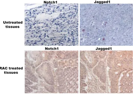
Immunohistochemistry
In renal carcinoma tissues the positive staining rate of Notch1 and Jagged1 was 97 and 96%, respectively compared with 19.8 and 35% in normal tissues (Figure 6). Notch1 and Jagged1
exhibited a significantly higher expression in
RCC tissues than in normal kidney tissues. Treatment with 30 µM concentration of RAC
caused a significant decrease in the expression
of Notch1 and Jagged1 after 96 h. In renal carcinoma tissues treated with RAC the positive staining rate of Notch1 and Jagged1 was decreased to 23.4 and 41.5% respectively compared to 97 and 96% in untreated carcinoma tissues.
Discussion
The Notch pathway plays a dual role by either acting as tumor suppressive or oncogenic pathway depending on various factors involved [21]. It has a key role in controlling cell proliferation and apoptosis [22]. In the current study, we observed overexpression of the Notch1 and Jagged1 in neoplastic tissue compared to that in normal kidney tissue. The expression of Notch1 and Jagged1 was related to tumor size and grade indicating that Notch pathway has oncogenic role in the renal cell carcinoma. Renal carcinoma tissues on
treatment with RAC showed a significant
decrease in expression levels of Notch1 and Jagged1 in neoplastic tissue.
The results from our study showed that RAC
significantly inhibits the cell proliferation in the renal carcinoma cells, induces G2/M-phase
[image:6.612.92.296.96.423.2]cell cycle arrest and apoptosis. The oncogenic effect of Notch in renal cell carcinoma may involve diverse mechanisms. There are reports that Notch signaling plays a vital role in tumor angiogenesis [23-25]. It is also reported that Notch signaling suppresses expression of p21Cip1 and p27Kip1, two cyclin-dependent kinase inhibitory proteins regulating the cell cycle control [26]. This suggests that the inhibitory effect of RAC on cell proliferation and induction of apoptosis is due to inhibition of Notch signaling pathway. Although kinase inhibitors like sorafenib and sunitinib increase the progression-free survival rate for patients but the side effects of kinase inhibitors hinder their clinical application [27].
Table 1. Pathological characteristics of renal cell carcinoma cases
Characteristics (n = 100)IHC Western blotting (n = 10)
Gender, n (%)
Male 79 (79) 7 (70) Female 21 (21) 3 (30) Age, years
Mean 59.8 58.4 Range 23-89 47-79 Surgery, n (%)
Radical nephrectomy 95 (95) 9 (90) Partial nephrectomy 5 (5) 1 (10) Tumor size (cm)
Mean 6 5.4
Range 2-19 1.9-14 TNM stage, n (%)
I 65 (5) 3 (30) II 15 (15) 4 (40) III 15 (15) 2 (20) IV 5 (5) 1 (10) Fuhrman grade, n (%)
1 38 (38) 3 (30) 2 42 (42) 4 (40) 3 14 (14) 2 (20) 4 6 (6) 1 (10) Relapse, n (%)
The mechanism of these drugs is believed to involve suppression of hypoxia-inducible factor-mediated autocrine growth factor signaling and proangiogenic effects. The therapeutic effect of RAC inhibition on clear cell RCC tumor growth indicates that the inhibition of Notch signaling presents at least a complementary therapeutic approach for treatment of clear cell RCC. In the present study, inhibition of clear cell RCC cells with RAC led to a considerable decrease of cell proliferation and increased apoptosis.
In conclusion, the current study indicated that Notch signaling is important in the tumo- rigenesis of RCC. The RAC has the potential of being a novel therapeutic regimen towards RCC, although, further investigation is required. Disclosure of conflict of interest
None.
Address correspondence to: Dr. Hong-Jun Li, Center
of Physical Examination, China-Japan Union Hospital of Jilin University, 126 Xiantai Street, Changchun 130033, Jilin, China. Tel: 0086-431-84995222; Fax: 0086-431-84995222; E-mail: hongjunli2004@ gmail.com
References
[1] Ferlay J, Shin HR, Bray F, Forman D, Mathers C and Parkin DM. Estimates of worldwide burden
of cancer in 2008: GLOBOCAN 2008. Int J
Cancer 2010; 15: 2893-2917.
[2] Ljungberg B, Hanbury DC, Kuczyk MA,
Merseburger AS, Mulders PF, Patard JJ, Sinescu IC; European Association of Urology
Guideline Group for renal cell carcinoma.
Renal cell carcinoma guideline. Eur Urol 2007; 51: 1502-1510.
[3] Escudier B, Eisen T, Stadler WM, Szczylik C, Oudard S, Siebels M, Negrier S, Chevreau C,
Solska E, Desai AA, Rolland F, Demkow T, Hutson TE, Gore M, Freeman S, Schwartz B, Shan M, Simantov R, Bukowski RM; TARGET Study Group. Sorafenib in advanced clear-cell
renal-cell carcinoma. N Engl J Med 2007; 356: 125-134.
[4] Motzer RJ, Hutson TE, Tomczak P, Michaelson
MD, Bukowski RM, Rixe O, Oudard S, Negrier
S, Szczylik C, Kim ST, Chen I, Bycott PW, Baum CM, Figlin RA. Sunitinib versus interferon alfa in metastatic renal-cell carcinoma. N Engl J Med 2007; 356: 115-124.
[5] Kim WY and Kaelin WG. Role of VHL gene
mutation in human cancer. J Clin Oncol 2004; 22: 4991-5004.
[6] Wu K, Xu L, Zhang L, Lin Z and Hou J. High Jagged1 expression predicts poor outcome in clear cell renal cell carcinoma. Jpn J Clin Oncol 2010; 41: 411-416.
[7] Orfanos CE, Ehlert R and Gollnick H. The
retinoids. A review of their clinical pharmacology
and therapeutic use. Drugs 1987; 34: 459-503.
[image:7.612.91.521.73.300.2][8] Kligman AM. The growing importance of topical retinoids in clinical dermatology: A retrospective
and prospective analysis. J Am Acad Dermatol
1998; 39: S2-S7.
[9] Chandraratna RA. Current research and future developments in retinoids: oral and topical agents. Cutis 1998; 61: 40-45.
[10] Zheng Y, Kramer PM, Lubet RA, Steele VE,
Kelloff GJ and Pereira MA. Effect of retinoids
on AOM-induced colon cancer in rats: Modulation of cell proliferation, apoptosis and aberrant crypt foci. Carcinogenesis 1999; 20: 255-260.
[11] Liang JY, Fontana JA, Rao JN, Ordonez JV,
Dawson MI, Shroot B, Wilber JF and Feng P. Synthetic retinoid CD437 induces S-phase
arrest and apoptosis in human prostate cancer cells LNCaP and PC-3. Prostate 1999; 38: 228-236.
[12] Weber E, Ravi RK, Knudsen ES, Williams JR,
Dillehay LE, Nelkin BD, Kalemkerian GB,
Feramisco JR and Mabry M. Retinoic acid-mediated growth inhibition of small cell lung cancer cells is associated with reduced myc and increased p27Kip1 expression. Int J Cancer 1999; 80: 935-943.
[13] Mologni L, Ponzanelli I, Bresciani F, Sardiello
G, Bergamaschi D, Gianni M, Reichert U, Rambaldi A, Terao M and Garattini E. The novel
synthetic retinoid 6-[3-adamantyl-4-hydroxy-
phenyl]-2-naphtalene carboxylic acid (CD437)
causes apoptosis in acute promyelocytic leukemia cells through rapid activation of caspases. Blood1999; 93: 1045-1061. [14] Irving H, Lovat PE, Hewson QC, Malcolm AJ,
Pearson AD and Redfern CP. Retinoid induced
differentiation of neuroblastoma: comparison
between LG69, an RXR-selective analogue and
9-cis retinoic acid. Eur J Cancer 1998; 34: 111-117.
[15] Dirks PB, Patel K, Hubbard SL, Ackerley C,
Hamel PA and Rutka JT. Retinoic acid and the cyclin dependent kinase inhibitors synergi- stically alter proliferation and morphology of U343 astrocytoma cells. Oncogene1997; 15: 2037-2048.
[16] Yung WK, Kyritsis AP, Gleason MJ and Levin VA.
Treatment of recurrent malignant gliomas with high-dose 13-cis-retinoic acid. Clin Cancer Res 1996; 2: 1931-1935.
[17] Chen Q and Ross AC. Retinoic acid regulates cell cycle progression and cell differentiation in human monocytic THP-1 cells. Exp Cell Res 2004; 297: 68-81.
[18] Kaba SE, Kyritsis AP, Conrad C, Gleason MJ,
Newman R, Levin VA and Yung WK. The treatment of recurrent cerebral gliomas with all-trans-retinoic acid (tretinoin). J Neurooncol 1997; 34: 145-151.
[19] Bouterfa H, Picht T, Keb D, Herbold McSC, Noll
E, Black PM, Roosen K, Tonn JC. Retinoids inhibit human glioma cell proliferation and migration in primary cell cultures but not in established cell lines. Neurosurgery 2000; 46: 419-430.
[20] Rotan R. Retinoids as modulators of tumor cell invasion and metastasis. Semin Cancer Biol 1991; 2: 197-208.
[21] Weng AP and Aster JC. Multiple niches for Notch in cancer: context is everything. Curr
Opin Genet Dev 2004; 14: 48-54.
[22] Artavanis-Tsakonas S, Rand MD and Lake RJ.
Notch signaling: cell fate control and signal integration in development. Science 1999; 284: 770-776.
[23] Qi R, An H, Yu Y, Zhang M, Liu S, Xu H, Guo Z,
Cheng T, Cao X. Notch1 signaling inhibits growth of human hepatocellular carcinoma through induction of cell cycle arrest and apoptosis. Cancer Res 2003; 63: 8323-8329. [24] Ridgway J, Zhang G, Wu Y, Stawicki S, Liang
WC, Chanthery Y, Kowalski J, Watts RJ, Callahan C, Kasman I, Singh M, Chien M, Tan
C, Hongo JA, de Sauvage F, Plowman G, Yan M. Inhibition of Dll4 signalling inhibits tumour
growth by deregulating angiogenesis. Nature 2006; 444: 1083-1087.
[25] Zeng Q, Li S, Chepeha DB, Giordano TJ, Li J,
Zhang H, Polverini PJ, Nor J, Kitajewski J, Wang CY. Crosstalk between tumor and endothelial cells promotes tumor angiogenesis by MAPK activation of Notch signaling. Cancer Cell 2005; 8: 13-23.
[26] Sjölund J, Johansson M, Manna S, Norin C, Pietras A, Beckman S, Nilsson E, Ljungberg B, Axelson H. Suppression of renal cell carcinoma growth by inhibition of Notch signaling in vitro and in vivo. J Clin Invest 2008; 118: 217-228. [27] Park SJ, Lee JL, Park I, Park K, Ahn Y, Ahn JH,
Lee DH, Ahn S, Song C, Hong JH, Kim CS, Ahn H. Comparative efficacy of sunitinib versus sorafenib as first-line treatment for patients