Original Article
Hyperbaric oxygenation promotes neural stem cell
proliferation and protects the learning and memory
ability in neonatal hypoxic-ischemic brain damage
Lixia Wei1,2, Jinshen Wang2, Yuntao Cao3, Qing Ren2, Lili Zhao2, Xingang Li4, Jiwen Wang5
1Department of Neurology, Children’s Medical Center, Qilu Hospital of Shandong University, Jinan 250012, China; 2Department of Pediatrics, Liaocheng People’s Hospital, Liaocheng 252000, China; 3Department of Neonatal,
Affiliated Hospital of Zunyi Medical College, Zunyi 563003, China; 4Brain Science Research Institute, Shandong
University, Jinan 250012, China; 5Department of Neurology, Children’s Medical Center, Qilu Hospital of Shandong
University, Brain Science Research Institute, Shandong University, Jinan 250012, China
Received October 26, 2014; Accepted December 22, 2014; Epub February 1, 2015; Published February 15, 2015
Abstract: The aim of our study was to evaluate whether hyperbaric oxygenation (HBO) was an effective therapy for neonatal hypoxic ischemic brain damage (HIBD). Seven-day-old rat pups were divided into 3 groups: sham, hypoxia-ischemia (HI) control and HI-HBO group. HBO was administered for HI rats daily. The pathologic changes in brain tissues were observed by hematoxylin-eosin (H-E) staining. The immunohistochemical staining was applied to detect the Nestin and 5-bromo-2-deoxyuridine (BrdU) positive cells in hippocampal dentate gyrus region. The learning and memory function of rats was examined by Morris water maze. The HI rats showed obvious pathologic changes ac-companied by levels decreasing and disorder arrangement of pyramidal cells, glial cells proliferation in postopera-tive, and nerve nuclei broken, while pathologic changes of rats in sham group was approximate to that in the HI + HBO group that was opposite to the HI group. Compared with the sham group, the Nestin and BrdU positive cells in HBO + HI group at different time points increased significantly (P < 0.01). Learning and memory function of rats in HI group was poor compared with the sham/HI + HBO group (P < 0.01), while that in HI + HBO group was approxi-mate to that in sham group (P > 0.05). HBO treatment improved the learning and memory ability of the HI rats. HBO therapy may be effective for neonatal HIBD treatment.
Keywords:Hypoxic ischemic brain damage, hyperbaric oxygenation, hematoxylin-eosin staining, Nestin, 5-bromo-2-deoxyuridine, Morris water maze
Introduction
Hypoxic ischemic brain damage (HIBD) is a common and severe complication resulting from reduced supply of oxygen in brain [1]. The neonatal HIBD may cause metal impairment, seizures, motor deficits, cognize and learning disability [2, 3]. Besides, HI brain damage is always associated with high rate of morbidity and mortality in newborns [4]. The HIBD espe-cially in newborns is a heath concern highlight-ed all over the world.
Hyperbaric oxygenation (HBO) has been applied in neonatal disease treatment for many years [5]. Recently, there is an issue on debate whether hyperbaric oxygenation (HBO) is an effective therapy for HIBD treatment. Some studies reported that HBO stimulated the
prolif-eration and migration of neural stem cells (NSCs) in brain tissues, which contributed to central nervous system recovery and repaired the brain damage [6]. While others are anxious about the safety and validity of HBO treatment due to the oxygen toxicity, which may increase the infract area [7]. However, there is no con-firming verdict about this issue.
Hyperbaric oxygenation and neonatal hypoxic-ischemic brain damage
and memory disability of HBO treatment rats were evaluated by Morris water maze (MWM) test. The purpose of this work was to explore the therapeutic effect of HBO on HIBD neonates.
Materials and methods
Animals and group
Unsexed 7-day-postnatal Sprague-Dawley (SD) rats (purchased from the Animal Department of the Third Military Medical University, Chongqing, China) were randomly divided into 3 groups: sham group (underwent surgery but no carotid ligation and no hypoxia) (n = 40), HI control
[image:2.612.90.521.71.500.2]group (carotid ligation and hypoxia, no interven-tion) (n = 40) and HI-HBO group (carotid ligation and hypoxia, HBO treatment) (n = 40). In each group, the pups were confirmed to be from each litter for parity. After surgery for 15-30 min, the pups in HI-HBO group were placed in the baby HBO chamber (YLC0.5/1A, Wuhan, China) and treated with HBO. The HBO treat-ment was performed daily as previously described [9]. All the pups were maintained with food and water ad libitum normally throughout the study. At postnatal day 30, 8 pups from each group underwent Morris water maze behavior measurement and same num-bers of rats were used at different time points
for hematoxylin-eosin (H-E) and immunohisto-chemical staining.
Hypoxia-ischemia (HI) animal model construc-tion
The protocol was approved by the local Animal and Ethics Review Committee. The 7-day-post-natal (SD) rats were subjected to the construc-tion of HIBO animal model based on the modi-fied Rice-Vannucci procedure [8]. Briefly, neo-natal rats were administrated with ether inhala-tionally. At a temperature of 37°C, the left com-mon carotid artery of the pups were exposed and double ligatured with 0 surgical sutures. The pups were placed in self-controlled hypoxic device perfused with a mixed gas of 8% O +
92% N2 for 2 h. After hypoxia exposure, the pups were returned to their dams with food and water available ad libitum.
BrdU (5-bromo-2’-deoxyuridine) labeling
All the rats were administrated with BrdU (Sigma, USA) (50 µg/g) intradermally at one day before being killed per 4 h for 3 times. At 12 h after the last injection, the rats were put to death for further study.
Tissue preparation for microscopy and H-E staining
[image:3.612.91.521.70.491.2]Animals (n = 8) were death by lethal injection with 4% paraformaldehyde at 4 sequential time
Hyperbaric oxygenation and neonatal hypoxic-ischemic brain damage
intervals after surgery (4 d, 7 d, 14 d and 21 d). The brain was removed and post-fixed in para-formaldehyde for 24 h, followed by embedding in paraffin wax. The dentate gyrus (DG) areas were cut into 4 μm sections coronally for HE and immumohistochemical staining. The paraf-fin sections were stained with H-E for cell and observed under microscope (Olympus, Japan).
Immunohistochemical staining
The immunohistochemical staining was per-formed using the modified streptavidin-biotin complex (SABC) method previously reported [10]. Briefly, sections were de-waxed, steeped in 3% H2O2-methyl alcohol solution for 15 min and blocked in 10% goat serum albumin for 20 min. Then the sections were incubated with pri-mary antibodies: Mouse anti-Nestin monoclo-nal antibody (1:100, Chemicon, USA) and Mouse anti-BrdU monoclonal antibody (1:300, Chemicon, USA) at 37°C for 90 min. For BrdU processing, antigens were retrieved by high pressure repairing (0.01 M Tris-EDTA, PH: 9.0) for 2 min. Sections were incubated with the second antibodies: Biotin-conjugated goat anti-mouse (1:100, Shenzhen Jingmei, China ) for Nestin and biotin-conjugated goat anti-rabbit (1:100, Shenzhen Jingmei, China) for BrdU, at 37°C for 30 min. Sections were treated with 3, 3’-duaminobenzidine (DAB) for 2-5 min, rinsed and analyzed by DM2500 microscope (Leica, Germany) equipped with Leica Qwin cell image analysis software.
BrdU and Nestin cells that colored into brown colored-nucleus or brown colored-cytoplasm cells were considered as positive cells, respec-tively. The 4-5 discontinued sections from dif-ferent kinds brain tissues at each time point (dentate gyrus granular cell layer and hyperpla-sia of the grain belt) of each rat were selected for positive cells counted with magnifications up to 400-fold.
ized tracking system (MT-200, Chengdou Taimeng, China). The tank with white interior was filled with milk powder solution with a tem-perature of 23-25°C. The rats were labeled with picric acid at the head. The tank and surround-ing environment remained the same through-out the MWM test.
In the spatial navigation test, the animals were released in the water from 4 different initial points: N (north), E (east), S (south), W (west) to locate the hidden platform (11.5 cm diameter, 30 cm depth, 2 cm below the water surface) in the NE (Northeast) of the tank. Before the test, the rats were allowed to swim freely in the circu-lar tank for 2 min to be familiar with the maze environment. Every rat was subjected to 4 swimming trials in morning and afternoon time period for 5 consecutive days. The rats started the test randomly from four positions and the escape latency time (from starting point to plat-form) was recorded for each trial. If the rats failed to reach the hidden platform within 120 s, they were guided to the platform and the escape latency time was recorded as 120 s for each rat in this trail. The time interval was set as 60 s between two trails.
On day 6 of the MWM test, the spatial probe trial was carried out to evaluate the spatial memory. The platform was removed before the rats were released to the water from random starting points at the same time. The number of times that one rat crossed the former platform area within 2 min was recorded as the platform crossing frequencies. In the test process, the computerized tracking system was applied for automatic video and data acquisition.
Data analysis
[image:4.612.87.383.97.168.2]All the data were displayed as mean ± standard deviation (SD) and analyzed by SPSS 16.0 soft-ware. The immunohistochemical data was examined by one-way ANOVA combined with Fisher’s Least Significant Difference (LSD) test.
Table 1. Numbers of Nestin positive cells in hippocampal DG region
at different time points
Groups 4 d 7 d 14 d 21 d
Sham 30.50 ± 2.45 20.38 ± 1.69 8.63 ± 1.60 2.88 ± 1.25 HI control 40.00 ± 4.81* 29.50 ± 2.45* 16.63 ± 1.69* 4.75 ± 1.28* HI-HBO 51.25 ± 6.04*,# 40.00 ± 4.90*,# 24.75 ± 3.11*,# 8.00 ± 1.31*,#
*P < 0.01 compared with sham group; #P < 0.01 compared with HI group; DG: dentate
gyrus.
Morris water maze (MWM) test
computer-Differences among groups at different time points in the escape latency time and platform crossing frequencies were evaluated by the two-way ANOVA with repeated measures and one-way ANOVA, respectively. P < 0.05 was considered as significant.
[image:5.612.91.521.70.491.2]after surgery, among which, 2 ones died from bites of the mother rat and 3 ones for disability to suck. The dead animals were replenished to maintain the number of rats as 40. But during the experimental period, no animal died unexpectedly.
Figure 3. Immunohistochemical staining demonstrated BrdU labeled cells in the hippocampus of each group (× 400).
Table 2. Numbers of BrdU labeled cells in hippocampal DG region at
different time points
Group 4 d 7 d 14 d 21 d
Sham 50.00 ± 4.54 34.50 ± 2.45 15.13 ± 2.03 2.13 ± 0.64 HI control 64.88 ± 4.67* 46.00 ± 4.07* 24.50 ± 3.34* 6.75 ± 1.28* HI-HBO 80.00 ± 4.34*,# 56.88 ± 4.29*,# 35.00 ± 3.12*,# 12.75 ± 1.83*,#
Note: *P < 0.01 compared with sham group; #P < 0.01 compared with HI control group;
DG: dentate gyrus.
Results
HI animal model construc-tion
[image:5.612.89.388.558.611.2]Hyperbaric oxygenation and neonatal hypoxic-ischemic brain damage
Table 4. Changes in platform crossing frequencies
among three groups
Groups Sham HI-HBO HI control Frequencies 8.13 ± 0.64 7.38 ± 1.06 5.00 ± 0.76*,#
*P < 0.01 compared with Sham group; #P < 0.01 compared
with HBO group.
HE staining
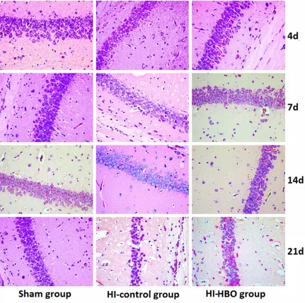
The pathologic changes in brain tissues were observed by HE staining. As shown in Figure 1, the pyramidal cells of DG region in sham group grew in multiple layers and arranged in neat rows. The cell contour was normal and nucleoli in the center of cells were observed clearly. In the HI group, the lesions were mainly occurred in cortex, CA1 and CA3 region of DG. The cell layers in brain tissues reduced and cells arranged in disorder. At day 4 and day 7 after surgery, the gliocyte proliferation, karyopykno-sis and cell fragmentation were found in brain tissues. In addition, the neurons degenerated and glial scar formed. Compared with the brain tissues in HI group, the lesion was alleviated after HBO treatment. The pyramidal cells arranged loosely and reduced inconspicuously. The phenomena of karyopyknosis and frag-mentation were relatively less.
Expression of Nestin in hippocampal DG re-gion
Nestin protein is expressed by the differentiat-ing neural stem cell (NSC) [11] and commonly considered as the marker for NSC [12]. Nestin positive cells in sham group displayed thinner and shorter protuberances compared with those in HI and HI-HBO group (Figure 2). The difference in the number of Nestin positive cells was significant (P < 0.01) between sham and HI group at different time points. And the Nestin positive cells in HI-HBO group increased significantly among groups (P < 0.01). The num-ber of Nestin positive cells peaked 4 days after surgery in the three groups, then decreased
significantly at 7 d and 21 d after surgery (P < 0.01) (Table 1).
Expression of BrdU in hippocampal DG region
The positive-BrdU cells mainly distributed in the polymorphous cell layer and adopt round shape, fusiform shape and pole shape (Figure 3). The BrdU positive cells peaked 4 days after surgery and began to decrease at 7 d after sur-gery in all of the three groups and there was significant difference in the number of BrdU positive cells between different time points (P < 0.01). A significant difference of the number of BrdU positive cells was observed at different time points compared the sham group with HI group. And there were significant increases of BrdU positive cells at different time points between HI group and HI-HBO group (P < 0.01) (Table 2).
MWM test analysis
The learning and memory abilities of the rats were manifested by delayed escape latency time and platform crossing frequencies. In the spatial navigation test, all the animals were likely to find the platform more quickly after the first trial constructed. Among the three groups, rats in HI group spent the statistically longest time to search for the platform hidden in water (P < 0.05). Compared with HI-HBO group and sham group, difference was not observed in the escape latency time of the rats (P > 0.05) (Table 3). In the spatial probe trial, rats exposed to the HBO showed significant higher platform cross-ing frequencies compared with HI rats (P <
0.05). And sham-surgery rats showed similar memory ability with HBO-treated rats (P > 0.05) (Table 4).
Discussion
Currently, an important understanding of HIBD pathogenesis is the strong activation of neuro-nal apoptosis in brain [13]. NSCs have potential of self-renewal, migration and multiple
differen-Table 3. Changes in escape latency time
Groups 1 d 2 d 3 d 4 d 5 d
Sham 44.75 ± 2.82 32.25 ± 2.12 19.00 ± 2.00 15.50 ± 2.45 12.75 ± 2.31 HI-HBO 48.63 ± 3.78 34.13 ± 3.60 21.30 ± 2.12 18.13 ± 3.14 15.63 ± 2.33 HI control 72.00 ± 2.00*,# 64.88 ± 2.23*,# 59.00 ± 3.12*,# 50.00 ± 2.83*,# 42.25 ± 2.38*,#
tiations into neurons, astrocyte and oligoden-drocyte for injury repair. There is a hypothesis that the increasing renewal capability of the endogenous NSCs contributes to the repair in brain damage [6]. Since, the NSCs in mamma-lian brain persist mainly in hippocampal DG region [14] and the hippocampus is susceptible to HI injury [15], we chose the hippocampal DG region for endogenous NSCs analysis. Nestin is an intermediate filament protein which is known as the biological marker for NSCs in central ner-vous system. BrdU served as a synthetic nucle-oside, can be incorporated into the newly syn-thesized DNA during the S phase of cell cycle. BrdU is widely used to detect the proliferation cells in living tissues [16]. In the present study, we used the Nestin and BrdU staining to evalu-ate the NSC proliferation.
Our results showed that the number of Nestin positive cells was significantly larger in HI and HBO group than that in sham group. It is report-ed that the HIBD can induce the proliferation of NSCs in situ [17, 18]. We speculated that the proliferation of NSCs in situ was involved with the Nestin expression after HIBD. Figure 2 indi-cated that the shape of Nestin positive cells was similar with astrocyte. A previous report revealed that a majority of Nestin expression was observed in astrocytes following brain inju-ry [19]. The Nestin expression in astrocytes consisted with the embryonal protein played key role in brain remodeling and recovery [20, 21]. In this study the Nestin positive cells observed may be reactive hyperplasia astro-cytes differentiated by SNC after HIBD. Our results also showed that HBO treatment pre-dominately increased the BrdU labeled cells in DG region of pups, which suggested that HBO promoted the proliferation and differentiation in HI rats. Besides, reports documented that the optimum time of HBO treatment was at 6h after injury [22] and in contrast, HBO treatment aggravated the injury after 12 h [23, 24]. We found that the both Nestin and BrdU positive cells peaked 4 days after surgery and then decreased significantly. The most effect of HBO on HIBD treatment may be for 4 days. Thus, there is a problem which is the best treatment of HBO for HIBD that needs to be further investigated.
Furthermore, hippocampus has close associa-tion with learning and memory ability [25]. Then
HIBD can affect the hippocampal function and declined the learning and memory ability. Our paper designed MWM test for pups at 30 days after surgery to test HBO effect on the learning and memory abilities by delayed escape laten-cy time and platform crossing frequencies. The MWM test showed that the HIBD significantly induced the cognitive dysfunction evidenced by long escape latency time and less platform crossing frequencies. The rats exposed to HBO retained similar learning and memory function compared with those in sham group. Therefore, HBO therapy can improve the learning and memory ability following HIBD.
In summary, HBO treatment promoted the repair and regeneration of the nervous system and contributed to the self-recovery and pro-tection of the damaged brain. The improve-ments in learning and memory function were observed by HBO treatment. HBO may be a promising therapy for HIBD in newborn based on the appropriate treatment time and pres-sure. However, the optimum treatment condi-tion needs to be further studies.
Acknowledgements
This work is supported by Special foundation for Taishan Scholars No. ts20110814.
Disclosure of conflict of interest
None.
Address correspondence to: Dr. Jiwen Wang, De- partment of Neurology, Children’s Medical Center, Qilu Hospital of Shandong University, and Brain Science Research Institute, Shandong University, 107 Wenhuaxi Road, Jinan 250012, China. Tel: +86-18561810900; Fax: +86-531-82169114; E-mail: [email protected]
References
[1] Mattiesen WR, Tauber SC, Gerber J, Bunkowski S, Brück W, Nau R. Increased neurogenesis af-ter hypoxic-ischemic encephalopathy in hu-mans is age related. Acta Neuropathol 2009; 117: 525-534.
[2] Gulczyńska E, Gadzinowski J. Therapeutic hy -pothermia for neonatal hypoxic-ischemic en-cephalopathy]. Ginekol Pol 2012; 83: 214. [3] Tagin MA, Woolcott CG, Vincer MJ, Whyte RK,
system-Hyperbaric oxygenation and neonatal hypoxic-ischemic brain damage
atic review and meta-analysis. Arch Pediatr Adolesc Med 2012; [Epub ahead of print]. [4] Shah P, Riphagen S, Beyene J, Perlman M.
Multiorgan dysfunction in infants with post-as-phyxial hypoxic-ischaemic encephalopathy. Arch Dis Child Fetal Neonatal Ed 2004; 89: F152-F155.
[5] Rudge LCFW. Carbon monoxide poisoning in infants: treatment with hyperbaric oxygen. South Med J 1993; 86: 334-337.
[6] Yang Y, Wang X, Yu X, Wang X, Xie M, Liu C. Hyperbaric oxygen induces endogenous neural stem cells to proliferate and differentiate in hypoxic-ischemic brain damage in neonatal rats. Undersea Hyperb Med 2008; 35: 113-29. [7] Badr A, Yin W, Mychaskiw G, Zhang J. Dual ef-fect of HBO on cerebral infarction in MCAO rats. Am J Physiol Regul Integr Comp Physiol 2001; 280: R766-R770.
[8] Rice JE, Vannucci RC, Brierley JB. The influence of immaturity on hypoxic-ischemic brain dam-age in the rat. Ann Neurol 1981; 9: 131-141. [9] Yujia LL. Relationship of Dose-Effect and
Time-Effect to Hyperbaric Oxygen Therapy on Hypoxic-Ischemic Brain Damage in the Neonatal Rat. Zhongguo Dang Dai Er Ke Za Zhi 2001; 3: 355-358.
[10] Santini D, Ceccarelli C, Mazzoleni G, Pasquinelli G, Jasonni VM, Martinelli GN. Demonstration of cytokeratin intermediate filaments in oo -cytes of the developing and adult human ova-ry. Histochemistry 1993; 99: 311-319. [11] Strojnik T, Røsland GV, Sakariassen PO,
Kavalar R, Lah T. Neural stem cell markers, nestin and musashi proteins, in the progres-sion of human glioma: correlation of nestin with prognosis of patient survival. Surg Neurol 2007; 68: 133-143.
[12] Suzuki S, Gerhold LM, Böttner M, Rau SW, Dela Cruz C, Yang E, Zhu H, Yu J, Cashion AB, Kindy MS. Estradiol enhances neurogenesis following ischemic stroke through estrogen re-ceptors α and β. J Comp Neurol 2007; 500: 1064-1075.
[13] Kilicdag H, Daglioglu YK, Erdogan S, Zorludemir S. Effects of caffeine on neuronal apoptosis in neonatal hypoxic-ischemic brain injury. J Matern Fetal Neonatal Med 2014: 1-21. [14] Komitova M, Mattsson B, Johansson BB,
Eriksson PS. Enriched environment increases neural stem/progenitor cell proliferation and neurogenesis in the subventricular zone of stroke-lesioned adult rats. Stroke 2005; 36: 1278-1282.
[15] Calvert JW, Zhou C, Nanda A, Zhang JH. Effect of hyperbaric oxygen on apoptosis in neonatal hypoxia-ischemia rat model. J Appl Physiol 2003; 95: 2072-2080.
[16] Lehner B, Sandner B, Marschallinger J, Lehner C, Furtner T, Couillard-Despres S, Rivera FJ, Brockhoff G, Bauer HC, Weidner N. The dark side of BrdU in neural stem cell biology: detri-mental effects on cell cycle, differentiation and survival. Cell Tissue Res 2011; 345: 313-328. [17] Kokaia Z, Lindvall O. Neurogenesis after isch -aemic brain insults. Curr Opin Neurobiol 2003; 13: 127-132.
[18] He Z, Cui L, Meschia JF, Dickson DW, Brott TG, Simpkins JW, Day AL, Mckinney M. Hippo- campal progenitor cells express nestin follow-ing cerebral ischemia in rats. Neuroreport 2005; 16: 1541-1544.
[19] Douen A, Dong L, Vanance S, Munger R, Hogan M, Thompson C, Hakim A. Regulation of nestin expression after cortical ablation in adult rat brain. Brain Res 2004; 1008: 139-146. [20] Duggal N, Schmidt-Kastner R, Hakim AM.
Nestin expression in reactive astrocytes follow-ing focal cerebral ischemia in rats. Brain Res 1997; 768: 1-9.
[21] Sahin Kaya S, Mahmood A, Li Y, Yavuz E, Chopp M. Expression of nestin after traumatic brain injury in rat brain. Brain Res 1999; 840: 153-157.
[22] Liu X, Zhao Y, Ma Q, Zhou X, Wang Y. Optimal therapeutic window of hyperbaric oxygenation in neonatal rat with hypoxic-ischemic brain damage]. Zhonghua Er Ke Za Zhi 2006; 44: 177.
[23] Lou M, Eschenfelder CC, Herdegen T, Brecht S, Deuschl G. Therapeutic window for use of hy-perbaric oxygenation in focal transient isch-emia in rats. Stroke 2004; 35: 578-583. [24] Northington FJ, Ferriero DM, Graham EM,
Traystman RJ, Martin LJ. Early neurodegenera-tion after hypoxia-ischemia in neonatal rat is necrosis while delayed neuronal death is apop-tosis. Neurobiol Dis 2001; 8: 207-219. [25] Sakimura K, Kutsuwada T, Itot I, Manabel T,