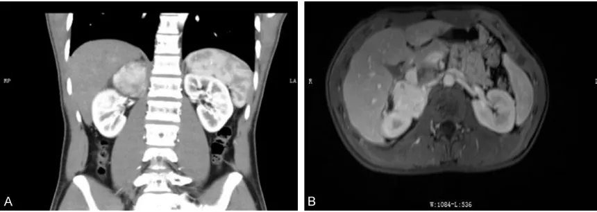
Case Report Indolent T-lymphoblastic proliferation associated with Castleman disease and low grade follicular dendric cell sarcoma: report of a case and review of literature
Full text
Figure
Related documents
We present here the effects of iron and/or zinc treatment on cognitive function of Mexican school- children who were exposed to lead from a metal foundry.. These results are part of
This study reports the neurodevelopmental outcomes of a cohort of school-aged children with HLHS who under- went either primary heart transplantation or staged re- construction with
Following bisphosphonate therapy, there was no improvement in existing osteolytic disease, but there was gradual healing and union of her left distal femoral fracture over time..
Higher baseline Vitamin D levels associated with slower decline in verbal fluency but no association across other domains.. Mean Age (years) Mean Follow- up Inclusion
In this regards, the application of an electrical power source to produce active radicals is one of the most important restrictions of application of advanced
Nubian goats showed a greater (P < 0.10) preference for Agave lechuguilla compared with Granadina goats, whereas Granadina goats showed a greater (P < 0.05) preference for
individual differences particularly cognitive styles influence users interaction with.. information
In Figures 5-6, samples of tracking results for two frames is depicted where the upper image shows the 3D coordinates of the objects, the lower left image is the
