Evaluation of clinical signs and causes of lower
urinary tract disease in Polish cats
S. Lew-Kojrys*, E. Mikulska-Skupien, A. Snarska, W. Krystkiewicz,
A. Pomianowski
Faculty of Veterinary Medicine, University of Warmia and Mazury, Olsztyn, Poland
*Corresponding author: [email protected]
ABSTRACT: This paper describes the results of a retrospective study performed on 385 cats with feline lower urinary tract disease. The study was conducted to obtain epidemiological data and to evaluate clinical symptoms and the results of laboratory tests in a population of Polish cats with symptoms of lower urinary tract disease. The analysed population comprised feline patients of the veterinary clinic at the University of Warmia and Mazury in Olsztyn who had not been treated prior to admission. Medical history was obtained for all patients. Urine samples were collected mostly, but not solely, by cystocentesis. Feline idiopathic cystitis was diagnosed in most cats (60.7%), while urinary tract infections were noted in only 7.8% of patients. Urethral obstruction caused by mucus plugs was observed in 17.4% of animals. Urolithiasis was observed in 13% of cats, 5% of whom were also diagnosed with urinary tract infections. Hyperplastic changes were identified in only 1% of the studied popula-tion. In 59% of cats, feline lower urinary tract disease was accompanied by urethral obstrucpopula-tion. Cats with feline idiopathic cystitis were the youngest animals in the analysed population, and the risk of urinary tract infections and neoplasia increased with age. Our results, obtained over a period of six years in a Polish feline population, show that sex, neutering, age, living conditions and diet influence the type of urinary tract disease, data which are consistent with those obtained in other countries.
Keywords: feline lower urinary tract disease; feline idiopathic cystitis; urethral obstruction; bacterial cystitis
Feline lower urinary tract disease (FLUTD) in-volves dysfunctions of the urinary bladder or ure-thra in cats. Pollakiuria without polyuria is one of the first symptoms of FLUTD. Other symptoms include painful urination (stranguria) and blood in urine (haematuria). Periuria, which involves pres-sure on the bladder associated with interstitial cystitis and urethritis, is also a characteristic sign of the disease. The disease may also be accompa-nied by urination in inappropriate places (periuria) due to increased pressure in the bladder caused by interstitial cystitis and urethritis. The affected animals are restless, and they vocalise excessively (Gunn-Moore 2003; Forrester and Towell 2015). Lower urinary tract infections are often
of FLUTD in 55–69% of patients (Lekcharoensuk et al. 2001; Gerber et al. 2005; Westropp et al. 2006; Dorsch et al. 2014).
In the literature, significant differences can be found in the reported prevalence of FLUTD. In German, Swiss and Norwegian studies, bacte-rial infections of the urinary tract accounted for up to 20% of FLUTD cases (Gerber et al. 2005; Eggertsdottir et al. 2011; Saevik et al. 2011; Dorsch et al. 2014). The prevalence of FLUTD was consid-erably lower in the work of Kruger et al. (1991) and Lekcharoensuk et al. (2001). The above discrepan-cies could be attributed to differences in inclusion criteria and methodology. Nevertheless, most re-searchers agree that the prevalence of urinary tract infections increases with animal age.
The above studies also demonstrate that the risk of disease increases with age. Young cats are most often diagnosed with feline idiopathic cystitis, and the risk of urinary tract infection, urolithiasis and neoplasia increases with age (Lekcharoensuk et al. 2001). Feline idiopathic cystitis is usually diagnosed by exclusion. Around 60% of cats with symptoms of FLUTD are diagnosed with feline idiopathic cys-titis. Feline lower urinary tract disease is gener-ally diagnosed by eliminating other urinary tract disorders such as urolithiasis, bacterial infection, anatomical defects or tumours. Accurate diagno-ses of urinary tract diseadiagno-ses require access to the patient’s medical history and the results of imaging and laboratory tests. Feline idiopathic cystitis is diagnosed based on the results of extensive tests after other disorders symptomatic of dysuria have been ruled out.
The aim of this retrospective study was to evalu-ate epidemiological data, clinical symptoms and causes of lower urinary tract disease in a popula-tion of Polish cats based on the data supplied by the veterinary clinic at the University of Warmia and Mazury in Olsztyn, and to compare those data with published research findings.
MATERiAl And METhodS
The data for analyses were collected from patients admitted to the clinic of the Faculty of Veterinary Medicine at the University of Warmia and Mazury in Olsztyn in 2008–2014. A total of 385 patients with clinical symptoms of dysuria were selected for the study. Patients that had undergone previous
treatment or urethral marsupialisation in private clinics were not taken into consideration. Animals with concomitant diseases, including kidney dis-eases, diabetes or hyperthyroidism, were excluded from the study. Only patients displaying urinary tract symptoms were tested. The patients’ medi-cal history and symptoms were thoroughly ana-lysed before clinical examination. The following data were acquired: the animal’s age, breed, sex, body weight, diet, access to water and type of clini-cal symptoms. The owners provided information about the duration and persistence of symptoms, urination frequency, the animal’s reproductive status, number of animals in the household and possible stressors. All cats were subjected to de-tailed clinical examinations with special emphasis on the genitourinary system and the nervous sys-tem. The urinary bladder was palpated before and after urination, and most patients were observed during voiding. The neurological exam involved evaluations of the anal wink and irregularities in extremity reflexes. In more than 90% of patients, urine was sampled by bladder puncture, and the remaining animals were subjected to urethral cath-eterisation. However, since many feline idiopathic cystitis patients experience pressure on the blad-der and urinate frequently, in some animals, the bladder contained very small amounts of urine that could not be sampled by puncture.
RESulTS
A total of 385 cats with symptoms of urinary tract disease were examined in accordance with the de-scribed protocol. The patients’ detailed medical history was obtained. All patients were subjected to clinical examinations, imaging tests, blood and urine tests.
The studied population comprised 361 (354 short-hair and seven long-(354 short-hair) domesticated cats (93.7%) and 24 pedigree cats (6.3%). The pedigree popula-tion was represented by nine Persian cats (37.5%), five Maine coons (21%), four Siamese cats (16%), four Norwegian forest cats (16%), one Burmese cat (4.2%) and one Russian blue cat (4.2%). The exam-ined population consisted of 335 males (87.01%) and 50 females. A total of 298 males (88.9%) and 39 females (78%) had been neutered.
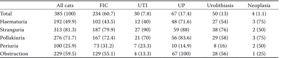
The majority (73%) of owners declared that this was the first time their cat had been admitted to a veterinary clinic due to urinary tract problems, while 27% of the animals had a previous history of urination problems. The owners were asked to describe the animals’ behaviour and position dur-ing voiddur-ing, vocalisation and urination outside the litter box. Clinical symptoms were similar in the evaluated groups. More than 75% of animals suf-fered from pollakiuria and dysuria. Macroscopic haematuria was observed in around 50% of patients (Table 1).
Bacterial growth in urine cultures was observed in 30 cats. Escherichia coli was isolated from 16 pa-tients, Staphylococcus spp. – from six patients,
Streptococcus spp. – from five patients, Enterococcus
spp. – from one patient, Citrobacter spp. – from one patient, and a mixed infection with group D
Streptococcus spp. and Staphylococcus spp. was
noted in one patient. Haematuria (> 10 erythro-cytes in the field of view) was observed in all cats cells (pyuria was diagnosed when more than five
cells were present in the field of view). The pres-ence and type of crystal-like formations was eval-uated. Microbiological analyses were performed on agar with 5% sheep blood, MacConkey agar and mannitol salt agar. Plates were incubated un-der aerobic conditions at a temperature of 37 °C, and were analysed 24 h and 48 h after incubation. Urine samples for microbiological analyses were collected by cystocentesis and, in isolated cases, by catheterisation. All patients with a suspicion of urolithiasis, developmental defects or neoplasia were examined by ultrasonography, radiology and contrast radiography. Cystotomy was performed in cats with urinary bladder stones. The chemical composition of stones was evaluated by infrared spectroscopy. Cystoscopy was performed in female cats with proliferative changes in the bladder wall, and cystotomy was performed in males. Tissue samples collected by biopsy and laparotomy were subjected to histopathological examinations.
The patients were divided into groups based on the underlying cause of urinary tract disease. Cats were diagnosed with feline idiopathic cystitis when other comorbidities associated with FLUTD had been ruled out. Bacterial infections were diag-nosed based on positive results of urine cultures. Urolithiasis was confirmed in ultrasound and/or ra-diologic examinations. In selected patients, urolith-iasis was accompanied by urinary tract infections. Cats with urethral plugs and hyperplastic changes in the bladder constituted a separate group. The patients were additionally divided into groups with and without urethral obstruction.
[image:3.595.64.536.639.737.2]The results were processed statistically in the Statistica v. 12.5 program (StatSoft, Inc., www. statsoft.com) using analysis of variance. The sig-nificance of differences between means was deter-mined in the Newman-Keuls test at P < 0.01.
Table 1. Clinical symptoms of all cats with feline lower urinary tract disease and cats diagnosed with five different conditions (n (%) of cats)
All cats FIC UTI UP Urolithiasis Neoplasia
Total 385 (100) 234 (60.7) 30 (7.8) 67 (17.4) 50 (13) 4 (1.1) Haematuria 192 (49.9) 102 (43.5) 12 (40) 48 (71.6) 27 (54) 3 (75) Stranguria 313 (81.3) 187 (79.9) 27 (90) 59 (88) 38 (76) 2 (50) Pollakiuria 276 (71.7) 167 (72.4) 21 (70) 56 (83.6) 29 (58) 3 (75) Periuria 100 (25.9) 73 (31.2) 7 (23.3) 10 (14.9) 8 (16) 2 (50) Obstruction 229 (59.5) 129 (55.1) 4 (13.3) 67 (100) 28 (56) 1 (25)
with urolithiasis and in around 90% of patients with the remaining diseases. Pyuria (> 5 leukocytes in the field of view) was noted in 80% of animals with urinary tract infection, in 38% of cats with feline idiopathic cystitis andurethral plugs, and in 67% of patients with urolithiasis. In the group of cats with urolithiasis, 19 patients were also diagnosed with urinary tract infection. In this group, the infection was caused by E. coli in 84.2% of cats (16), and by
Enterococcus spp. in 10.5% of patients, whereas a
mixed E. coli and Streptococcus spp. infection was noted in one cat. The average urine pH was 6.2, which was elevated to 6.9 in the urine of patients with bladder inflammation and urinary tract stones. The urine of patients with bladder inflammation and proliferative changes was characterised by lower specific gravity. These samples were obtained mostly from older animals. Glycosuria was observed in 64% of patients with urethral obstruction. Protein levels in urine were higher in feline idiopathic cystitis pa-tients than in the remaining animals, possibly due to urothelial damage and the penetration of acute phase proteins into urine (Lemberger et al. 2011).
Ultrasound scans were performed in all cats, and animals with a suspicion of urolithiasis and neo-plasia were additionally referred for radiographic tests and cystoscopy. Urolithiasis was diagnosed in 50 cats, and in this group, nine animals had de-veloped oxalate stones and struvite stones were
observed in seven cats. Bladder stones were not analysed in the remaining animals. In the studied population, 32% cats were catheterised, but urine samples were collected by cystocentesis before catheterisation in all patients.
A total of 36.66% patients with urinary tract in-fection and 75% of cats with neoplasms were fe-male. The prevalence of other diseases was higher in males, and all patients with urethral plugs were male. The average age of cats with FLUTD was 5.6 years. Feline idiopathic cystitis was observed in the youngest cats, and neoplasms were noted in the oldest animals. The age, body weight and sex proportions of the five groups diagnosed with FLUTD in the studied population of 385 cats are presented in Table 2.
Cats with urinary tract infection and neoplasms were significantly older than cats with feline idi-opathic cystitis, urolithiasis and urethral plugs (P < 0.005). In cats older than 10 years, urinary tract infections and neoplasms were diagnosed more fre-quently than feline idiopathic cystitis (P < 0.001). Cats with neoplasms were characterised by lower body weight than animals with feline idiopathic cystitis, urethral obstruction and urinary tract in-fection (P < 0.005). Significant differences were not noted between the remaining groups.
[image:4.595.64.532.127.214.2]Among all patients diagnosed with FLUTD, 322 (83.6%) were strictly indoor cats. The prevalence
Table 2. The age, body weight and sex proportions of the five groups diagnosed with feline lower urinary tract disease in the studied population of 385 cats
All cats
(n = 385) (n = 234)FIC (nUTI = 30) (n = 67)UP Urolithiasis (n = 50) Neoplasia (n = 4) Age (year) (0.7–16)5.67 4.35 ± 2.15 (0.5–7) 8.92 ± 3.11 (5–16) 4.9 ± 2.08 (1–7) 5.1 ± 2.99 (3–8) 12.5 ± 3.41 (8–16) Weight (kg) 5.23 5.48 ± 1.65 5.1 ± 1.39 5.36 ± 1.32 4.98 ± 1.48 4.11 ± 1.01 Males (%) 335 (87.01) 213 (91.02) 19 (63.33) 67 (100) 35 (70) 1 (25) Females (%) 50 (13) 21 (8.98) 11 (36.66) 0 15 (30) 3 (75)
FIC = feline idiopathic cystitis, UP = urethral plugs, UTI = urinary tract infections
Table 3. Diets of all cats with feline lower urinary tract disease and cats diagnosed with five different conditions (n
(%) of cats)
All cats (%) FIC UTI UP/urolithiasis Urolithiasis/UTI Neoplasia Dry food only 34 71 (30.4) 8 (25.8) 22 (32.8) 30 (60) 1 (25)
Wet food only 3.9 15 (6.4) 2 (6.5) 6 (9) 1 (2) 0
Dry and wet food 42 125 (53.4) 17 (54.8) 33 (49.2) 14 (28) 1 (25)
Special diet 18.3 23 (9.8) 4 (12.9) 6 (9) 5 (10) 2 (50)
[image:4.595.64.535.664.737.2]of FLUTD was not correlated with the presence of other pets in the household. Most cats with FLUTD were fed a combination of wet food and dry food. Approximately 50% of cats with feline idiopathic cystitis, urinary tract infection and urethral plugs, and 60% of cats with urolithiasis were fed only dry food. Cats fed only wet food constituted the small-est group (Table 3).
FLUTD was accompanied by urethral obstruction in 229 cats. Urethral obstructions were more com-mon in cats with feline idiopathic cystitis (129 cats) and urolithiasis (67 cats) than in animals with uri-nary tract infection (P < 0.005). The majority (204) of patients with urethral obstruction was male, and only 25 were female. All cats with urethral plugs were classified as animals with urethral obstruc-tion.
The percentage distribution of diseases associat-ed with FLUTD is presentassociat-ed in Figure 1. Feline idi-opathic cystitis was diagnosed in 234 cats (60.7%), urinary tract infections – 49 cats (12.7%), urethral obstruction caused by mucus plugs – in 67 cats (17.4%), urolithiasis – in 50 cats (13%), of which nearly 5% had also developed hyperplasia in their urinary bladders.
diSCuSSion
In this study and in published research, feline idiopathic cystitis has emerged as the most com-mon cause of FLUTD. In our study, 60% of cats
with FLUTD were diagnosed with feline idiopathic cystitis. The prevalence of feline idiopathic cystitis was estimated at 57% in Switzerland by Gerber et al. (2005), at 55% by Kruger et al. (1991) and at 51% by Barsanti (1996). In the work of Lekcharoensuk (2001), 63% of evaluated cats were diagnosed with feline idiopathic cystitis. In a Norwegian study performed in the period 2003–2008 (Saevik et al. 2011), the prevalence of feline idiopathic cystitis in cats with FLUTD was similar to that reported by Kruger (55.5%) and by Dorsch (55%; Kruger et al. 2009; Dorsch et al. 2014). In the present study, urethral plugs were the second most common cause of FLUTD (17.4%). Unfortunately, obstructive uropathy is difficult to distinguish from obstruc-tions caused by urethral plugs or urethral calculi (Gerber et al. 2005). Feline idiopathic cystitis may involve functional obstruction due to urethral in-flammation, contraction of the urethral sphincter and bladder sphincter dyssynergia. Urethral plugs composed of organic matter and crystals (mainly struvite crystals) mechanically block the flow of urine through the urethra, which often leads to complete obstruction and postrenal azotaemia. During veterinary procedures such as catheterisa-tion, urethral plugs can be pushed into the bladder and lost, which often contributes to an incorrect diagnosis. The presence of urethral plugs should be confirmed by contrast radiography or ureter-oscopy (Chew et al. 1996). According to Kruger, cats with feline idiopathic cystitis and crystalluria are susceptible to the formation of urethral plugs, which can lead to obstruction (Kruger et al. 2009). Those results indicate that the prevalence of feline idiopathic cystitis can be higher than determined in research studies.
[image:5.595.65.269.96.265.2]The greatest discrepancies were noted in the prevalence of urinary tract infection as the under-lying cause of FLUTD. In a US study, the percentage of cats with urinary tract infection was determined to be below 3% (Kruger et al. 1991). Saevik et al. (2011) determined the prevalence of urinary tract infection to be 11.8%. According to other research-ers, urinary tract infection affects 18.9% of cats (Dorsch et al. 2014) or even 22.2% of cats with a primary infection (Kraijer et al. 2003). Urinary tract infection was observed to be common in cats with chronic kidney disease, hyperthyroidism and diabe-tes (Mayer-Roenne et al. 2007; Martinez-Ruzafa et al. 2012). The above results were used by other au-thors to exclude patients with comorbidities from
Figure 1. Percentage distribution of diseases associated with feline lower urinary tract disease
FIC = feline idiopathic cystitis, UP = urethral plugs, UTI = urinary tract infections
their studies, which could explain the significant discrepancies in the prevalence of urinary tract infection in the literature. In our study, nearly 8% of cats had primary infections of the urinary tract, and this percentage increased to 13% when patients with both urolithiasis and urinary tract infection were included in that group.
According to research findings from the United States cited in textbooks, around 3% of young cats suffer from urinary tract infection, and the preva-lence of infections increases with age (Lekcharoensuk et al. 2001). European studies indicate that the per-centage of cats with urinary tract infection is much higher and that infections should always be consid-ered when diagnosing FLUTD (Gerber et al. 2005; Kruger et al. 2009; Saevik et al. 2011; Dorsch et al. 2014).
Urine samples for bacteriological tests should be collected by cystocentesis to eliminate the risk of contamination from the lower urinary tract (Kruger et al. 1996b; Bartges 2004; van Duijkeren et al. 2004; Barsanti 2006). Researchers differ in their opinions regarding bacterial counts that have a positive pre-dictive value for urinary tract infection. The critical bacterial count is usually established at 1000 CFU/ml, and higher values should be regarded as suspect (Barsanti 2006; Pressler and Bartges 2010).
In our study, the average age of cats with uri-nary tract infection was 8.9 years, which is higher than the 5.1, 5.6 and 6.7 years reported by other authors (Buffington et al. 1997; Saevik et al. 2011; Dorsch et al. 2014; respectively). Bartges (2004) and Barsanti (2006) found a correlation between the age of feline patients and the prevalence of uri-nary tract infection. In their study, uriuri-nary tract infection was diagnosed in 45% of cats older than 10 years with symptoms of FLUTD, and 17% of cats were diagnosed with both urolithiasis and urinary tract infection (Bartges 2004). In most reports, fe-males are more susceptible to urinary tract infec-tion (Lekcharoensuk et al. 2001; Bailiff et al. 2008; Litster et al. 2009). In our study, females repre-sented only 36.7% of the population with urinary tract infection. Our results could be attributed to the fact that animals with systemic diseases and previously treated animals were not included in the study. Escherichia coli is most frequently iso-lated from urine, and it was noted in 44% of posi-tive samples in Norway (Saevik et al. 2011) and in 65% of samples in Germany (Dorsch et al. 2014). In our study, E. coli was isolated in 53% of cases,
and similar results were reported by Passmore et al. (2008) and Litster et al. (2009).
Urolithiasis was observed in 13% of cats with FLUTD, but not all calculi were subjected to chemi-cal composition analysis. Nearly 40% of patients with urolithiasis were also diagnosed with urinary tract infection. Based on the results of urine sedi-ment analysis, haematuria was noted in more than 90% of cases and crystalluria was observed in 78% of cases. The majority of crystal-like formations were struvite crystals.
In our study, marked haematuria in cats with FLUTD was reported by 46% of owners (macro-scopic evaluation), and in micro(macro-scopic analyses, hae-maturia was diagnosed in more than 94% of cases. In the literature, the prevalence of haematuria was determined to be 97% by Kruger et al. (1991), 91% by Dorsch et al. (2014) and only 83% by Saevik et al. (2011). Such high rates of prevalence could be attrib-uted to the fact that cystocentesis was the method of choice for collecting urine samples. Research indicates that cystocentesis can lead to mild and transient microscopic haematuria which cannot be distinguished from pathological haematuria in cats with urinary tract infection (Kruger et al. 1996b).
Pyuria was observed in 80% of cats with urinary tract infection and in 42% of patients with feline idiopathic cystitis. In general, pyuria was noted in 48% of animals with FLUTD. In the literature, the prevalence of pyuria in patients with FLUTD was determined in the range of 43–54% (Bailiff el al. 2008; Litster et al. 2009; Saevik et al. 2011; Dorsch et al. 2014).
In our study, the majority of patients with FLUTD were castrated male European shorthairs. Most au-thors did not observe a correlation between breed and the risk of FLUTD (Gerber et al. 2005; Saevik et al. 2011; Dorsch et al. 2014). In our study, males ac-counted for 87% of the studied population. Similar observations were made in other studies where the female to male ratio was estimated to be 1 : 4. Male cats are more often admitted for treatment due to a higher risk of obstructive uropathy which requires quick medical intervention. In the present study, all cats with urethral plugs were male. Symptoms of feline idiopathic cystitis without urethral obstruc-tion may be very subtle, and they can subside by the time the owner decides to consult a veterinary practitioner.
levels of physical activity, indoor confinement and lit-ter boxes that are too small for the animal (Cameron et al. 2004; Defauw et al. 2011). In our study, 83% of patients were strictly indoor cats, and more than 53% of the animals shared the household with other pets. Most cats with FLUTD (42%) were fed a combination of commercial wet food and dry food, more than 30% of animals were fed dry food only, and less than 4% of cats received wet food only. More than 60% of cats with urolithiasis were fed only dry food. Previous research demonstrated that diets consisting of dry food only increased the risk of FLUTD (Willeberg and Priester 1976; Jones et al. 1997).
In our research, the study protocol had to be some-what modified for selected patients. Cystocentesis could not be performed in cats with normal ure-thral patency and ureure-thral syndrome whose blad-ders were empty during the examination. In those animals, small amounts of urine were collected by catheterisation. Ultrasound scans were performed in all patients, but radiological and cystoscopic examinations were conducted in selected patients based on specific indications.
Our findings confirm the results of American and European studies which demonstrated that feline idiopathic cystitis is the most common un-derlying cause of FLUTD. The prevalence of uri-nary tract infection in Polish cats is lower than in other European populations but higher than in the American population. Feline idiopathic cystitis is most often diagnosed in young cats, whereas the risk of urinary tract infection, urolithiasis and neo-plasms increases with age.
REfEREnCES
Bailiff NL, Westropp JL, Nelson RW, Sykes JE, Owens SD, Kass PH (2008): Evaluation of urine specific gravity and urine sediment as risk factors for urinary tract infections in cats. Veterinary Clinical Pathology 37, 317–322. Barsanti JA (2006): Genitourinary infections. In: Greene
CE (ed.): Infectious Diseases of the Dog and Cat. 3rd edn. Saunders Elsevier, St Louis. 935–961.
Barsanti JA, Brown J, Marks A, Reece L, Greene CE, Fingo DR (1996): Relationship of lower urinary tract signs to seropositivity for feline immunodeficiency virus in cats. Journal of Veterinary Internal Medicine 10, 34–38. Bartges JW (2004): Diagnosis of urinary tract infections.
Veterinary Clinics of North America: Small Animal Prac-tice 34, 923–933.
Buffington CA, Chew DJ, Kendall MS, Scrivani PV, Thomp-son SB, Blaisdell JL, Woodworth BE (1997): Clinical evaluation of cats with nonobstructive urinary tract dis-eases. Journal of the American Veterinary Medical As-sociation 210, 46–50.
Cameron ME, Casey RA, Bradshaw JW, Waran NK, Gunn-Moore DA (2004): A study of environmental and behav-ioural factors that may be associated with feline idiopathic cystitis. Journal of Small Animal Practice 45, 144–147. Chew DJ, Buffington CAT, Kendall M, Osborn SD,
Woods-worth BE (1996): Urethroscopy, cystoscopy and biopsy of the feline lower urinary tract. Veterinary Clinics of North America: Small Animal Practice 26, 441–462. Defauw PA, Van de Maele I, Duchateau L, Polis IE, Saunders
JH, Daminet S (2011): Risk factors and clinical presenta-tion of cats with feline idiopathic cystitis. Journal of Feline Medicine and Surgery 13, 967–975.
Dorsch R, Remer C, Sauter-Louis C, Hartmann K (2014): Feline lower urinary tract disease in a German cat popu-lation. A retrospective analysis of demographic data, causes and clinical signs. Tieraerztliche Praxis Ausgabe K: Kleintiere/Heimtiere 42, 231–239.
Eggertsdottir AV, Lund HS, Krontveit R, Sorum H (2007): Bacteriuria in cats with feline lower urinary tract disease: a clinical study of 134 cases in Norway. Journal of Feline Medicine and Surgery 9, 458–465.
Eggertsdottir AV, Saevik BK, Halvorsen I, Sorum H (2011): Occurrence of occult bacteriuria in healthy cats. Journal of Feline Medicine and Surgery 13, 800–803.
Forrester SD, Towell TL (2015): Feline idiopathic cystitis. Veterinary Clinics of North America: Small Animal Prac-tice 45, 783–806.
Gerber B, Boretti FS, Kley S, Laluha P, Muller C, Sieber N, Unterer S, Wenger M, Fluckiger M, Glaus T, Reusch CE (2005): Evaluation of clinical signs and causes of lower urinary tract disease in European cats. Journal of Small Animal Practice 46, 571–577.
Gunn-Moore D (2003): Feline lower urinary tract disease. Journal of Feline Medicine and Surgery 5, 133–138. Jones BR, Sanson RL, Morris RS (1997): Elucidating the risk
factors of feline lower urinary tract disease. New Zealand Veterinary Journal 45, 100–108.
Kraijer M, Fink-Gremmels J, Nickel RF (2003): The short-term clinical efficacy of amitriptyline in the management of idiopathic feline lower urinary tract disease: a con-trolled clinical study. Journal of Feline Medicine and Surgery 5, 191–196.
Kruger JM, Osborne CA, Venta PJ, Sussman MD (1996a): Viral infections of the feline urinary tract. Veterinary Clinics of North America: Small Animal Practice 26, 281–296.
Kruger JM, Osborne CA, Ulrich LK (1996b): Cystocentesis. Diagnostic and therapeutic considerations. Veterinary Clinics of North America: Small Animal Practice 26, 353–361.
Kruger JM, Osborne CA, Lulich JP (2009): Changing para-digms of feline idiopathic cystitis. Veterinary Clinics of North America: Small Animal Practice 39, 15–40. Lekcharoensuk C, Osborne CA, Lulich JP (2001):
Epide-miologic study of risk factors for lower urinary tract dis-eases in cats. Journal of the American Veterinary Medical Association 218, 1429–1435.
Lemberger SI, Deeg CA, Hauck SM, Amann B, Hirmer S, Hartmann K, Dorsch R (2011): Comparison of urine pro-tein profiles in cats without urinary tract disease and cats with idiopathic cystitis, bacterial urinary tract infection, or urolithiasis. American Journal of Veterinary Research 72, 1407–1415.
Litster A, Moss S, Platell J, Trott DJ (2009): Occult bacterial lower urinary tract infections in cats-urinalysis and cul-ture findings. Veterinary Microbiology 136, 130–134. Martinez-Ruzafa I, Kruger JM, Miller R, Swenson CL,
Bo-lin CA, Kaneene JB (2012): CBo-linical features and risk fac-tors for development of urinary tract infections in cats. Journal of Feline Medicine and Surgery 14, 729–740.
Mayer-Roenne B, Goldstein RE, Erb HN (2007): Urinary tract infections in cats with hyperthyroidism, diabetes mellitus and chronic kidney disease. Journal of Feline Medicine and Surgery 9, 124–132.
Passmore CA, Sherington J, Stegemann MR (2008): Efficacy and safety of cefovecin for the treatment of urinary tract infections in cats. Journal of Small Animal Practice 49, 295–301.
Pressler B, Bartges JW (2010): Urinary tract infections. In: Ettinger SJ, Feldman EC (eds): Textbook of Veterinary Internal Medicine. 7th edn. Saunders Elsevier, St Louis. 2036–2047.
Saevik BK, Trangerud C, Ottesen N, Sorum H, Eggertsdot-tir AV (2011): Causes of lower urinary tract disease in Norwegian cats. Journal of Feline Medicine and Surgery 13, 410–417.
van Duijkeren E, van Laar P, Houwers DJ (2004): Cystocen-tesis is essential for reliable diagnosis of urinary tract infections in cats. Tijdschrift Voor Diergeneeskunde 129, 394–396.
Westropp JL, Kass PH, Buffington CA (2006): Evaluation of the effects of stress in cats with idiopathic cystitis. American Journal of Veterinary Research 67, 731–736. Willeberg P, Priester WA (1976): Feline urological
syn-drome: associations with some time, space, and indi-vidual patient factors. American Journal of Veterinary Research 37, 975–978.