Original Article
VEGF-C is positively associated with lymphangiogenesis
and lymphatic metastasis in rectal cancer
Zhubin Pan1, Xianying Lu1, Jindu Zhao1, Qun Gao1, Jian Wang2
1Department of General Surgery of Anhui Provincial Children’s Hospital, Hefei, Anhui, China; 2Children’s Hospital of Soochow University, Suzhou, China
Received December 12, 2017; Accepted February 6, 2018; Epub March 1, 2018; Published March 15, 2018
Abstract: Rectal cancer is a common malignancywith a less than 5-year postoperative survival rate. Although it often metastasizes via the lymph and blood, the detailed mechanism of this process remains unclear. This study investi-gated the relationship between vascular endothelial growth factor-C (VEGF-C) expression and lymphangiogenesis, as well as its relation to lymphatic metastasis of rectal cancer. To address this question, VEGF-C expression in rectal cancer and normal tissue adjacent to tumor was assessed by immunohistochemistry. The lymphatic endothelial
cell-specific marker D2-40 was used to label lymphatic endothelial cells and the lymphatic vessel density (LVD) was subsequently quantified. As expected, the expression of VEGF-C in rectal cancer (75%) was significantly higher than in normal adjacent tissue (25%), and this level correlated with differentiation, Dukes stage, and lymph node metas -tasis, though not with sex or age. The LVD was higher in VEGF-C positive rectal cancer than in VEGF-C negative rectal cancer, and was also higher in lymphatic metastases than in non-lymphatic metastases. These results indicate that expression of VEGF-C may impact the prognosis of rectal cancer via its effect on the formation of new lymphatic
vessels. This represents a significant advance in the study of the genesis and development of rectal cancer, and
may have value in clinical care.
Keywords: Rectal cancer, vascular endothelial growth factor-C, lymphatic vessel density, lymphangiogenesis, and lymphatic metastasis
Introduction
Rectal cancer is a common malignancy [1-3]. Its development is a complex multi-factor, multi- stage process, involving numerous genes [4, 5]. Though improvements in its diagnosis and treatment have led to greatly improved survival rate and functional recovery, efficacy of treat-ment is still unsatisfactory. The study of colorec-tal cancer etiology, pathogenesis, diagnosis, treatment, and prevention remains a vital topic of research [6].
The lymphatic system is one of the main routes of metastasis of rectal cancer, which correlates with a poor prognosis [7, 8]. Lymphangiogenesis can promote lymph node metastasis, further facilitating distant metastasis that involves multiple genes and products [9, 10]. The vascu-lar endothelial growth factor family includes vascular endothelial growth factor-A-F (VEGF-A-F), and placental growth factor, of which VEGF-
lymphangiogen-esis and lymph node metastasis, in order to determine new approaches for diagnosis, treat-ment, and prognosis of rectal cancer.
were enrolled based on their surgical resection and clinical diagnosis. None of the patients had received chemotherapy, radiotherapy, or bio-therapy prior to surgery. The mean patient age was 59.3 ± 6.35 (range: 3087) years. Twenty patients had well-differentiated adenocarcino-ma, while 20 had moderately-or poorly-differ-entiated adenocarcinoma. Tumors of 22 patien- ts were classified as the Dukes stage (A + B), while 18 patients as Dukes stage (C + D); 16 patients had lymph node metastasis, while 24 did not have metastasis. The normal tissue adjacent to carcinoma (> 8 cm from the tumor edge) was selected as control. The study pro- tocol was approved by the Research Ethics Committee of Children’s Hospital of Soochow University and all patients provided written informed consent prior to their enrollment into the study.
Immunohistochemistry (IHC)
[image:2.612.91.523.72.233.2]The aforementioned tissues were fixed and sectioned in order to perform IHC staining according to the manufacturer’s instructions. Endogenous peroxidase was inactivated by
Figure 1. VEGF-C expression level assay by immunohistochemistry (IHC) in rectal cancer and normal adjacent
tis-sue. A: VEGF-C is expressed in rectal cancer; B: VEGF-C is negative in normal adjacent tistis-sue. VEGF-C expression in
[image:2.612.87.360.319.373.2]rectal cancer was higher than that of the normal adjacent tissue (**: P < 0.01, 100×).
Table 1. VEGF-C* expression by immunostaining in rectal can-cer and normal adjacent tissue
Types Cases VEGF-C Positive rate (%) P-value - + ++ +++
Rectal cancer 40 11 12 9 8 72.5 0.00025 Para-carcinoma tissue 40 35 4 1 0 12.5
*Note: Vascular endothelial growth factor-C.
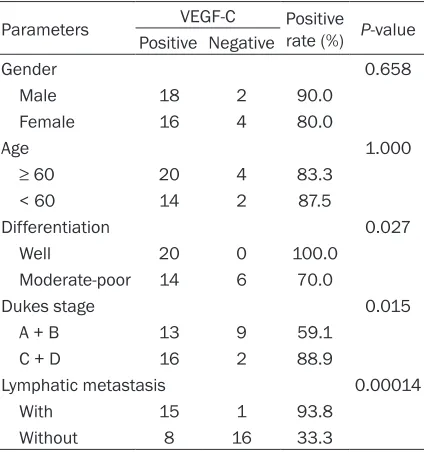
Table 2. Relationship of VEGF-C* with clinical features
Parameters VEGF-C Positive rate (%) P-value Positive Negative
Gender 0.658
Male 18 2 90.0 Female 16 4 80.0
Age 1.000
≥ 60 20 4 83.3
< 60 14 2 87.5
Differentiation 0.027 Well 20 0 100.0
Moderate-poor 14 6 70.0
Dukes stage 0.015
A + B 13 9 59.1
C + D 16 2 88.9
Lymphatic metastasis 0.00014 With 15 1 93.8
Without 8 16 33.3
*VEGF-C: Vascular endothelial growth factor-C.
Materials and methods
Patients
[image:2.612.91.303.427.653.2]incubating the sections in 3% H2O2 for 30 min. The sections were subjected to sequential incu-bations with 10% normal goat serum in 0.01 M PBS for 30 min at room temperature, and then respectively incubated in rabbit-derived anti-VEGFC antibody (ab135506, Abcam, USA), and mouse-derived anti-D2-40 antibody [D2-40] (ab77854, Abcam, USA) in PBS containing 0.3% Triton X-100 overnight at 4°C. The sections were washed 3 times (5 min each) with PBS, and then incubated in peroxidase-conjugated goat anti-rat IgG (1:200; Zymed, South San Francisco, CA, USA) for 1 hr at room tempera-ture. Finally, the sections were developed with diaminobenzidine (DAB) in 0.1 M Tris-buffered saline (TBS) containing 0.001% H2O2 for 30 min. The sections were observed by
microsco-py (Olympus, Tokyo, Japan), images were captur- ed, and the VEGF-C and D2-40 expression lev-els were detected as described below.
VEGF-C was identified in the cytoplasm and cell membrane as tan/brown particles, and the VEGF-C signal was evaluated according to the staining intensity: 0: No color, 1: Light yellow, 2: Claybank, 3: Tawny, and the positive cells num-ber: 0: < 5%; 1: 5%-25%; 2: 26%-50%; 3: 51%-75%; 4: > 75%, and defined as the immunohis-tochemical score: -: 0-2 score; +: 3-5 score; ++: 6-8 score; +++: > 8 score.
D2-40 was specifically expressed in the cyto-plasm of lymphatic vessel endothelial cells and stained brown, and the lymphatic vessel
densi-Figure 2. D2-40 expression level assay by immunohistochemistry (IHC) in rectal cancer and normal adjacent tissue.
A: The D2-40 expressed in rectal cancer; B: The D2-40 expressed in normal adjacent tissue. D2-40 expression in
[image:3.612.92.525.73.235.2]rectal cancer was obviously higher than in normal adjacent tissue (**: P < 0.01, 100×).
[image:3.612.91.287.297.448.2]Figure 3. Histogram analysis of LVD in rectal cancer and normal adjacent tissue. LVD was higher in rectal cancer than in normal adjacent (Para-carcinoma) tis-sue (**: P < 0.01).
[image:3.612.323.523.297.449.2]ty (LVD), was quantified according to the aver-age number of lymphatic vessels per area, and a histogram was generated using Origin 9.0 software (http://www.originlab.com/; Northam- pton, MA, USA).
Statistical analysis
All data were expressed as the mean ± stan-dard deviation (SD). Statistical analyses were performed via one-way ANOVA using SPSS software (version 21.0, http://spss.en.softonic. com/), and Student′s t-tests were performed in a group of two samples, with P < 0.05 and P < 0.01 indicated significant differences and high-ly significant differences, respectivehigh-ly.
Results
VEGF-C expression was increased in rectal cancer compared to normal adjacent tissue
As shown in Figure 1A, VEGF-C is localized to the cytoplasm and cell membrane, with the brown particles representing positive signal. VEGF-C was weak in normal tissue (Figure 1B). The VEGF-C positive rate in rectal cancer (72.5%) was significantly higher than in normal adjacent tissue (12.5%, Table 1, **: P < 0.01).
VEGF-C expression is closely associated with differentiation, Dukes staging, and lymph node metastasis
VEGF-C was associated with the differentiation, Dukes staging, and lymph node metastasis (Ta- ble 2, **: P < 0.01), but not with either sex or age.
D2-40 is specifically expressed in lymphatic
endothelial cells
As shown in Figure 2A and 2B, the D2-40 pro-tein is specifically expressed in lymphatic endo-thelial cells of rectal cancer. The expression of D2-40 was significantly higher in the lymphatic endothelial cells of rectal cancer than in the normal adjacent tissue (**: P < 0.01).
LVD is higher in VEGF-C positive rectal cancer than in normal adjacent tissue
LVD of rectal cancer tissue demonstrated a sig-nificantly greater proportion of VEGF-C positive cells than VEGF-C negative cells (Figure 3). Similarly, in normal adjacent tissue, increased
VEGF-C expression was noted with more LVD. In comparing the two LVD expressions, significant- ly higher VEGF-C expression was present in the rectal cancer tissue than that of normal adja-cent tissue.
LVD was higher in lymphatic metastasis than that of non-lymphatic metastasis
As shown in Figure 4, LVD is higher in lymphatic metastasis than non-lymphatic metastasis.
Discussion
This study demonstrated that enhanced VEGF-C expression is located in the cytoplasm and cell membrane of rectal cancer tissue, and these levels were significantly higher than that of nor-mal adjacent tissue. In addition, VEGF-C expres-sion was closely associated with differentia-tion, Dukes staging, and lymph node metasta-sis, but not with patient sex or age. Moreover, D2-40 protein was specifically expressed in lymphatic endothelial cells of rectal cancer, at a higher level than lymphatics of normal adjacent tissue. The LVD was higher in VEGF-C positive tissue than in VEGF-C negative tissue in rectal cancer, and correlated with the expression of VEGF-C in rectal cancer but not normal tissue.
Lymphatic metastasis is an important predictor of clinical prognosis, in which lymphangiogene-sis is an essential step. Many studies have sho- wn that VEGF-C can promote lymphangiogene-sis in tumors and increase tumor invasiveness, thus facilitating distant metastasis [28, 29]. Yang et al. applied immunohistochemistry to investigate VEGF-C expression in 64 patients with esophageal cancer, and found that VEGF-C expression in esophageal squamous carcino-ma tissue was 59.4%, which was higher than in adjacent normal tissue [30]. Furthermore, its positive expression was related to lymph no- de metastasis and TNM stage, suggesting that VEGF-C is associated with lymph node metas-tasis in esophageal cancer and is a potential marker of esophageal cancer lymphatic metas-tasis. Kostis et al. identified overexpression of VEGF-C expression in prostate cancer tissue. They also found that its expression was asso- ciated with lymphatic metastasis. High LVD counts showed elevated VEGF-C, indicating that VEGF-C played an important role in pros-tate cancer lymphatic metastasis [31]. Liu et al. explored VEGF-C expression in 109 cases of primary non-small cell lung cancer patients. VEGF-C expression was found in 65.14% of cases, significantly higher than in normal tis-sue. LVD counting was higher in VEGF-C posi-tive cells than in VEGF-C negaposi-tive cells, sug-gesting that VEGF-C can promote non-small cell lung cancer growth and lymph node metasta-sis. This provides a new direction for non-small cell lung cancer targeted therapy [32].
Xu et al. found that VEGF-C expression in co- lorectal cancer tissue was 78.2%, which was associated with Dukes staging and lymph no- de metastasis [33]. Our study confirmed that VEGF-C was highly expressed in colorectal can-cer (positive rate 72.5%) and related to differ-entiation, Dukes stage, and lymph node metas-tasis, in accordance with the results of Xu et al. We further illustrated that VEGF-C had a critical role in colorectal cancer and lymphatic metas-tasis and can be used as anindicator of poor prognosis. Liu et al. found that LVD counting in VEGF-C positive lung cancer cells was clearly higher than that in VEGF-C negative cancers. Our investigation revealed a similar result, fur-ther illustrating that VEGF-C is an important factor in lymphangiogenesis. LVD counting was higher in patients with lymph node metastasis than in patients without lymph node
metasta-sis, indicating that VEGF-C is closely related to lymphangiogenesis and lymphatic metastasis in rectal cancer. These results provide new pathways for early diagnosis, treatment, and prognosis of rectal cancer [32]. Limits of our study include limited sample size, warranting an increase the sample size and use of multiple methods for systemic investigation in the fu- ture. Our study provides a significant advance on the genesis and development of rectal can-cer and represents a significant advance in clinical practice.
Acknowledgements
This research project was sponsored by the National Natural Science Foundation of China (Grant No. 81420108002).
Disclosure of conflict of interest
None.
Address correspondence to: Dr. Qun Gao, Depart- ment of General Surgery of Anhui Provincial
Children’s Hospital, No. 39, Wang Jiang Dong Street, Bao He District, Hefei 230000, Anhui, China. Tel: 62237468; Fax:
+86-05551-62237468; E-mail: superjcwx@163.com; Dr. Jian
Wang, Children’s Hospital of Soochow University, No. 92, Zhong Nan Street, Industrial Park, Suzhou 215025, China. Tel: +86-0512-80693588; Fax: +86-0512-80693599; E-mail: Shuaicai8888@sina.
com
References
[1] Buchs NC, Koessler T, Vitali GC, Zilli T, Puppa G, Breguet R, Bichard P, Roth A, Morel P and
Ris F. [Multidisciplinary management of local-ized rectal cancer]. Rev Med Suisse 2017; 13: 1229-1235.
[2] Fu C and Han J. [Strategy and technique for si-multaneous resection of rectal cancer and liv-er metastasis]. Zhonghua Wei Chang Wai Ke Za Zhi 2017; 20: 618-620.
[3] Xu P and Xu J. [Application of robotic surgical system in sphincter-preserving surgery for low rectal cancer]. Zhonghua Wei Chang Wai Ke Za Zhi 2017; 20: 606-609.
[4] Joye I, Debucquoy A, Deroose CM,
Vandecav-eye V, Cutsem EV, Wolthuis A, D’Hoore A,
Sagaert X, Zhou M, Gevaert O and Hauster-mans K. Quantitative imaging outperforms
mo-lecular markers when predicting response to
[5] Korphaisarn K, Loree JM, Nguyen V, Coulson R,
Holla V, Litzenburger BC, Chen K, Mills GB, Maru DM, Meric-Bernstan F, Mills Shaw KR
and Kopetz S. Genomic analysis of exceptional responder to regorafenib in treatment-refracto-ry metastatic rectal cancer: a case report and review of the literature. Oncotarget 2017; 8: 57882-57888.
[6] Papp G, Besznyak I, Porneczi B, Saftics G and Bursics A. [Advantages of transanal approach
in low rectal cancer resections]. Magy Seb 2017; 70: 119-124.
[7] Hoshino A and Lyden D. Metastasis: lymphatic detours for cancer. Nature 2017; 546: 609-610.
[8] Wang Y, Han C, Teng F, Bai Z, Tian W and Xue F.
Predictive value of serum HE4 and CA125 con-centrations for lymphatic metastasis of endo-metrial cancer. Int J Gynaecol Obstet 2017; 136: 58-63.
[9] Kinashi H, Falke LL, Nguyen TQ, Bovenschen N, Aten J, Leask A, Ito Y and Goldschmeding R. Connective tissue growth factor regulates fibro -sis-associated renal lymphangiogenesis. Kid-ney Int 2017; 92: 850-863.
[10] Guc E, Briquez PS, Foretay D, Fankhauser MA, Hubbell JA, Kilarski WW and Swartz MA. Local
induction of lymphangiogenesis with
engi-neered fibrin-binding VEGF-C promotes wound healing by increasing immune cell trafficking and matrix remodeling. Biomaterials 2017;
131: 160-175.
[11] Yang GH, Zhou X, Ji WJ, Liu JX, Sun J, Dong Y, Jiang TM and Li YM. VEGF-C-mediated cardiac
lymphangiogenesis in high salt intake acceler -ated progression of left ventricular remodeling in spontaneously hypertensive rats. Clin Exp Hypertens 2017; 39: 740-747.
[12] Peng FW, Liu DK, Zhang QW, Xu YG and Shi L. VEGFR-2 inhibitors and the therapeutic appli-cations thereof: a patent review (2012-2016). Expert Opin Ther Pat 2017; 27: 987-1004. [13] Lee S, Rho SS, Park H, Park JA, Kim J, Lee IK,
Koh GY, Mochizuki N, Kim YM and Kwon YG.
Carbohydrate-binding protein CLEC14A regu-lates VEGFR-2- and VEGFR-3-dependent sig-nals during angiogenesis and lymphangiogen-esis. J Clin Invest 2017; 127: 457-471. [14] Wang XL, Zhao J, Qin L and Qiao M. Promoting
inflammatory lymphangiogenesis by vascular
endothelial growth factor-C (VEGF-C)
aggravat-ed intestinal inflammation in mice with experi
-mental acute colitis. Braz J Med Biol Res 2016;
49: e4738.
[15] Franc M, Kachel-Flis A, Michalski B, Fila-Danilow A, Mazurek U, Michalski M, Michalska A, Kuczerawy I and Skrzypulec-Plinta V. Lym -phangiogenesis in cervical cancer evaluated by expression of the VEGF-C gene in clinical
stage IB-IIIB. Prz Menopauzalny 2015; 14:
112-117.
[16] Zhao G, Zhu G, Huang Y, Zheng W, Hua J, Yang S, Zhuang J and Ye J. IL-6 mediates the sig- nal pathway of JAK-STAT3-VEGF-C promoting growth, invasion and lymphangiogenesis in gastric cancer. Oncol Rep 2016; 35: 1787-1795.
[17] Starek I, Salzman R, Kucerova L, Skalova A and
Hauer L. Expression of VEGF-C/-D and lym-phangiogenesis in salivary adenoid cystic car-cinoma. Pathol Res Pract 2015; 211: 759-765. [18] Ji H, Cao R, Yang Y, Zhang Y, Iwamoto H, Lim S,
Nakamura M, Andersson P, Wang J, Sun Y,
Dissing S, He X, Yang X and Cao Y. TNFR1 me-diates TNF-alpha-induced tumour lymphangio-genesis and metastasis by modulating VEGF-C-VEGFR3 signalling. Nat Commun 2014; 5: 4944.
[19] Hall MA, Robinson H, Chan W and Sevick-Mu -raca EM. Detection of lymphangiogenesis by
near-infrared fluorescence imaging and re -sponses to VEGF-C during healing in a mouse
full-dermis thickness wound model. Wound
Repair Regen 2013; 21: 604-615.
[20] Xu Z, Han K, Chen J, Wang C, Dong Y, Yu M, Bai
R, Huang C and Hou L. VEGF is neuroprotective against ischemic brain injury by inhibiting scav-enger receptor A expression on microglia. J Neurochem 2017; 142: 700-709.
[21] Matsui T, Shigeta T, Umeda M and Komori T. Vascular endothelial growth factor C (VEGF-C) expression predicts metastasis in tongue can-cer. Oral Surg Oral Med Oral Pathol Oral Radiol 2015; 120: 436-442.
[22] Visuri MT, Honkonen KM, Hartiala P, Tervala TV, Halonen PJ, Junkkari H, Knuutinen N, Yla-Herttuala S, Alitalo KK and Saarikko AM.
VEGF-C and VEGF-VEGF-C156S in the pro-lymphangiogenic growth factor therapy of lymphedema: a large animal study. Angiogenesis 2015; 18: 313-326.
[23] Xu L, Sun K, Xia M, Li X and Lu Y. MMP13 regu-lates aggressiveness of pediatric multiple
my-eloma through VEGF-C. Cell Physiol Biochem
2015; 36: 509-516.
[24] Tan Y, Hu Y, Wu J, Cheng Q and Lei X. Massive retiform hemangioendothelioma that express-es D2-40. Indian J Dermatol Venereol Leprol 2017; 83: 360-363.
[25] Tajima S and Fukayama M. Possibility of D2-40
as a diagnostic and tumor
differentiation-sug-gestive marker for some of phosphaturic mes -enchymal tumors. Int J Clin Exp Pathol 2015; 8: 9390-9396.
[26] Herwig MC, Munstermann K, Klarmann-Schulz
U, Schlereth SL, Heindl LM, Loeffler KU and Muller AM. Expression of the lymphatic marker
[27] Wang L, Gao T and Wang G. Expression of Prox1, D2-40, and WT1 in spindle cell heman-gioma. J Cutan Pathol 2014; 41: 447-450. [28] Chitale S, Al-Mowallad AF, Wang Q, Kumar S
and Herrick A. High circulating levels of VEGF-C
suggest abnormal lymphangiogenesis in sys-temic sclerosis. Rheumatology (Oxford) 2008; 47: 1727-1728.
[29] Sun P, Gao J, Liu YL, Wei LW, Wu LP and Liu ZY. RNA interference (RNAi)-mediated vascular endothelial growth factor-C (VEGF-C) reduction interferes with lymphangiogenesis and en-hances epirubicin sensitivity of breast cancer
cells. Mol Cell Biochem 2008; 308: 161-168.
[30] Yang Z, Wang YG and Su K. C and VEGF-D expression and its correlation with lymph node metastasis in esophageal squamous cell cancer tissue. Asian Pac J Cancer Prev 2015; 16: 271-274.
[31] Kostis G, Ioannis L, Helen K and Helen P. The expression of vascular endothelial growth fac-tor-C correlates with lymphatic microvessel density and lymph node metastasis in prostate carcinoma: an immunohistochemical study. Urol Ann 2014; 6: 224-230.
[32] Liu J, Liu C, Qiu L, Li J, Zhang P and Sun Y. Over-expression of both platelet-derived growth
fac-tor-BB and vascular endothelial growth
factor-C and its association with lymphangiogenesis in primary human non-small cell lung cancer. Diagn Pathol 2014; 9: 128.