Original Article
Identification of gene expression
profiles associated with doxorubicin
resistance in paired doxorubicin-resistant
and doxorubicin-sensitive osteosarcoma cell lines
Xiao-Long Ma*, Kun-Peng Zhu*, Chun-Lin Zhang*
Department of Orthopaedic Surgery, The Tenth People’s Hospital Affiliated to Tongji University, Shanghai, PR China. *Equal contributors and co-first authors.
Received February 2, 2017; Accepted February 26, 2017; Epub June 1, 2017; Published June 15, 2017
Abstract: By investigating the differences in the gene expression patterns among paired doxorubicin-resistant and doxorubicin-sensitive human osteosarcoma cell lines and seeking novel associated genes using a human mRNA
microarray, we identified that 3,278 mRNAs (1,607 up-regulated and 1,671 down-regulated) were aberrantly ex -pressed in three sets of doxorubicin-resistant MG63/DXR cells and their corresponding parental MG63 cells
(fold-change >2.0, P<0.05 and FDR<0.05). Eleven randomly selected mRNAs were confirmed by qRT-PCR and WB detec -tion in three paired doxorubicin-resistant and doxorubicin-sensitive osteosarcoma cell lines (MG63 vs MG63/DXR, KH-OS vs KH-OS/DXR, U2-OS vs U2-OS/DXR), and the results were consistent with our microarray data.
Bioinformat-ics analysis identified some novel genes and pathways related to the development of chemoresistance, including
the RUNDC3B, ADAM22, ARMCX2, CRYAB and NOD-like receptor signaling pathway, the RIG-I-like receptor signaling
pathway and more classical genes and pathways such as ABCB1, apoptosis-related pathways, the TNF signaling
pathway, and chemokine signaling pathways. In addition, we found that RUNDC3B was distinctly increased in speci-mens of OS patients with a poor response to chemotherapy and that patients with reduced expression may survive longer than those with elevated expression, which suggests that RUNDC3B may be a possible biomarker to predict
chemotherapeutic response and prognosis of osteosarcoma patients. These results provided valuable clues for
future studies to discover possible novel targets to combat multi-drug resistance.
Keywords: Osteosarcoma, mRNA, drug-resistant, chemoresistance
Introduction
Osteosarcoma (OS) is the primary malignant bone tumor which has the highest incidence among children and adolescents [1]. The intro-duction of multi-agent chemotherapy followed by surgical resection and postoperative chemo-therapy has improved the long-term survival of osteosarcoma patients from 20% to nearly 70%. However, the survival of patients who develop local recurrence, metastasis or multi-drug resistance has largely dropped to less than 20% [2]. Of them, patients who are resis-tant to common multi-agent chemotherapy regimens always present chemoresistance to the second-line chemotherapy agents and tar-geted agents, such as Sorafenib and Apatinib [3].
researchers have reported the presence of cross-drug resistance in established single-drug-resistant cell lines, which laid the found- ation of multi-drug resistance (MDR) [6-8]. Previous studies have focused on the most important molecules, such as the ABC trans-porter family which controls the entry and efflux of drugs [9-12] or other genes and pathways related to the vital cell processes, including Bcl-2, CyclinD1 [13], IL-6 [14] or the MAPK, Notch, Wnt, and mTOR signaling pathways [15-17]. However, few studies have focused on the com-prehensive changes of gene expression profile in cell lines during the process of acquiring multi-drug resistance.
The development of advanced technologies, including serial analysis of gene expression using microarrays or second-generation se- quencing, have provided the means to identify integral gene expression patterns for a large number of tumor tissues and cell lines [18]. These approaches have been used to charac-terize genes whose altered expression is key in tumor development and behavior. In addition, extensive GO analysis can be used to identify the molecular signature of the differentially expressed genes based on the gene expres-sion array profile. Subsequent pathway analy-ses with the resulting gene lists can reveal dis-tinct signaling pathways that might account for the observed biological properties [19, 20]. In this study, we conducted a comprehensive analysis of mRNAs in the human
doxorubicin-(advanced DMEM, Gibco, Cat. 12491-015, California, USA) supplemented with 10% fetal bovine serum (Gibco, Gran Island, NY, USA). The doxorubicin-resistant osteosarcoma cell line MG63/DXR, which was kindly provided by Dr. Yoshio Oda [21], was selected in a step-wise manner by exposing drug-sensitive MG63 cells to increasing doses of doxorubicin (DXR). The paired KH-OS and KH-OS/DXR cells were kindly donated by Dr. Efstathios S. Gonos (National Hellenic Research Foundation, Athens, Greece), which used the same method for selection. The paired U2-OS and U2-OS/DXR were a generous gift from Dr. Duan ZF [22] (Massachusetts General Hospital and Harvard Medical School, Boston, USA) and were produced using the same selection method. The surviving cells were then cultured in the conditioned medium containing with 1 μg/ml DXR (Sigma-Aldrich, Cat. No. D1515, Missouri, USA) to retain their drug-resistant phenotype.
Collection and histological evaluation of clini-cal samples
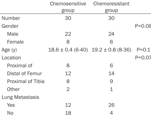
[image:2.612.90.341.98.293.2]A total 60 of patients’ specimens were used in this study (Table 1). All patients received the same multidrug chemotherapy before surgery. The 60 primary osteosarcoma tissues were obtained from patients who underwent com-plete resection at the Shanghai Tenth People’s Hospital between 2006 and 2014. Informed consents were obtained from all patients. After preoperative chemotherapy, the tumors were
Table 1. Clinical parameters of osteosarcoma patients enrolled in this study
Chemosensitive
group Chemoresistant group
Number 30 30
Gender P=0.08
Male 22 24
Female 8 6
Age (y) 18.6 ± 0.4 (6-40) 19.2 ± 0.8 (8-36) P=0.1
Location P=0.07
Proximal of 8 6 Distal of Femur 12 14
Proximal of Tibia 8 9
Other 2 1
Lung Metastasis
Yes 12 26
No 18 4
Follow-up time (m) 25.0 ± 1.2 (6-96) 22.4 ± 1.4 (3-80) P=0.55
resistant osteosarcoma cell line MG63/DXR and its corresponding MG63 parental cell line using micro-array analysis. We then validated the expression of some randomly select-ed differentially expressselect-ed genes and predicted their possible functions or molecular mechanism using bioinfor-matics. These findings may provide critical understanding to the mecha-nism of drug-resistance in osteosar-coma, and may reveal new therapeu-tic targets.
Materials and methods
Cell lines and cell culture
6256 Int J Clin Exp Pathol 2017;10(6):6254-6267 resected, and an expert panel of pathologists
reviewed the histology. If the percentage of tumor necrosis was greater than 90%, the patients were defined as good responders, and if the percentage of tumor necrosis was less than 90%, the patients were classified as poor responders.
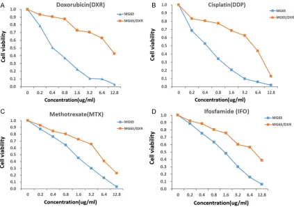
CCK-8 assay
Cell suspensions were prepared, and the num-ber of cells was counted using a cell counting board. Cells were plated in 96-well plates at a density of 2,000 cells per well. After 24 hours, doxorubicin (DXR), cisplatin (DDP), methotrex-ate (MTX), orifosfamide (IFO) was added at
dif-ferent concentrations. Each concentration was repeated in triplicate. Meanwhile, the same vol-ume of conditioned medium was added to the control wells. After a 48-h incubation at 37°C, 10 μL of CCK-8 was added to each well for another 2-h. The absorbance value (OD) at 450 nm was measured using an enzyme-linked immunological detector to calculate the viabili-ty rate and draw the related curves. The results shown are representative results from three independent experiments (Table 3).
RNA isolation, labeling and array hybridization
Total RNA was extracted from the three groups of paired doxorubicin-resistant osteosarcoma MG63/DXR cells and the parental MG63 cells with TRIzol reagent (Invitrogen) according to the manufacturer’s protocols. The concentration, purity, and integrity of the RNA samples were determined by the ratio of UV absorbance at 260 nm to 280 nm and by electrophoresis. Total RNA was repurified with an RNeasy MinElute Kit according to manufacturer’s proto-cols (Qiagen, Dalian, China), and 5 µg of RNA was then used for GeneChip analysis. cDNA pre- paration, hybridization to the human mRNA GeneChips (Arraystar Inc., MD, USA) and scan-ning (Axon GenePix 4000B) of the arrays were conducted according to manufacturer’s proto-cols (https://www.arraystar.com). The microar-ray analysis was performed by KangChen Bio-tech (Shanghai, China).
Bioinformatics analysis
GO analysis was applied to analyze the primary functions of the differentially expressed genes according to the GO database (www.geneontol-ogy.org). Pathway analysis was used to deter-mine the significant pathways of the different genes according to the KEGG database (Kyoto Encyclopedia of Genes and Genomes, http:// www.genome.jp/kegg).
Real-time RT-PCR
[image:3.612.91.288.85.417.2]Total RNA from cultured cell lines and human tissues was isolated using TRIzol reagent (Invitrogen) according to the manufacturer’s protocols. Primers (Table 2) for real-time RT- PCR were designed using Primer Express v 2.0 software (Applied Biosystems, Foster, CA, USA). RT was performed using the SuperScript First-Strand Synthesis System for RT-PCR
Table 2. Primers used for PCR validation
mRNA Forward and Reverse primer
RUNDC3B F: 5’ ACCAGTTATCAGCAGAAGTTAGC 3’ R: 5’ GGCCAAGTAATGAGGGAGTATCT 3’
ABCB1 F: 5’ GGGATGGTCAGTGTTGATGGA 3’ R: 5’ GCTATCGTGGTGGCAAACAATA 3’
RASGRP2 F: 5’ CAGCCTAATCGACATAGACAGC 3’ R: 5’ GGACATCTTGCGCTTTTTCTG 3’
ARMCX2 F: 5’ GCGGGGATAGTGATAGGGG 3’ R: 5’ TCGATTGTGAATCCGGCTCTTA 3’
CRYAB F: 5’ AGGTGTTGGGAGATGTGATTGA 3’ R: 5’GGATGAAGTAATGGTGAGAGGGT 3’
C3orf14 F: 5’ AGAAAAGGCATCTCAACTCCAAA 3’ R: 5’ GAAGTGGGTGAATCCTGGTCT 3’ C8orf48 F: 5’ CAACAGAAATCACTCGGGTATCA 3’
R: 5’ CCTCTACATCCAATCCAAGATGC 3’
MAGEH1 F: 5’GAGATCCGAAGAAGATCGTCAC 3’ R: 5’ AGCATCGGTTACGAACCCTTG 3’
LSP1 F: 5’ AGGACCGAGTCCCTAAACCG 3’ R: 5’ CTGGGTGTATTGTTCCAGCCA 3’
FABP5 F: 5’ TGGCCAAGCCAGATTGTATCAT 3’ R: 5’ GATGCTGAACCAATGCACCA 3’
ADAM22 F: 5’ GCAGACGCCTCATTGATGGA 3’ R: 5’ GTCGTGCCGACTTTCGTCTT 3’
GAPDH F: 5’ CATGAGAAGTATGACAACAGCCT 3’ R: 5’ AGTCCTTCCACGATACCAAAGT 3’
Table 3. IC50 for MG63 and MG63/DXR
IC50 (ug/ml) MG63 MG63/DXR R factor Doxorubicin (DXR) 0.4 10.2 23.2 Cisplatin (DDP) 0.4 5.2 13
Methotrexate (MTX) 1.2 4.4 3.7
[image:3.612.92.287.449.518.2](Invitrogen) according to the manufacturer’s protocol. Real-time PCR was performed using SYBR Green I (Applied Biosystems). The data were normalized to the arithmetic mean of housekeeping gene GAPDH and calculated using the 2-ΔΔCT method. The primer sequenc-es are listed in Table 2.
Western blot
Cells were collected, and cell lysis buffer (ThermoFisher Scientific, California, USA) sup-plemented with the appropriate protease inhib-itors (Invitrogen Inc, CA, USA) was used accord-ing to the manufacturer’s protocol to extract total protein. After the protein concentration was determined using a NanoDrop 2000 micro-spectrophotometer, 5 × loading buffer was added and mixed to the appropriate volume of sample. Then, the samples were boiled for 10 min at 100°C until the protein was fully dena-tured, after which samples were stored at -20°C. Equal quality of proteins as well as
[image:4.612.96.519.73.370.2]pro-tein markers was separated using sodium dodecyl sulfate polyacrylamide gel electropho-resis (SDS-PAGE) (Beyotime Biotechnology Co., Shanghai, China), after which the proteins in the gel were transferred onto a poly-vinylidene fluoride (PVDF) membrane (Merck Millipore Corporation, Billerica, Massachusetts, USA). After blocking with 5% skim milk for 1 h, the PVDF membranes were incubated with primary antibodies (Abcam, MA, USA) overnight at 4°C. The following day, the supernatant was removed, and the samples were washed three times with Tris-buffered saline containing tween (TBST). Then, the membranes were incu-bated with secondary antibodies (Abcam, MA, USA) at room temperature for 1 h and washed three times with TBST. Finally, the PVDF mem-branes were incubated with an enhanced che-miluminescence (ECL) working solution in a darkroom. The light emission was detected by X-ray to determine the presence of a protein band, which can reflect the relative expression of the protein.
Figure 1. CCK-8 assay. The CCK-8 assay was performed to verify that MG63/DXR cells were more resistant to doxo
6258 Int J Clin Exp Pathol 2017;10(6):6254-6267
Statistical analysis
The expression levels of mRNAs that were dif-ferentially expressed between human doxorubi-cin-resistant osteosarcoma MG63/DXR cells
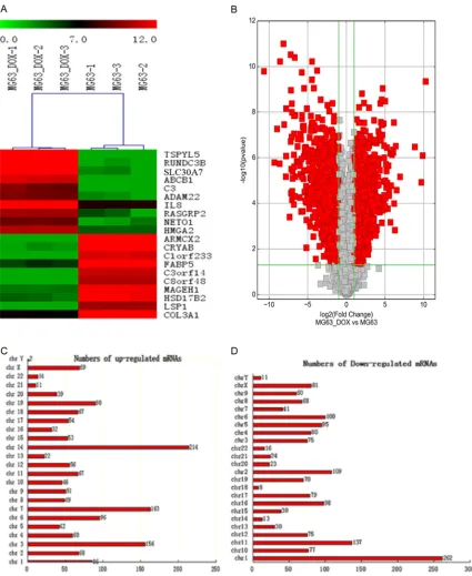
and the parental MG63 cells were compared by the paired, two-tailed t-test using the SPSS 20.0 software package (SPSS, Chicago, IL). Overall survival was calculated by Kaplan-Meier survival analysis and compared using the log-Figure 2. Differentially expressed mRNAs and their chromosome distribution. A. Differentially expressed mRNAs shown in the heat map. “Red” indicates high relative expression, and “green” indicates low relative expression. B.
Differentially expressed mRNAs as shown in the volcanic map. The scatter plot is a visualization method used for
[image:5.612.92.517.74.593.2]rank test. All of the data are shown as the means ± SD of three independent experiments. P<0.05 was considered statistically significant.
Results
CCK-8 assay
Drug resistance of the MG63/DXR cell line was identified by comparing the IC50 values in MG63/DXR cells with those in the MG63 cell line. As shown in Figure 1, when exposed to doxorubicin for 48 h, the IC50 value in MG63/ DXR cells was 10.2 μg/ml, whereas that in MG63 cells was 0.4 μg/ml. In addition, when exposed to cisplatin, methotrexate or ifos-famide for 48 h, the IC50 values in MG63/DXR cells were 5.2 μg/ml, 4.4 μg/ml, and 8.6 μg/ml respectively, whereas those values in MG63 cells were 0.4 μg/ml, 1.2 μg/ml, and 1.5 μg/ml respectively. The resistance factors (R factor) were 23.2, 13, 3.7, and 5.7 for doxorubicin, cis-platin, methotrexate, and ifosfamide, respec-tively (Table 3). Obviously, the MG63/DXR cells were more resistant to the four common che-motherapy drugs than MG63 cells, laying a solid foundation for further study.
Differentially expressed mRNAs and their chro-mosome distribution
In total, 3,278 mRNAs in the paired MG63/ DXR and MG63 cells showed statistically sig-nificant differences in expression (P<0.05; fold-change >2). Among these differentially express- ed mRNAs, 1,607 mRNAs were found to be up-regulated more than 2-fold in MG63/DXR cells than in MG63 cells, while 1,671 were down-regulated more than 2-fold in MG63/DXR cells (P<0.05). We used a hierarchical cluster-ing analysis to arrange the samples into groups based on their expression levels, which allowed us to hypothesize the relationships among the identified genes (Figure 2A). The result- ing volcanic map shows the relationships of the differently expressed mRNAs (Figure 2B) between the samples. TSPYL5 (fold change: 1217.0948879) was the most up-regulated mRNA, and ARMCX2 (fold change: 1624.4826- 651) was the most down-regulated mRNA. Chromosomal localization was established to determine the chromosomal patterns of the overall differentially expressed mRNAs based on the microarray data (Figure 2C and 2D). The differentially regulated 3,278 mRNAs were dis-tributed to every chromosome. Among them,
chromosome 14 had the most up-regulated mRNAs (214) and chromosome 1 had the most down-regulated mRNAs (262). However, chromosomes Y and 8 contained the fewest number of up-regulated and down- regulated mRNAs, respectively (2 and 8), which implies that mRNAs transcribed from these chromo-somes could play a vital role in the occurrence of chemoresistance in OS.
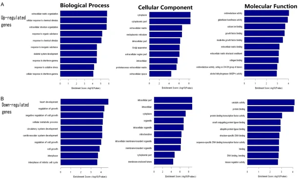
GO and pathway analysis
Based on the primary data, GO enrichment analysis further classified the differentially expressed mRNAs into three categories: bio-logical processes, cellular components and molecular functions. We found that the dysreg-ulated transcripts were associated with heart development and extracellular matrix organiza-tion (ontology: biological processes), intracellu-lar molecules and cytoplasm (ontology: celluintracellu-lar components), and catalytic activity and oxido-reductase activity (ontology: molecular func-tion) (Figure 3). There were 278 dysregulated genes involved in biological processes, 152 genes involved in cellular components and 125 genes involved in molecular functions.
Further pathway analysis, which is based on the differential genes compared to the KEGG database, found that 31 pathways were significantly enriched among the up-regulated mRNAs and that 25 pathways were enriched among the down-regulated mRNAs. ‘Glutathi- one metabolism-Homo sapiens (human)’ and ‘Transcriptional dysregulation in cancer-Homo sapiens (human)’ were the most enriched networks, respectively. In addition, some of these pathways, such as the apoptosis-related pathway, TNF signaling pathway, and chemo-kine signaling pathway, have been reported to be involved in the development of drug resis-tance in osteosarcomas [27-30] (Figure 4). In addition, some novel pathways have been correlated to the occurrence of chemoresis-tance, including the NOD-like receptor signaling pathway and the RIG-I-like receptor signaling pathway.
Real-time RT-PCR validation and Western blot validation
6260 Int J Clin Exp Pathol 2017;10(6):6254-6267 Figure 3. GO analysis of the dysregulated mRNAs. The results of GO analysis of up-regulated mRNAs from our dataset are shown in (A). The results of GO analysis
Figure 4. Pathway analysis of the differentially expressed profiles. The ‘Apoptosis related pathway’ (A) and “NOD-like receptor signaling pathway” (B) are associ
-ated with chemoresistance of osteosarcoma. The genes labeled “yellow” are differentially expressed between MG63/DXR and MG63 cells and are involved in the
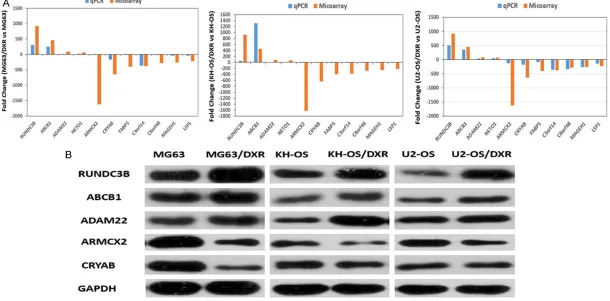
6262 Int J Clin Exp Pathol 2017;10(6):6254-6267 Figure 5. Differential expression of mRNAs was validated by qPCR and WB. Comparison of the microarray data and quantitative real-time PCR results. A. Eleven dif
vs MG63/DXR, KH-OS vs KH-OS/DXR, U2-OS vs U2-OS/DXR); these methods are the gold standards for data verification. The selected mRNAs included 4 up-regulated and 7 down-regulated mRNAs in MG63/DXR cells. As shown in Figure 5A, these data supported a strong consistency between the qRT-PCR results and microarray data. The results showed that RUNDC3B, ABCB1, ADAM22 and NETO1 were up-regulated and that ARMCX2, CRYAB, FAB- P5, C3orf14, C8orf48, MAGEH1, and LSP1 were down-regulated in the doxorubicin-resis-tant cells compared with their corresponding parental cells (MG63 vs MG63/DXR, KH-OS vs KHOS/DXR, U2-OS vs U2-OS/DXR).
Western blotting was used to verify the accu-racy of the microarray at the protein level. We randomly choose up-regulated mRNAs, including RUNDC3B, ABCB1, and ADAM22, and down-regulated mRNAs, including ARMCX2 and CRYAB. The results showed that the expression of the five selected genes (including the classi-cal multi-drug resistance related gene ABCB1) were all significantly dysregulated in the paired resistant and sensitive osteosarcoma cell lines, which reflects the results of the microarray (Figure 5B).
Potential clinical significance of RUNDC3B in osteosarcoma
To explore the potential clinical significance of these selected genes in osteosarcoma, the
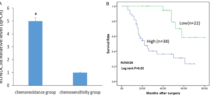
expression level of RUNDC3B was examined using real-time RT-PCR in 60 primary osteosar-coma tissue samples, which were divided into two groups: the chemosensitivity group (N=30) and chemoresistance group (N=30). The base-line between the two groups was comparable (Table 1). We used the median expression lev-els of RUNDC3B as a cut-off to stratify the 60 patients into the RUNDC3B-high group (n=38) and the RUNDC3B-low group (n=22). As shown in Figure 6A, our data indicated that RUNDC3B expression in the chemoresistance group was approximately five fold greater than that in the chemosensitivity group. In addition, Figure 6B
shows that patients with lower expression of RUNDC3B may survive longer than those with higher expression (52.6 ± 1.4 months vs 22.2 ± 1.8 months), which suggests that RUNDC3B may be a biomarker to predict the chemothera-peutic response and prognosis of osteosarco-ma patients. However, this theory should still be examined and verified with more methods and more clinical samples of osteosarcoma.
Discussion
[image:10.612.95.522.70.266.2]6264 Int J Clin Exp Pathol 2017;10(6):6254-6267 osteosarcoma has been extensively
investigat-ed from different aspects in molecular biology [3]. Multidrug resistance mechanism in OS include the following [25]: 1) increased efflux of drugs through ATP-binding cassette transport-ers, 2) decreased drug accumulation in the cell mediated by lower RFC, 3) contribution of PKC in drug efflux, 4) APE1- or ERCC-mediated repair of DNA damage, 5) inhibition of the activity and structural changes of DNA topoisomeraseII, 6) detoxification in the cell by GSTP1, 7)inhibition of apoptosis by p53, Bcl-2, or miRNAs, and 8) osteosarcoma stem cells.
Many important genes have been reported to be involved in the drug resistance of osteosar-coma. For example, Jia M et al. [26] found that Trps1 was associated with the multidrug resis-tance of osteosarcoma by regulating MDR1 gene expression. He Z et al. [27] found the overexpression of MRP4 was related to multi-drug resistance in osteosarcoma cells. Ron- cuzzi L et al. [28] found that HIF-1α activation was involved in the doxorubicin resistance of human osteosarcoma cells. Wang JJ et al. [29] reported a relationship between RFC gene expression and the intracellular drug concen-tration in methotrexate-resistant osteosarco-ma cells. Wang D et al. [30] found that huosteosarco-man apurinic endonuclease 1 (APE1) could en- hance the sensitivity of osteosarcoma to DNA-damaging agents.
To overcome and reverse multidrug resistance in OS, many methods targeting key molecules or signal transduction pathways have been established, including siRNA, small bioactive molecules, and extracts based on Chinese medicine. For example, Fanelli M et al. [31] found that targeting ABCB1 and ABCC1 with their specific inhibitor CBT-1 can overcome drug resistance in osteosarcoma. Duan Z et al. [32] found that A-770041, a small biological mole-cule, could reverse paclitaxel and doxorubicin resistance in osteosarcoma cells. Wang Z et al. [33] found that diallyltrisulfide (DATS) has sig-nificant anti-cancer effects on human osteo-sarcoma cells by reversion of P-glycoprotein-mediated drug resistance. Liu T et al. [34] found that targeting ABCB1 (MDR1) in multi-drug-resistant osteosarcoma cells using the CRISPR-Cas9 system could reverse drug resistance. To explore changes in the comprehensive gene profile between the doxorubicin-resistant
and doxorubicin-sensitive human osteosarco-ma cell lines MG63/DXR and MG63, the mRNA expression profiles of these cells were investi-gated using a microarray analysis. We analyzed three sets of human primary MG63/DXR osteosarcoma drug-resistant cells and their MG63 parental drug-sensitive cells and identi-fied (fold change >2.0) that a total of 3,278 mRNAs were differently expressed in drug-resistant MG63/DXR cells, including 1,607 up-regulated and 1,671 down-up-regulated mRNAs relative to the expression in MG63 cells. Ad- ditionally, the 3,278 differentially regulated mRNAs were distributed along every chromo-some. There were 11 mRNAs (4 up-regulated and 7 down-regulated) randomly chosen to undergo qRT-PCR and Western blotting to vali-date the consistency of our results in three paired resistant and doxorubicin-sensitive osteosarcoma cell lines (MG63 vs MG63/DXR, KH-OS vs KH-OS/DXR, U2-OS vs U2-OS/DXR). In addition, we examined the expression of RUNDC3B in 30 pairs of osteo-sarcoma patient samples to explore the poten-tial clinical significance of this gene.
that were differentially expressed in three dif-ferent drug-resistant malignant melanoma cell lines and that CRYAB is a strong influence in developing resistance against DNA-damaging drugs. However, there are few reports about the function of these genes in the chemoresistance of osteosarcoma. In our study, we found that these genes were stably dysregulated in the paired drug-resistant and drug-sensitive osteo-sarcoma cell lines and further revealed the potential clinical significance of RUNDC3B in osteosarcoma patients. It is possible that these genes are also important for the development of drug resistance in OS.
By using bioinformatics analyses such as GO and pathway analyses, we identified the biologi-cal functions that are enriched among the differentially expressed mRNAs. GO analysis revealed that these genes were primarily involved in cell components and basic meta-bolic processes, which may suggest the impor-tance of metabolic processes in regulating the chemoresistance of osteosarcoma. Pathway analysis showed that there were 31 pathways corresponding to all the up-regulated tran-scripts and 25 pathways corresponding to all the down-regulated transcripts. Among these pathways, classical signaling pathways such as apoptosis-related pathways, the TNF-signaling pathway, and chemokine signaling pathways have been reported to be involved in the acqui-sition of drug resistance in osteosarcoma, which may indicate the reliability of our results. In addition, we have identified some obscure pathways that may be involved in drug resis-tance of osteosarcoma, such as the NOD-like receptor signaling pathway and RIG-I-like recep-tor signaling pathway, which may provide novel directions in researching the underlying mecha-nism of resistance.
Our present study shows a set of mRNAs with differential expression in the human doxorubi-cin-resistant osteosarcoma cell line MG63/ DXR and its sensitive parental cell line MG63. Furthermore, our study revealed that the up-regulated genes RUNDC3B and ADAM22 as well as the down-regulated genes ARMCX2 and CRYAB may be involved in the regulation of drug sensitivity of osteosarcoma cell lines. In addition, we identified some novel pathways related to the development of chemoresis-tance, including the NOD-like receptor signaling
pathway. Furthermore, RUNDC3B may be a potential biomarker to predict the chemothera-peutic response and prognosis of osteosarco-ma patients. These results provide the ground-work for future studies. A deeper understand- ing of these transcripts and their role in the for-mation of osteosarcoma chemoresistance are necessary to discover possible novel targets for reversing multi-drug resistance.
Acknowledgements
This work was supported by a Grant from The National Natural Science Foundation of China (NSFC No. 81572630).
Disclosure of conflict of interest
None.
Address correspondence to: Dr. Chun-Lin Zhang,
Department of Orthopaedic Surgery, The Tenth People’s Hospital Affiliated to Tongji University, 301
Yanchang Middle Road, Shanghai 200072, PR
China. Tel: +86 13761904091; Fax: +86
13761-904091; E-mail: shzhangchunlin123@163.com References
[1] Luetke A, Meyers PA, Lewis I, Juergens H. Os-teosarcoma treatment-where do we stand? A
state of the art review. Cancer Treat Rev 2014;
40: 523-32.
[2] Anderson ME. Update on survival in
osteosar-coma. Orthop Clin North Am 2016; 47: 283-92.
[3] Ferrari S, Serra M. An update on chemotherapy for osteosarcoma. Expert Opin Pharmacother 2015; 16: 2727-36.
[4] Wu Q, Yang Z, Nie Y, Shi Y, Fan D.
Multi-drug resistance in cancer chemotherapeutics: mechanisms and lab approaches. Cancer Lett 2014; 347: 159-66.
[5] Vtorushin SV, Khristenko KY, Zavyalova MV, Perelmuter VM, Litviakov NV, Denisov EV,
Dule-sova AY, Cherdyntseva NV. The phenomenon of
multi-drug resistance in the treatment of ma-lignant tumors. Exp Oncol 2014; 36: 144-56. [6] Avner BS, Fialho AM, Chakrabarty AM.
Over-coming drug resistance in multi-drug resistant cancers and microorganisms: a conceptual framework. Bioengineered 2012; 3: 262-70. [7] Krasnov GS, Dmitriev AA, Sadritdinova AF,
6266 Int J Clin Exp Pathol 2017;10(6):6254-6267 [8] Hall MD, Handley MD, Gottesman MM. Is
resis-tance useless? Multidrug resisresis-tance and
col-lateral sensitivity. Trends Pharmacol Sci 2009;
30: 546-56.
[9] Ween MP, Armstrong MA, Oehler MK, Ric
-ciardelli C. The role of ABC transporters in ovar -ian cancer progression and chemoresistance. Crit Rev Oncol Hematol 2015; 96: 220-56. [10] Silverton L, Dean M, Moitra K. Variation and
evolution of the ABC transporter genes ABCB1,
ABCC1, ABCG2, ABCG5 and ABCG8: implica -tion for pharmacogenetics and disease. Drug Metabol Drug Interact 2011; 26: 169-79. [11] Robey RW, Massey PR, Amiri-Kordestani L,
Bates SE. ABC transporters: unvalidated thera-peutic targets in cancer and the CNS. Antican-cer Agents Med Chem 2010; 10: 625-33. [12] Chen Z, Shi T, Zhang L, Zhu P, Deng M, Huang
C, Hu T, Jiang L, Li J. Mammalian drug efflux transporters of the ATP binding cassette (ABC)
family in multidrug resistance: a review of the past decade. Cancer Lett 2016; 370: 153-64. [13] Zhang C, Zhao Y, Zeng B. Enhanced
chemosen-sitivity by simultaneously inhibiting cell cycle progression and promoting apoptosis of drug-resistant osteosarcoma MG63/DXR cells by targeting Cyclin D1 and Bcl-2. Cancer Biomark 2012; 12: 155-67.
[14] Duan Z, Lamendola DE, Penson RT, Kronish
KM, Seiden MV. Overexpression of IL-6 but not
IL-8 increases paclitaxel resistance of U-2OS
human osteosarcoma cells. Cytokine 2002; 17: 234-42.
[15] McManus MM, Weiss KR, Hughes DP. Under -standing the role of Notch in osteosarcoma.
Adv Exp Med Biol 2014; 804: 67-92.
[16] Li C, Shi X, Zhou G, Liu X, Wu S, Zhao J. The canonical Wnt-beta-catenin pathway in devel -opment and chemotherapy of osteosarcoma.
Front Biosci 2013; 18: 1384-91.
[17] Li L, Liang S, Wasylishen AR, Zhang Y, Yang X, Zhou B, Shan L, Han X, Mu T, Wang G, Xiong S.
PLA2G16 promotes osteosarcoma metastasis and drug resistance via the MAPK pathway.
Oncotarget 2016; 7: 18021-35.
[18] Yuryev A. Gene expression profiling for target -ed cancer treatment. Expert Opin Drug Discov 2015; 10: 91-9.
[19] Nannini M, Pantaleo MA, Maleddu A, Astolfi A, Formica S, Biasco G. Gene expression profiling
in colorectal cancer using microarray
technolo-gies: results and perspectives. Cancer Treat
Rev 2009; 35: 201-9.
[20] Kwiatkowski P, Wierzbicki P, Kmieć A, Godlews -ki J. DNA microarray-based gene expression
profiling in diagnosis, assessing prognosis and
predicting response to therapy in colorectal cancer. Postepy Hig Med Dosw 2012; 66:
330-8.
[21] Zhao Y, Zhang CL, Zeng BF, Wu XS, Gao TT, Oda
Y. Enhanced chemosensitivity of drug-resistant
osteosarcoma cells by lentivirus-mediated Bcl-2 silencing. Biochem Biophys Res Commun 2009; 390: 642-7.
[22] Susa M, Iyer AK, Ryu K, Hornicek FJ, Mankin H, Amiji MM, Duan Z. Doxorubicin loaded poly-meric nanoparticulatedelivery system to over-come drug resistance in osteosarcoma. BMC Cancer 2009; 9: 399.
[23] Yamamoto N, Tsuchiya H. Chemotherapy for osteosarcoma-where does it come from? What is it? Where is it going? Expert Opin Pharmaco
-ther 2013; 14: 2183-93.
[24] Xiao X, Wang W, Wang Z. The role of chemo -therapy for metastatic, relapsed and refractory osteosarcoma. Paediatr Drugs 2014; 16: 503-12.
[25] Li S, Sun W, Wang H, Zuo D, Hua Y, Cai Z. Re -search progress on the multidrug resistance mechanisms of osteosarcoma chemotherapy
and reversal. Tumour Biol 2015; 36: 1329-38.
[26] Jia M, Hu J, Li W, Su P, Zhang H, Zhang X, Zhou G. Trps1 is associated with the multidrug resis -tance of osteosarcoma by regulating MDR1
gene expression. FEBS Lett 2014; 588:
801-10.
[27] He Z, Hu B, Tang L, Zheng S, Sun Y, Sheng Z, Yao Y, Lin F. The overexpression of MRP4 is re -lated to multidrug resistance in osteosarcoma
cells. J Cancer Res Ther 2015; 11: 18-23. [28] Roncuzzi L, Pancotti F, Baldini N. Involvement
of HIF-1alpha activation in the doxorubicin re-sistance of human osteosarcoma cells. Oncol
Rep 2014; 32: 389-94.
[29] Wang JJ, Li GJ. Relationship between RFC gene
expression and intracellular drug concentra-tion in methotrexate-resistant osteosarcoma cells. Genet Mol Res 2014; 13: 5313-21. [30] Wang D, Luo M, Kelley MR. Human apurinic en
-donuclease 1 (APE1) expression and
prognos-tic significance in osteosarcoma: enhanced
sensitivity of osteosarcoma to DNA damaging agents using silencing RNA APE1 expression
inhibition. Mol Cancer Ther 2004; 3: 679-86.
[31] Fanelli M, Hattinger CM, Vella S, Tavanti E, Mi -chelacci F, Gudeman B, Barnett D, Picci P,
Ser-ra M. Targeting ABCB1 and ABCC1 with their specific inhibitor CBT-1(R) can overcome drug
resistance in osteosarcoma. Curr Cancer Drug
Targets 2016; 16: 261-74.
[32] Duan Z, Zhang J, Ye S, Shen J, Choy E, Cote G, Harmon D, Mankin H, Hua Y, Zhang Y, Gray NS, Hornicek FJ. A-770041 reverses paclitaxel and doxorubicin resistance in osteosarcoma cells.
BMC Cancer 2014; 14: 681.
[33] Wang Z, Xia Q, Cui J, Diao Y, Li J. Reversion of
P-glycoprotein-mediated multidrug resistance
by diallyl trisulfide in a human osteosarcoma
cell line. Oncol Rep 2014; 31: 2720-6. [34] Liu T, Li Z, Zhang Q, De Amorim Bernstein K,
Z. Targeting ABCB1 (MDR1) in multi-drug resis -tant osteosarcoma cells using the CRISPR-Cas9 system to reverse drug resistance.
Onco-target 2016; 7: 83502-13.
[35] Balaguer TM, Gómez-Martínez A, García-Mo -rales P, Lacueva J, Calpena R, Reverte LR,
Riquelme NL, Martinez-Lacaci I, Ferragut JA,
Saceda M. Dual regulation of P-glycoprotein expression by trichostatin A in cancer cell lines. BMC Mol Biol 2012; 30: 13-25.
[36] McCartan D, Bolger JC, Fagan A, Byrne C, Hao Y, Qin L, McIlroy M, Xu J, Hill AD, Gaora PÓ, Young LS. Global characterization of the SRC-1
transcriptome identifies ADAM22 as an ER-in -dependent mediator of endocrine-resistant breast cancer. Cancer Res 2012; 72: 220-9. [37] Kirby TJ, Walton RG, Finlin B, Zhu B, Unal R,
Rasouli N, Peterson CA, Kern PA. Integrative mRNA-microRNA analyses reveal novel inter-actions related to insulin sensitivity in human
adipose tissue. Physiol Genomics 2016; 48:
145-53.
[38] Bolger JC, Young LS. ADAM22 as a prognostic and therapeutic drug target in the treatment of endocrine-resistant breast cancer. Vitam Horm 2013; 93: 307-21.
[39] Zeller C, Dai W, Steele NL, Siddiq A, Walley AJ, Wilhelm-Benartzi CS, Rizzo S, van der Zee A,
Plumb JA, Brown R. Candidate DNA
methyla-tion drivers of acquired cisplatin resistance in ovarian cancer identified by methylome and expression profiling. Oncogene 2012; 31:
4567-76.
[40] Wittig R, Nessling M, Will RD, Mollenhauer J,
Salowsky R, Münstermann E, Schick M, Helm-bach H, Gschwendt B, Korn B, Kioschis P, Lich-ter P, Schadendorf D, Poustka A. Candidate genes for cross-resistance against