Polycomb response element-binding sites in the MDR of
CLL: Potential tumor suppressor regulation
Christine E. Cutucache1,Javeed Iqbal2,Philip J. Bierman3, Robert Gregory Bociek3, Dennis D. Weisenburger2, Shantaram S. Joshi4
1Department of Biology, University of Nebraska at Omaha, Omaha, USA
2Department of Pathology and Microbiology, University of Nebraska Medical Center, Omaha, USA
3Department of Internal Medicine Oncology/Hematology, University of Nebraska Medical Center, Omaha, USA 4Department of Genetics, Cell Biology and Anatomy, University of Nebraska Medical Center, Omaha, USA Email: [email protected]
Received 1 November 2012; revised 5 December 2012; accepted 10 January 2013
ABSTRACT
Chronic lymphocytic leukemia [CLL] is the most com- mon adult leukemia and is heterogeneous in clinical presentation. CLL cases present with various chro- mosomal aberrations, including 11q23, 14q32, 17p, and trisomy 12, with the most common abnormality being deletion of 13q14 [1]. Although monoallelic de- letion of 13q14 is common, there is a subset of pa- tients who have complete nullisomy at 13q14, a locus that has been hypothesized to contribute to CLL pa- thogenesis [2] due to loss of tumor suppressors [DLEU and miR-15a/16-1]. We hypothesized that deletion of both copies of 13q14 would lead to uncontrollable proliferation of CLL cells and a poor prognosis. We examined our 13q14 nullisomy for survival, treat- ment-free survival, lymphocyte doubling time, and the presence of lymphadenopathy. Furthermore, we compared the gene expression profiles between pa- tients with 13q14 monosomy, nullisomy, or normal karyotype. Our results suggest that patients with 13q nullisomy have a higher incidence of bulky lympha- denopathy [16.6% compared to 10% of monosomy patients], a higher frequency of lymphocyte doubling time [27.7% compared to 7.4% of monosomy pa- tients], and a higher rate of needing treatment [50% compared to 18.5% of monosomy patients]. We ob- served deletion of DLEU1 and HTR2A, consistent with a gene dosage effect, and observed PRE-binding sites on DLEU1. Patients with homozygous deletion of 13q14 had a worse prognosis compared to heterozy- gotes. Lastly, the DLEU1 locus is a possible “second hit” loss for CLL progression.
Keywords:Chronic Lymphocytic Leukemia; Gene
Expression; 13q14; Nullisomy; DLEU; Tolerogenic
1. INTRODUCTION
Chronic lymphocytic leukemia [CLL] is the most com- mon adult leukemia and is heterogenous in clinical pres- entation. Previous studies have focused on identifying prognostic markers such as CD38− and ZAP70-expres- sion, immunoglobulin mutational status, lymphocyte dou- bling time, presence of lymphadenopathy, and chromo- somal abnormalities to better group patients for therapy [1,2]. Patients with CLL may have none, one, or several of the following chromosomal abnormalities: del [11q23], del [13q14], del [17p], trisomy 12, and, less frequently, del [14q32]. However, there exists a cohort of patients with loss of both alleles at 13q14 [13q null] that is dis- tinct of patients with monosomy at this region [13q mono] and wild type [WT]. The 13q14 locus was hypothesized to contribute to CLL pathogenesis due to the loss of tu- mor suppressors, DLEU and miR-15a/16-1 [3-6]. We hypothesized that deletion of both copies of 13q14 would lead to uncontrollable proliferation of CLL cells and a poor prognosis. To test this hypothesis, we performed retrospective analyses with the data we accumulated from CLL patients cared for at the University of Ne-braska Medical Center [UNMC].
2. PATIENTS AND METHODS
2.1. Patient Samples
serial sections of frozen lymph node biopsies were ob- tained from the UNMC tissue bank. Immunohistochem- istry was used to stain each section for CD5 and CD19 to identify areas of >90% CLL cells. The CLL cells were microdissected out for the analyses.
2.2. Fluorescent in Situ Hybridization [FISH] and Microarray Analyses
Fluorescent in situ hybridization [FISH] was used to de- termine chromosomal abnormalities in CLL cells as pre- viously described [7]. The annotated patient data was examined to determine the association between time to treatment, treatment-free survival, lymphocyte doubling time, and the presence of lymphadenopathy. Microarray analyses were used to determine differential expression of genes in a region of 13q suggested to be deleted in CLL [8,9].
Previously, a tolerogenic signature was associated with poor prognosis in CLL [9-12]. Genes from the tolero- genic signature were examined among patients with 13q null, 13q mono, and WT cases. Next, the genes within the 13q locus that were included in our array were clus- tered by CLL patients WT at this locus compared to 13q mono and 13q null. These results would describe whe- ther there was a gene copy effect between CLL cases with losses at this region, which would identify a candi- date tumor suppressor gene[s].
2.3. Examination of Regulatory Elements
The sequence for DLEU1 was examined using Vista
[pipeline.lbl.gov/cgi-bin/gateway2] and the University of California, Santa Cruz [UCSC] Genome Bioinformatics [genome.ucsc.edu] Programs. The entire coding and non- coding sequence of DLEU1 was blasted for the poly- comb response elements [PRE], including: GAF, G10, PHO, and Z binding sites.
3. RESULTS AND DISCUSSION
3.1. Prognosis for CLL Patients with 13q Nullisomy
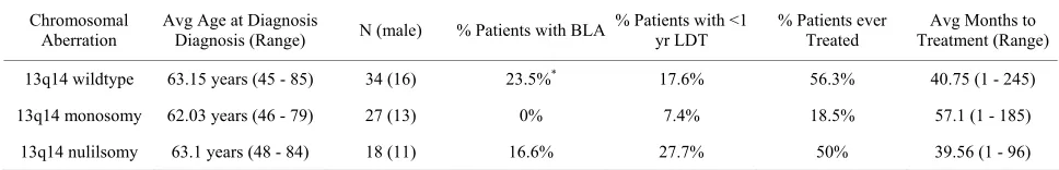
Our cohort of 13q null patients [n = 34] had divergent clinical characteristics from that of 13q mono CLL cases [n = 27]. 13q null patients had higher incidences of bulky
lymphadenopathy [16.6% null compared to 0% mono], shorter lymphocyte doubling time [27.7% null compared to 7.4% mono], and greater occurrences of needing treat- ment (50% null, 18.5% mono; Table 1). CLL patients
were broadly defined as having a poor prognosis if there was lymph node-involvement, a short lymphocyte dou- bling time [defined as less than a year for doubling], and if they required treatment. Although CLL patients with 13q deletion are generally classified as having a favor- able outcome, we suggest a divergence in prognosis be- tween patients with a heterozygous, compared to a ho- mozygous, deletion at this locus. Accordingly, patients with homozygous deletion of 13q14 might benefit from earlier treatment.
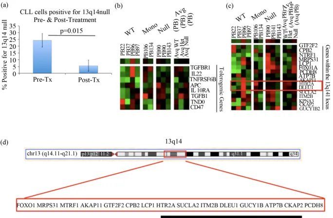
We next examined the percentage of CLL cells with 13q null to determine their susceptibility to treatment. The FISH results from pre-treatment and post-treatment for CLL patients showed a decrease in the number of CLL cells positive for 13q14 nullisomy from 18% pre- treatment to only 5% patients after treatment (p = 0.015;
Figure 1(a)). This 73% decline preliminarily suggests
that early treatment of patients with 13q null could be beneficial.
3.2. Gene Expression Profiles [GEP] of Patients with 13q Nullisomy
In order to determine whether there was a divergence in GEP between WT, 13q null, and 13q mono cases, mi- croarray data were examined. Previously, we reported that a tolerogenic signature is indicative of patients with an unfavorable outcome compared to those with a more indolent disease [8]. Furthermore, based on clinical pro- gnostic indicators, our results suggested that CLL pa- tients with 13q14 null had a less favorable prognosis compared to those mono or WT. As a consequence, we examined the tolerogenic GEP between CLL cases WT, mono, or null at 13q14 to determine if this expression signature correlated with clinical outcome. We hypothe- sized that patients with 13q14 null would have higher expression of the tolerogenic signature compared to 13q mono or WT cases.
[image:2.595.55.539.646.724.2]Changes in the GEP were compared between 13 null, 13q mono, and patients WT at the 13q14 locus (Figure 1(b)). There was an increase in the immunosuppressive
Table 1. Characteristics of CLL patients with 13q14 nullisomy compared to 13q14 heterozygous deletion.
Chromosomal Aberration
Avg Age at Diagnosis
Diagnosis (Range) N (male) % Patients with BLA
% Patients with <1 yr LDT
% Patients ever Treated
Avg Months to Treatment (Range)
13q14 wildtype 63.15 years (45 - 85) 34 (16) 23.5%* 17.6% 56.3% 40.75 (1 - 245)
13q14 monosomy 62.03 years (46 - 79) 27 (13) 0% 7.4% 18.5% 57.1 (1 - 185)
13q14 nulilsomy 63.1 years (48 - 84) 18 (11) 16.6% 27.7% 50% 39.56 (1 - 96)
Figure 1. Clinical and molecular characteristics of CLL patients with 13q14 nullisomy: (a) The change in the percentage of CLL cells positive for nullisomy at 13q14 pre- and post-treatment. The percentage of CLL cells positive for deletion at 13q14 as analyzed by FISH before and af- ter therapy. N = 5, P = 0.015; (b) Tolerogenic gene expression profiles between CLL patients WT at the 13q14 locus, heterozygous (13q mono), or bearing a homozygous deletion (13q null). The genes in our microarray within this region were compared between patients who are WT, have a heterozygous deletion, or with a homozygous deletion at this region. Cluster analysis was performed using Cluster 3.0 and Treeview software; (c) GEP of genes at the 13q14 locus among CLL patients who are WT, have a heterozygous deletion, or with a homozygous dele- tion at this region. The genes in our microarray within this region were compared between pa- tients who are WT, have a heterozygous deletion, or with a homozygous deletion at this region. Cluster analysis was performed using Cluster 3.0 and Treeview software; (d) Genes on 13q14 that were on our microarray chip (not an exhaustive list). Of the genes in the 13q14 region commonly deleted in CLL, the genes listed above were on our microarray. We were able to de- termine the gene expression profile for these select genes for CLL patients with both copies of 13q14 compared to those that were missing either one or both copies at this locus.
molecules APC and TGFβ1 in 13q null CLL cases when compared to 13q mono cases. APC, a member of the Wnt signaling pathway, was shown to regulate cell migration and adhesion and colorectal cancer is associated with mutations in this gene [13,14]. TGFβ1has been impli- cated in immunosuppression, in addition to cellular pro- liferation, differentiation, adhesion, and migration [15- 20].
3.3. Gene Expression in CLL Cases Nullisomy, Monosomy, or WT at 13q14
After determining a prognostic significance for complete loss of 13q14, the next objective was to identify whether a tolerogenic/immunosuppressive signature was differen- tially expressed in these cells. Supervised cluster analy- ses were performed on the genes included in our mi- croarray platform that were within the 13q14 locus [8; MWG Biotech, Germany, Human 10K oligo set A; Fig-
ure 1(c)]. The expected result was to observe a dosage
effect of genes within the deleted region at 13q14. To determine critical genes in the region of interest, we ex- pected patients WT at the locus to have high expression [red], those missing one copy [+/−] to have a lower ex- pression [lighter red or green], and individuals null at this region to have no expression of critical genes [black]. We anticipated the results of these analyses would de- termine a region that was commonly deleted in CLL that we could compare to other reports in the literature.
The expression of two genes was consistent with the predicted expression pattern; these were HTR2A and DL- EU1 (Figure 1(c)). The location of HTR2A and DLEU1
is depicted in Figure 1(d). 5-hydroxytryptamine [sero-
previously reported to participate in CLL [21-25]. Al- though there are only 15 published papers describing DLEU1 to date, early reports and observations show promise that the regulation of this lnRNA appears to have a great influence on CLL progression.
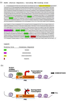
Although the role of DLEU1 in CLL is still elusive, we searched the sequence of this region to gain greater insight. A polycomb response element [PRE] conserved
sequence was identified, known to serve as an antagonist for epigenetic regulation of gene expression [26] within this region (Figure 2(a)). This region is a regulatory
switchable element that influences the architecture of chromatin and the expression of nearby genes [27,28]. In this regard, DLEU1 is potentially a docking site for the polyhomeotic [PHO] protein. This evolutionarily con- served, regulatory system was identified in Drosophila. The mammalian homolog of PHO in Drosophila is the
(a)
[image:4.595.169.432.200.606.2](b)
genome likely contribute to CLL progression.
transcription factor YY1[29]. Recently, a lnRNA was the link between copy number variation and a polycomb/ trithorax epigenetic switch in muscular dystrophy [26], and we suspect a similar switch may be occurring in CLL following the loss of regulation of the region con- taining DLEU1 (Figure 2(b)). This finding suggests a
potential negative feedback loop with the binding of YY1 to DLEU1thereby regulating gene expression. Therefore, this finding would explain the over decade-long conun- drum of observing a deletion in this region, but being unable to identify a clear point mutation or tumor sup- pressor gene.
In summary, this report describes an unfavorable prognosis for patients with biallelic deletion of 13q14, compared to 13q mono patients. However, the percent- age of CLL cells with 13q null decreased after treatment, suggesting that chemotherapy was effective at killing these malignant cells. GEP of patients with 13q null, compared to 13q mono, identified the overexpression APC and TGFβ1. Therefore, the upregulation of these immunoregulatory molecules in 13q null patients might lead to a greater immunosuppression in these cases.
The limitation of this study is the number of cases examined. A larger cohort is needed to conclusively de- termine the biological relevance of DLEU1 in CLL cases with 13q14 null, but hopefully this study will serve as a basis for additional in-depth analyses. Previously, a similar study with a large sample size [n = 323] showed that a higher percentage of 13q null cells were associated with a significantly shorter time to treatment [6]. Simi- larly, we suggest that CLL patients with 13q null have a worse prognosis than patients that are mono or WT at 13q14. Contrastingly, these data contradict those de- scribed earlier this year [30]. Garg et al. observed that the baseline characteristics between CLL cases with mono- or bi-allelic deletion of 13q differed only by ZAP70-expression and albumin levels [31]. This study differs from ours in that solely fluorescent in situ hy- bridization [FISH] was used to assess the genetic abnor- malities in CLL cells. Patients included in our study were first screened using both FISH and gene expression pro- filing. Therefore, this study presents a comprehensive picture as to the genetic abnormalities present within this frequently deleted region.
It will be difficult to prove a role for the consensus sequence of DLEU1 in CLL without engineering a dele- tion at that region both in CLL samples and an in vivo animal model. While this manuscript was in preparation, Lia et al. (2012) published a report regarding this precise question [31]. Transgenic mice engineered to have loss of 13q14 had a shorter life expectancy, similar to our data from patients described in this report [31; Table 1].
The data presented herein suggest that, first, CLL cases
show a heterozygous deletion at 13q14, and then a “sec- ond hit” or loss of the second allele [13q null] is neces- sary to take CLL from an indolent to an aggressive stage. Prospective studies are necessary to conclusively deter- mine this switch causing CLL cells to drive a more ag- gressive disease.
4. SUMMARY
The findings presented herein are significant in that they suggest a different way of classifying CLL cases with 13q null. However, it is important to note the limitations with the sample size described in this study. Based on this small sample size, we suggest that patients with the deletion of both copies of 13q14 potentially need therapy initiated earlier than their heterozygous counterparts and, thus, might require more frequent monitoring of lym- phocyte counts. The influence of DLEU1 in CLL was reported previously, and the microRNAs in this region have been studied extensively [2-5]. However, this is the first report suggesting that regulatory elements, specifi- cally PRE-sequences, of DLEU1 might contribute as a “second hit” to lead to a more aggressive disease pro- gression for patients with CLL.
5. ACKNOWLEDGEMENTS
Thanks to the CLL patients who so willingly donated cells for our study. Also, thank you to the clinicians and nurses at the University of Nebraska Medical Center for collecting these samples.
REFERENCES
[1] Mraz, M., Pospisilova, S., Malinova, K., Slapak, I. and Mayer, J. (2009) MicroRNAs in chronic lymphocytic leu- kemia pathogenesis and disease subtypes. Leukemia &
Lymphoma, 50, 506-509.
doi:10.1080/10428190902763517
[2] Kasar, S., Salerno, E., Yuan, Y., et al. (2012) Systemic in vivo lentiviral delivery of miR-15a/16 reduces malignan- cy in the NZB de novo mouse model of chronic lympho- cytic leukemia. Genes and Immunity, 13, 109-119. doi:10.1038/gene.2011.58
[3] Lerner, M., Harada, M., Loven, J., et al. (2009) DLEU2, frequently deleted in malignancy, functions as a critical host gene of the cell cycle inhibitory microRNAs miR- 15a and miR-16-1. Experimental Cell Research, 315, 2941-2952. doi:10.1016/j.yexcr.2009.07.001
[4] Klein, U., Lia, M., Crespo, M., et al. (2010) The DLEU2/ miR-15a/16-1 cluster controls B cell proliferation and its deletion leads to chronic lymphocytic leukemia. Cancer
Cell, 17, 28-40. doi:10.1016/j.ccr.2009.11.019
[5] Palamarchuk, A., Efanov, A., Nazaryan, N., et al. (2010) 13q14 deletions in CLL involve cooperating tumor sup- pressors. Blood, 115, 3916-3922.
Smoley, S.A., Rabe, K.G., Schwager, S.M., Sonbert, J.C., Slager, S.L. and Kay, N.E. (2010) A comprehensive eva- luation of the prognostic significance of 13q deletions in patients with B-chronic lymphocytic leukemia. British
Journal of Haematology, 148, 544-550.
[7] Dickinson, J.D., Joshi, A.D., Iqbal, J., Sanger, W., Bier- man, P.J. and Joshi, S.S. (2006) Genomic abnormalities in chronic lymphocytic leukemia influencing gene ex- pression by a gene dosing effect. International Journal of
Molecular Medicine, 17, 769-778.
[8] Joshi, A.D., Hegde, G.V., Dickinson, J.D., et al. (2007) ATM, CTLA4, MNDA, and HEM1 in high versus low CD38 expressing B-cell chronic lymphocytic leukemia.
Clinical Cancer Research, 13, 5295-5304.
doi:10.1158/1078-0432.CCR-07-0283
[9] Gilling, C.E., Mittal, A.K., Chaturvedi, N.K., et al. (2012) Lymph node-induced immune tolerance in chronic lym- phocytic leukemia: A role for caveolin-1. British Journal
of Haematology, 158, 216-231.
doi:10.1111/j.1365-2141.2012.09148.x
[10] Mittal, A.K., Iqbal, J., Nordgren, T.M., et al. (2008) Mo- lecular basis of proliferation/survival and migration of CLL in peripheral blood, bone marrow, and lymph nodes.
Blood ASH Annual Meeting, 112, 546.
[11] Mittal, A.K., Gilling, C.E., Iqbal, J., et al. (2009) Clinical heterogeneity of CLL: Role for immune dysregulation me- diated by the lymph node microenvironment. Blood ASH
Annual Meeting, 114, 1243.
[12] Gilling, C.E., Mittal, A.K., Nganga, V., Palmer, V.L., Wei- senburger, D.D., Bierman, P.J., Bociek, R.G., Swanson, P.C. and Joshi, S.S. (2010) Molecular determinants of lymph node microenvironment induced host immune tol- erance in CLL: Role for CAV1, PTPN6, and PKCβ in the process. Blood ASH Annual Meeting, 116, 1367.
[13] Fang, Z., Xiong, Y., Li, J., Zhang, W., Zhang, C. and Wan, J. (2012) APC gene deletions in gastic adenocarci- nomas in a Chinese population: A correlation with tumor progression. Clinical and Translational Oncology, 14, 60- 65. doi:10.1007/s12094-012-0762-x
[14] Valvezan, A.J., Zhang, F., Diehl, J.A. and Klein, P.S. (2012) Adenomatous polyposis coli (APC) regulates mul- tiple signaling pathways by enhancing glycogen synthase kinase-3 (GSK-3) activity. The Journal of Biological Che-
mistry, 287, 3823-3832. doi:10.1074/jbc.M111.323337
[15] Bommireddy, R. and Doetschman, T. (2007) TGFbeta1 and Treg cells: Alliance for tolerance. Trends in Molecular
Medicine, 13, 492-501.
doi:10.1016/j.molmed.2007.08.005
[16] Fogel-Petrovic, M., Long, J.A., Misso, N.L., Foster, P.S., Bhoola, K.D. and Thompson, P.J. (2007) Physiological concentrations of transforming growth factor beta1 selec- tively inhibit human dendritic cell function. International
Immunopharmacology, 20, 1924-1933.
doi:10.1016/j.intimp.2007.07.003
[17] Diaz-Valdes, N., Basagoiti, M., Dotor, J., et al. (2011) Induction of monocyte chemoattractant protein-1 and in- terleukin-10 by TGFbeta1 in melanoma enhances tumor infiltration and immunosuppression. Cancer Research, 71,
[18] Bobr, A., Igyarto, B.Z., Haley, K.M., Li, M.O., Flavell, R.A. and Kaplan, D.H. (2012) Autocrine/paracrine TGF- β1 inhibits langerhans cell migration. Proceedings of the
National Academy of Sciences of the USA, 109, 10492-
10497. doi:10.1073/pnas.1119178109
[19] Luo, H., Zhang, Y., Zhang, Z. and Jin, Y. (2012) The pro- tection of MSCs from apoptosis in nerve regeneration by TGFβ1 through reducing inflammation and promoting VEGF-dependent angiogenesis. Biomaterials, 33, 4277- 4287. doi:10.1016/j.biomaterials.2012.02.042
[20] Miron, R.J., Saulacic, N., Buser, D., Iizuka, T. and Scu- lean, A. (2012) Osteoblast proliferation and differentia- tion on a barrier membrane in combination with BMP2 and TGFβ1. Clinical Oral Investigations.
doi:10.1007/s00784-012-0764-7
[21] Liu, Y., Corcoran, M., Rasool, O., et al. (1997) Cloning of two candidate tumor suppressor genes within a 10 kb region on chromosome 13q14, frequently deleted in chro- nic lymphocytic leukemia. Oncogene, 15, 2463-2473. doi:10.1038/sj.onc.1201643
[22] Migliazza, A., Bosch, F., Komatsu, H., et al. (2001) Nu- cleotide sequence, transcription map, and mutation analy- sis of the 13q14 chromosomal region deleted in B-cell chronic lymphocytic leukemia. Blood, 97, 2098-2104. doi:10.1182/blood.V97.7.2098
[23] Wolf, S., Mertens, D., Schaffner, C., et al. (2001) B-cell neoplasia associated gene with multiple splicing (BCMS): The candidate B-CLL gene on 13q14 comprises more than 560 kb covering all critical regions. Human Molecular Ge-
netics, 10, 1275-1285. doi:10.1093/hmg/10.12.1275
[24] Rondeau, G., Moreau, I., Bezieau, S., et al. (2001) Com- prehensive analysis of a large genomic sequence at the putative B-cell chronic lymphocytic leukaemia (B-CLL) tumour suppresser gene locus. Mutation Research/Muta-
tion Research Genomics, 458, 55-70.
doi:10.1016/S0027-5107(01)00219-6
[25] Rowntree, C., Duke, V., Panayiotidis, P., et al. (2012) Deletion analysis of chromosome 13q14.3 and charac- terization of an alternative splice form of LEU1 in B cell chronic lymphocytic leukemia. Leukemia, 16, 1267-1275. doi:10.1038/sj.leu.2402551
[26] Cabianca, D.S., Casa, V., Bodega, B., et al. (2012) A long ncRNA linkes copy number variation to a polyco- mb/trithorax epigenetic switch in FSHD muscular dys-trophy. Cell, 149, 819-831.
doi:10.1016/j.cell.2012.03.035
[27] Ringrose, L. and Paro, R. (2004) Epigenetic regulation of cellular memory by the polycomb and trithorax group pro- teins. Annual Review of Genetics, 38, 413-443.
doi:10.1146/annurev.genet.38.072902.091907
[28] Schuettengruber, B., Chourrout, D., Vervoort, M., Leb- lanc, B. and Cavalli, G. (2007) Genome regulation by po- lycomb and trithorax proteins. Cell, 128, 735-745. doi:10.1016/j.cell.2007.02.009
[29] Simon, J.A. and Kingston, R.E. (2009) Mechanisms of polycomb gene silencing: Knowns and unknowns. Nature
[30] Garg, R., Wierda, W., Ferrajoli, A., Abruzzo, L., Pierce, S., Lerner, S., Keating, M. and O’Brien, S. (2012) The prognostic difference of monoallelic versus biallelic dele- tion of 13q in chronic lymphocytic leukemia. Cancer, 118, 3531-3537. doi:10.1002/cncr.26593