RC2 EXPRESSION IN CENTRAL NERVOUS SYSTEM WHITE
MATTER; AN ANALYSIS OF NEURAL STEM CELL DISTRIBUTION
DURING DEVELOPMENT
Timothy H.W. Balfour†, Ahmed M. Kamil† and Dr. Denis S. Barry*
Department of Anatomy, School of Medicine, Trinity Biomedical Sciences Institute, Trinity
College Dublin, Dublin 2, Ireland
†Corresponding Authors
Email:[email protected], [email protected] Both authors contributed to this paper equally
ABSTRACT
Introduction
Radial glial cells have many pivotal roles in the developing central nervous system.
Currently, they are recognised to have four main functions: to direct neuronal migration, to
act as neuronal progenitors, to encourage angiogenesis, and to coordinate the formation of
axon tracts in the white matter. In this study, we will investigate the fourth of those functions,
as we seek to quantify the number of radial processes crossing the white matter in the
developing spinal cord. We will then compare the number of processes between wild-type
specimens and animals missing genes encoding for proteins essential for normal axon
guidance, specifically Semaphorin6A and PlexinA4. This may help to determine whether
these signalling pathways are relevant in the developing spinal cord.
Methods and Materials
We examined cryosections from mouse embryos of age E15.5, which were stained to show
the traversing radial glial processes. We used a series of 50μm reference lines spaced 25μm
apart to quantify the number of processes at each level.
Results
We showed that there were especially large numbers of glial processes in the lateral columns
of white matter in all three phenotypes described. However, we found no link between the
number of axons and Semaphorin6A or PlexinA4 deficiency.
Discussion
Our results support conclusions made by previous studies, that many radial glial cells can be
seen in the developing spinal cord at a time when axon tracts are developing, suggesting a
possible role for these processes in their development. The fact that they are seen in
especially great numbers in the lateral columns may suggest which axon tracts they have a
role in guiding the development of.
Key Words: axonogenesis, development, radial glia, white matter patterning
INTRODUCTION
The History of Radial Glial Cells
Our understanding of radial glial cells has expanded greatly since their discovery in the late
19th century. Giuseppe Magini is the first to have identified and outlined these processes, which he did in two papers in 1887 and 1888, but it was nearly a century until some insight
into their function was provided. Wilhelm His, a contemporary of Golgi and Magini, had
originally hypothesised that they facilitated the migration of immature neurons in the
developing CNS, but this was only confirmed by Pasko Rakic in 1971 after his experiments
on monkey cerebella, where he observed that immature neurons bound to radial fibres during
migration (Bentivoglio and Mazzarello, 1999). More recent findings, however, have shown
that aside from aiding migration, radial glial cells may have a role as stem cells,
differentiating into not just glial cells, but neurons as well. This was first reported by a group
led by Paolo Malatesta, in 2000, when they used fluorescence-activated cell sorting to
demonstrate the neuronal fate of radial glial cells (Malatesta, Hartfuss and Götz, 2000).
The Function of Radial Glial Cells
Radial glial cells seem to have four main roles. Their first function is to aid neuronal
migration in the developing CNS. They can thus travel from germinal zones to their target
destination, where they can develop into functional neurons. This is a vital role in the
cerebrum especially, as which lamina the neuron occupies plays a large part in determining
its function (Rakic 1972). Secondly, they have relatively recently been recognised as
neuronal progenitors, as they may generate some of the neurons that they aid the migration
of. This demonstrates that neuronal and glial lineages are not always separate. Cells
performing this role are situated in the ventricular zones, and only a minority of radial glia in
the CNS display this function (Malatesta, Hartfuss and Götz, 2000). Another recently
discovered function is their role in brain angiogenesis, by both directly interacting with blood
vessels and modulating canonical Wnt signalling (Ma et al 2013). Finally, the function that
will be further explored in this paper is facilitating the organisation of white matter in the
spinal cord and cerebrum. In the spinal cord, they seem to form boundaries between the
developing axon tracts, preventing abnormal decussation and allowing them to form along
The Timing of Axon Tract Formation
In the adult human, the white matter is split into 4 bilateral principal columns. These are the
dorsal column, lateral column, ventral column and the ventromedial column. The columns are
further divided into several functional axon tracts, which radial glial cells may play a role in
the development of. These tracts include the corticospinal tract and the posterior column
pathways (Altman and Bayer, 2001). Generally, axons form these tracts by migrating from
the ventricular zone of the spinal cord, outwards towards the pial surface, and thus the
arrangement of an outer layer of white matter surrounding the inner grey matter is formed.
The different functional pathways develop at very different stages of development. In the
human embryo, the posterior column pathways develop early, and they are apparent by the
7th gestational week. This can be investigated by immunological staining by proteins such as
GAP-43, an indicator of axon growth. Immunoreactivity of GAP-43 is greatest in the dorsal
columns between the 10th and 14th weeks, showing that this is the peak period of tract
development (Clowry, Moss and Clough, 2005). In contrast, the corticospinal tract forms far
later. The axons are still developing and forming synapses until late into prenatal, and even
into early postnatal, development (Martin). Myelination of all tract axons continues into
postnatal development, until around 82 postconceptional weeks (Gao et al 2009).
The role of Radial glia in Axon Tract Formation
In the developing spinal cord, radial glial cells form a consistent scaffold along the entire
rostrocaudal axis. They arrange themselves with their cell bodies in the ventricular zone,
adjacent to the central canal, and an endfoot extends to the pial surface. They are arranged
perpendicular to the axis of the cord. It has been proposed that this morphology and arrangement enables them to assist in the formation of axon tracts in the white matter. Their
proposed function is to both guide the axon tracts along the correct pathways and to prevent
abnormal decussation. For example, in rats the dorsal midline glial septum stops premature
decussation of the corticospinal tract. Evidence for this function comes from numerous
sources. Firstly, electron microscopy has revealed that adhesions do form between radial glia
and the migrating axons. Also, vimentin staining demonstrates that radial glia are especially
well organised between embryological days 14 and 18 in rats, which is when the axon tracts
are forming at their greatest rate. The density of vimentin staining decreases after this period,
providing evidence that radial glia have started to differentiate, as they are no longer needed
Disorders Involving the Malformation of CNS White Matter
Deficiencies in the development of white matter can lead to a number of debilitating
disorders. First, there is the group of disorders known as leukodystrophy characterised by
abnormal myelination in the developing child, leading to gradually deteriorating motor and
sensory function. Symptoms include gait disturbances, abnormal movement patterns, declines
in language and concentration, and finally an early death (Kehrer et al 2014). Another
condition to consider is lissencephaly, which is caused by a defect in neuronal migration
affecting both the grey and white matter. In the extreme case, this symptom is known as
agyria, as the gyri and sulci are completely absent (Perez et al 2013). The defect in migration
usually occurs between the 12th and 24th weeks of gestation. Diagnosis is usually during
infancy, and symptoms include epilepsy and severely retarded motor and cognitive
development (Sasaki et al 2012).
The aim of this study is to quantify the number of radial processes present in different areas
of the developing mouse spinal cord in normal specimens, and compare this to mice with
mutations in the gene encoding for Semaphorin6A, and the gene encoding for PlexinA4, a
Semaphorin receptor. These proteins are usually vital for normal axon tract development, so
this may help to reveal whether or not the signalling pathway they form a part of is relevant
in the developing mouse spinal cord. Also, the location of certain developing axon tracts in
the spinal cord is known, so the differences in number of radial glial cells present between
axon tracts can be investigated.
MATERIALS AND METHODS
Animals
Embryonic day E 15.5 (of a 21-day gestation period) mice were obtained from the
Bioresource Unit, Trinity College Dublin. Embryos were removed by laparotomy following
anaesthesia using halothane, and a terminal overdose of sodium pentobarbital was
administered to pregnant dams. Embryos were fixed in 4% paraformaldehyde (PFA) for 24 h
at 4 °C. They were then cryoprotected in a 30% sucrose solution and snap-frozen in liquid
Immunohistochemistry
15-μm cryosections were used for all analyses,
and these were obtained using a Leica cryostat.
Sections were blocked in 10 mM PBS containing
20% normal goat serum (NGS) plus 0.05%
Triton-X 100 (Sigma, UK),and then incubated in
primary antibody in 10 mM PBS containing 1%
NGS plus 0.2% Triton-X 100 (Sigma) for 1 h at
room temperature. The primary antibody
solution consisted of 0.2% RC2, 1% NGS and
the remainder PBS. These preperations were
then blotted and left overnight at 4°C. Primary
antibody binding was detected by incubation
with secondary antibodies (1% NGS, 0.2%
Alexaphor IgM 488nm, remainder PBS) for 3 h
at room temperature. The microscope used was
an Olympus Bx51 upright fluorescent
microscope, with a mercury lamp attached, and
the images were displayed using CellSeus
software.
Quantification of process density in the spinal cord WM
For fibre density analysis, 15-μm
cryosections were cut from the rostral and caudal regions of the spinal cord, using a
Leica cryostat, in a coronal plane. The
analysis of radial glial process density in the
spinal cord WM was carried out using the
line intersect stereological approach
(Mayhew, 1992). This involves assessing the
number of times immunoreactive processes
The images above show our method for quantifying the number of radial glia in
each area of the white matter as described above. Fig 1.1 is from a
wild-type specimen, Fig 1.2 is from a PlexinA4-deficient sample, and Fig 1.3
is from a Semaphorin6A-deficient sample. PS=Pial Surface, CC=Central Canal, GM=Grey Matter, WM=White Matter, DH=Dorsal Horn, VH=Ventral
Horn, D=Dorsal, V=Ventral, ML=Midline.
Fig 1.1
Fig 1.2
intersect with a series of 50-μm reference lines placed centrally in the ventral, lateral and
dorsal WM, spaced 25-μm apart, and oriented in planes parallel to the pial surface. To place
these reference lines accurately, the each image was printed onto an A4 sheet of paper with a
scale factor of 250. This means that 50μm in the spinal cord was equivalent to 20mm on the
page, so a series of 20mm reference lines were drawn. These lines were drawn upon an
acetate cover placed over the image. The first reference line was placed lateral to the
developing ventral floor plate, while the last was placed in the dorsal WM, lateral to the
developing dorsal funiculus. Sections were taken from multiple levels within each spinal
cord. The number of processes crossing each reference line was noted, and an average was
taken for each numbered section for each of the three phenotypes.
RESULTS
Quantification of Radial Glial Processes
As expected, many radial glial processes were seen traversing the spinal cord. They stretched
from the central canal to the pial surface but there was an especially large number in the
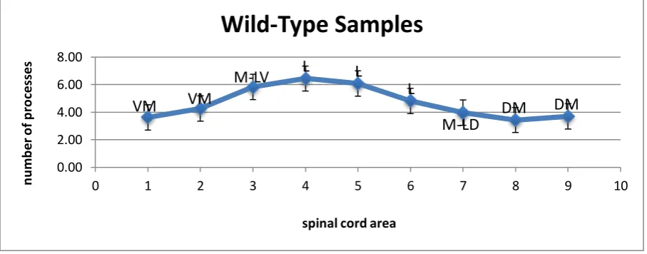
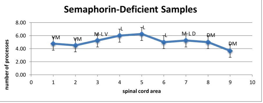
white matter. Figures 1.4, 1.5 and 1.6 below show the average number of glial processes seen
crossing the white matter in each section of the spinal cord for all three phenotypes. In each
case, the greatest number of processes was seen crossing in the lateral section. In the
wild-type specimens, the peak was seen in section four, with an average of 6.44 processes crossing
within the 50μm line. In the PlexinA4-deficient samples, the peak was in section 6 at 5.83
while in the Semaphorin6A-deficient specimens the peak was in section 5 at 6.25. The error
bars were derived by finding the standard deviation of the numbers used to generate the
average for each section of spinal cord. The average of these standard deviations was then
found to give a consistent size of error bar. For the wild type samples this came out to ±0.92,
for PlexinA4-deficient samples this was ±0.50, and for the Semaphorin6A-deficient
specimens this was ±0.84. Our full results can be seen summarised in table 1.1.
Comparison between Phenotypes
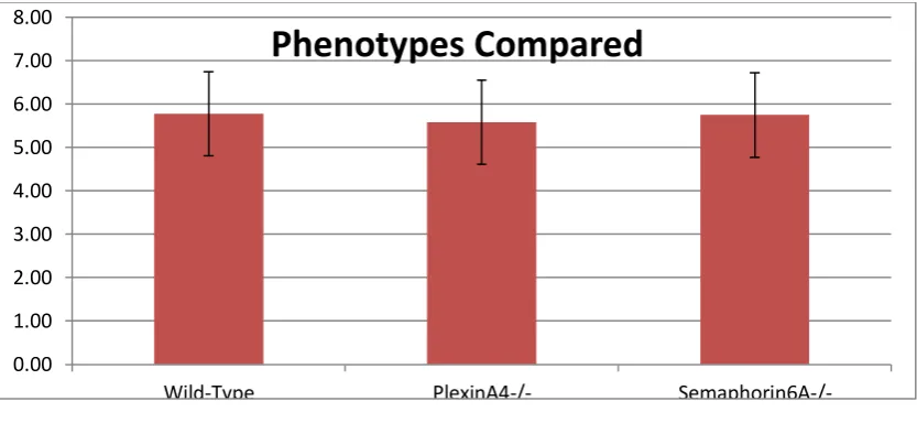
The number of processes crossing the white matter in each phenotype was directly compared in figure 1.7. Here, the average number of processes crossing the lateral column of white
matter in particular, consisting of sections 4, 5 and 6, are compared. In the wild-type samples,
Semaphorin6A-deficient samples it was 5.75. As can be seen, each result falls within the margin of
uncertainty of the other two, so no statistically significant difference can be found between
the three phenotypes. The margin of error was calculated similarly to above, and was found
to be ±0.97.
Section Number Wild Type (n=13)
(±0.92)
PlexinA4-/- (n=3) (±0.50)
Semaphorin6A -/-(n=4) (±0.84)
1 3.62 2.67 4.75
2 4.27 3.67 4.50
3 5.83 4.67 5.25
4 6.44 5.17 6.00
5 6.08 5.75 6.25
6 4.81 5.83 5.00
7 3.96 4.58 5.25
8 3.43 4.00 5.00
9 3.69 3.00 3.67
Table 1.1
This table shows the data used to generate figures 1.4, 1.5 and 1.6. It shows the average number of radial processes that were observed crossing each section of the white matter
[image:8.595.72.526.472.650.2]in each samples of each phenotype.
Fig 1.4
This line chart shows the average number of glial processes that were observed to be crossing each section of the wild-type mouse spinal cord. Each numbered area is
VM VM
M-LV L L
L
M-LD DM
DM 0.00 2.00 4.00 6.00 8.00
0 1 2 3 4 5 6 7 8 9 10
n u m b e r o f p ro ce ss e s
spinal cord area
labelled with its anatomical position. VM=Ventro-Medial, M-LV=Medio-Lateral Ventral, L=Lateral, M-LD=Medio-Lateral Dorsal, DM=Dorso-Medial.
Fig 1.5
This line chart shows the average number of glial processes that were observed to be crossing each section of the PlexinA4-deficient mouse spinal cord. For labels, see above.
Fig 1.6
This line chart shows the average number of glial processes that were observed to be crossing each section of the Semaphorin6A-deficient mouse spinal cord. For labels, see
above.
VM
VM M-L V
L L L
M-L D DM DM 0.00 2.00 4.00 6.00 8.00
0 1 2 3 4 5 6 7 8 9 10
n u m b er o f p ro ce ss es
spinal cord area
Plexin-Deficient Samples
VM VM M-L V
L L
L M-L D DM
DM 0.00 2.00 4.00 6.00 8.00
0 1 2 3 4 5 6 7 8 9 10
n u m b e r o f p ro ce ss es
spinal cord area
[image:9.595.73.526.420.598.2]Fig 1.7
This bar chart compares the average number of glial processes seen crossing the lateral sections of the three phenotypic categories of spinal cord. The lateral sections were
numbers 4, 5 and 6.
DISCUSSION
Context of our Findings
During the last 15 years, many advances have been made in the understanding of radial glial
cells, and their dual role as neuronal progenitors and aids to neuronal migration. Yet they
remain poorly characterised in the spinal cord when compared to the cerebral cortex. It had
been ascertained that they lack stem cell capabilities here, but it is only recently that some
insight has been gained into their potential function. It has been shown that the radial glial
scaffold, seen stretching from the ventricular zone to the pial surface, forms during the period
of development when the white matter tracts are forming, and then disappears as their
development completes. This was shown by vimentin and nestin staining. It therefore seems
possible that they could help to direct the development of these tracts, and in fact glial
scaffolds have been observed playing a similar role in other parts of the central nervous
system. For example, in the rostral migratory stream, the developing optic nerve and the
corpus callosum. It should be noted, however, that the scaffold in the spinal cord does not
express GLAST or BLBP, two markers typically expressed by radial glial cells, so these may
be of a slightly different phenotype (Barry et al 2013). 0.00
1.00 2.00 3.00 4.00 5.00 6.00 7.00 8.00
Wild-Type PlexinA4-/-
Discussing our Results
In our study, we examined the number of these processes crossing each column of white
matter in the developing mouse spinal cord at E15.5. As expected, we observed the processes
traversing the entire cord, from the ventricular zone to the pial surface, and there were
especially large numbers while crossing each region of the white matter, supporting the
previous conclusions. Our results show that the greatest number of processes was consistently
observed in the lateral column of the white matter. If their proposed function was confirmed,
this may suggest that axon tracts are developing particularly quickly in that column at this
stage of development. In humans, the lateral corticospinal tract and the spinocerebellar tracts
are known to occupy this area, so perhaps these glial processes are contributing towards the
formation of the mouse’s equivalent. Also, we compared the number of processes in normal
mice to numbers in both Semaphorin6A-deficient and PlexinA4-deficient specimens.
Semaphorins are proteins that have been shown to act as guides for the development of axons
in various contexts, but their effects on radial glia in the developing spinal cord have not yet
been shown conclusively. Plexins are the receptors which Semaphorins act on. Our results
did not show a significant difference between the three phenotypes. This shows that the role
of these proteins in the developing spinal cord remains uncertain, and is certainly an area that
warrants further study.
CONCLUSION
The many functions of radial glia continues to be an area of considerable research, and we
hope to have contributed a useful quantification of the glial processes present at one stage of
development to the discussion. Therefore, it is hoped that the full and precise role of these
glia in white matter patterning can be elucidated in the near future. Also, we have shown a
small glimpse at the possible involvement of Semaphorins and Plexins in the guiding of glial
formation.
ACKNOWLEDGEMENTS
The authors would like to thank Dr Denis S. Barry for his invaluable support, oversight and
technical assistance, and also Thomas McCartan and Eimear Duff for collaborating on the
REFERENCES
Barry D et al (2013). The spatial and temporal arrangement of the radial glial scaffold
suggests a role in axon tract formation in the developing spinal cord J. Anat. (2013) 222,
pp203—213
McDermott K, Barry D, McMahon S (2005). Role of radial glia in cytogenesis, patterning and
boundary formation in the developing spinal cord J. Anat. (2005) 207 pp241-250
Bentivoglio M, Mazzarello P (1999). The History of Radial Glia Brain Research Bulletin
(1999), Vol. 49, No. 5, pp. 305–315
Clowry G, Moss J, Clough R (2005). An immunohistochemical study of the development of
sensorimotor components of the early fetal human spinal cord J. Anat.(2005) 207, pp313–324
Gao W et al (2009). Temporal and Spatial Development of Axonal Maturation and
Myelination of White Matter in the Developing Brain AJNR Am J Neuroradiol (2009)
30:290–96
Kehrer C et al (2014) Language and cognition in children with metachromatic
leukodystrophy: onset and natural course in a nationwide cohort Orphanet Journal of Rare
Diseases (2014), 9:18
Malatesta P, Hartfuss E, Götz M (2000). Isolation of radial glial cells by fluorescent-activated
cell sorting reveals a neuronal lineage Development (2000) 127, 5253-5263
Martin J (2005). The Corticospinal System: From Development to Motor Control The
Neuroscientist (2005) 11, 2
Sasaki A et al (2012) A 14-year-old girl with lissencephaly and craniofacial dysmorphism
Neuropathology (2012); 32, 675–678
Rakic P (1972), Mode of cell migration to the superficial layers of fetal monkey neocortex
Journal of Comparative Neurology (1972) 145, 61-83
Barry D, Pakan J, McDermott K (2014). Radial glial cells: Key organisers in CNS
development The International Journal of Biochemistry & Cell Biology (2014) 46, 76-79
Ma S et al (2013). Radial glial neural progenitors regulate nascent brain vascular network
stabilization via inhibition of Wnt signalling PLoS Biology (2013) 11, e1001469
Altman J, Bayer S (2001). Development of the human spinal cord Oxford University Press pp
1-71
Pérez V et al (2013). Hereditary lissencephaly and cerebellar hypoplasia in Churra lambs
BMC Veterinary Research (2013) 9, no. 1, p1-12
Mayhew (1992). A review of recent advances in stereology for quantifying neural structure