http://www.scirp.org/journal/ojmn ISSN Online: 2163-0585
ISSN Print: 2163-0569
DOI: 10.4236/ojmn.2018.82016 Apr. 11, 2018 187 Open Journal of Modern Neurosurgery
Endoscopic Assisted Microscopic Skull Base
Surgery
Mohammed Attia, Islam Alaghoy, Magdy El Hawary, Maamon Abo-Shosha
Skull-Base Group, Neurosurgery Department, Faculty of Medicine, Al-Azhar University, Cairo, Egypt
Abstract
Background: Skull base tumors presented great challenge for neurosurgeons since decades due to their deep location, associated morbidity and limitation of operative field; however modern neurosurgery using the endoscope and/or the microscope served in minimizing peri-operative morbidities and improv-ing the clinical outcome. Objective: To demonstrate the value of endos-cope-assisted microsurgical technique for resection of skull base tumors. Pa-tients and Methods: 30 paPa-tients divided into 3 groups (10 paPa-tients had medi-al sphenoid wing meningioma constituted group 1, 10 patients had suprasel-lar meningioma constituted group 2 and 10 patients had Cerbello-Pontine Angle (CPA) epidermoids constituted group 3) were operated through En-doscope Assisted Microscopic Skull Base Surgery technique at Al-Azhar Uni-versity Hospitals during the period starting from January 2016 till the end of September 2017 using a rigid endoscope for inspection of tumor boundaries and neighboring vascularity in addition to confirm the extent of resection. Tumor resection was tried in all cases. Intra-operative resection rate and post-operative radiological outcomes were assessed. Results: Total gross re-section was possible in 27 patients (90%). Subtotal rere-section was done in the other 3 cases (recurrent medial sphenoid wing meningiomas) due to excessive bleeding and adhesions of the tumors with vascular structures. Total resection of the tumor in post operative radiology was obtained in 24 patients (80%) and post-operative residual tumor was noticed in 6 patients in early post operative radiology and in only 3 cases at 3 months follow up radiology. Con-clusion: Endoscopic-assisted microsurgical approach is a reliable, safe and ef-fective option for adequate surgical resection of skull base tumors. The tech-nique allowed proper inspection of the tumor relations and vascularity, detec-tion of any residual pordetec-tions, providing better chance for gross total resecdetec-tion with minimal tissue damage or vascular injury as well as convenient clinical outcome.
How to cite this paper: Attia, M., Alaghoy, I., El Hawary, M. and Abo-Shosha, M. (2018) Endoscopic Assisted Microscopic Skull Base Surgery. Open Journal of Modern Neurosurgery, 8, 187-200.
https://doi.org/10.4236/ojmn.2018.82016
Received: February 7, 2018 Accepted: April 8, 2018 Published: April 11, 2018
Copyright © 2018 by authors and Scientific Research Publishing Inc. This work is licensed under the Creative Commons Attribution International License (CC BY 4.0).
DOI: 10.4236/ojmn.2018.82016 188 Open Journal of Modern Neurosurgery
Keywords
Skull Base, Endoscopic Assisted
1. Introduction
DOI: 10.4236/ojmn.2018.82016 189 Open Journal of Modern Neurosurgery 94% of them, followed by meningiomas (3% - 10% of CPA tumors) and the epi-dermoids (2% - 4%) [7]. The approaches to the CPA are either posterior (through the posterior cranial fossa) or lateral (through the petrous bone). The most popular approach is retrosigmoid suboccipital [8]. Suprasellar meningi-omas originate most frequently from diaphragm sellae or anterior clinoid region [9]. Visual symptoms are still the most common presenting symptoms on ad-mission, and the most common first symptom followed by headache [10]. En-doscope-assisted microsurgery has shown to be particularly useful in the treatment of expansive lesions located in the anterolateral cisterns of the skull base and in the cerebellopontine cisterns. Endoscope Assisted Microscopic Sur-gery (EAM) provides visualization of critical deep-seated neurovascular struc-tures, allowing them to be dissected at an increased level of safety and preventing the superficial sectors from being exposed to inadvertent manipulation. EAM may be used effectively in the treatment of tumors located in the cerebellopon-tine angle (vestibular schwannomas, petroclival meningiomas, epidermoids, lower cranial nerve schwannomas, etc.) allowing visualization of critical neuro-vascular structures obscured by the lesion itself [11]. In the present study, we present our experience with endoscope-assisted microsurgical resection of skull base tumors through EAM approach in patients with medial sphenoid wing me-ningioma, CPA epidermoid and suprasellar meningioma regarding ability to achieve gross total resection and radiological outcome.
2. Patients and Methods
This study was conducted on 30 patients admitted and operated upon at Al-Azhar University Hospitals with a diagnosis of skull base tumor during the period starting from January 2016 till the end of September 2017. Skull base tu-mors involve all tutu-mors that originate from the base of the skull starting from the frontal region anteriorly until the foramen magnum posteriorly. In this study, Selection of only 3 types of skull base tumors was done through the clini-cal picture of each group and the irradiologiclini-cal appearance at Computed Tomo-graphy (CT) scan and Magnetic Resonant Imaging (MRI). The study cases we redivided into 3 groups; Group 1 (10 patients with medial sphenoid wing me-ningioma), Group 2 (10 patients harbouring suprasellar meningioma) and Group 3 (10 patients with CPA epidermoid). All patients were subjected to full clinical assessment, including history, complete neurological examination. Radi-ological assessment was performed by CT and MRI with Gadolinium for the brain demarcating the extent and nature of the tumor. Full laboratory investiga-tions were done for surgical preparation. No cerebral angiography was done in our series of cases since there was no encasement of large intracranial vessels in any case in this study (Table 1, Table 2).
2.1. Consent
DOI: 10.4236/ojmn.2018.82016 190 Open Journal of Modern Neurosurgery
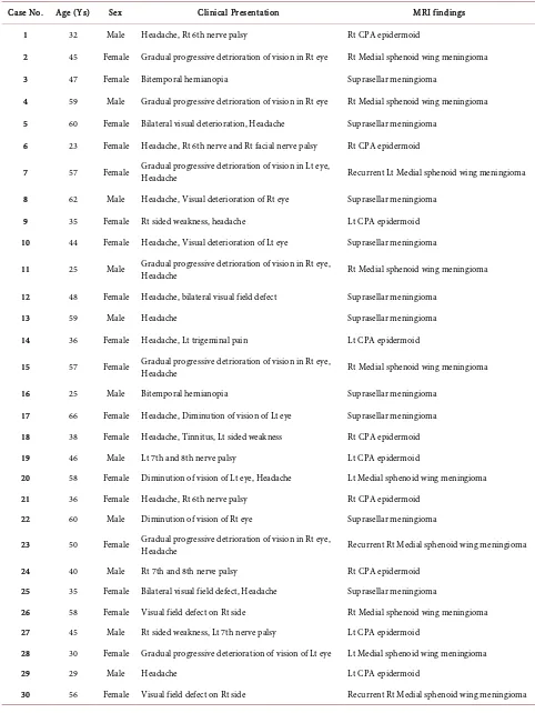
Table 1. Clinical and radiological data of the study patients.
MRI findings Clinical Presentation
Sex Age (Ys) Case No.
Rt CPA epidermoid Headache, Rt 6th nerve palsy
Male 32
1
Rt Medial sphenoid wing meningioma Gradual progressive detrioration of vision in Rt eye
Female 45 2 Suprasellar meningioma Bitemporal hemianopia Female 47 3
Rt Medial sphenoid wing meningioma Gradual progressive detrioration of vision in Rt eye
Male 59
4
Suprasellar meningioma Bilateral visual deterioration, Headache
Female 60
5
Rt CPA epidermoid Headache, Rt 6th nerve and Rt facial nerve palsy
Female 23
6
Recurrent Lt Medial sphenoid wing meningioma Gradual progressive detrioration of vision in Lt eye,
Headache Female
57 7
Suprasellar meningioma Headache, Visual deterioration of Rt eye
Male 62
8
Lt CPA epidermoid Rt sided weakness, headache
Female 35
9
Suprasellar meningioma Headache, Visual deterioration of Lt eye
Female 44
10
Rt Medial sphenoid wing meningioma Gradual progressive detrioration of vision in Rt eye,
Headache Male
25 11
Suprasellar meningioma Headache, bilateral visual field defect
Female 48 12 Suprasellar meningioma Headache Male 59 13
Lt CPA epidermoid Headache, Lt trigeminal pain
Female 36
14
Rt Medial sphenoid wing meningioma Gradual progressive detrioration of vision in Rt eye,
Headache Female 57 15 Suprasellar meningioma Bitemporal hemianopia Male 25 16 Suprasellar meningioma Headache, Diminution of vision of Lt eye
Female 66
17
Rt CPA epidermoid Headache, Tinnitus, Lt sided weakness
Female 38
18
Lt CPA epidermoid Lt 7th and 8th nerve palsy
Male 46
19
Lt Medial sphenoid wing meningioma Diminution of vision of Lt eye, Headache
Female 58
20
Rt CPA epidermoid Headache, Rt 6th nerve palsy
Female 36
21
Suprasellar meningioma Diminution of vision of Rt eye
Male 60
22
Recurrent Rt Medial sphenoid wing meningioma Gradual progressive detrioration of vision in Rt eye,
Headache Female
50 23
Rt CPA epidermoid Rt 7th and 8th nerve palsy
Male 40
24
Suprasellar meningioma Bilateral visual field defect, Headache
Female 35
25
Rt Medial sphenoid wing meningioma Visual field defect on Rt side
Female 58
26
Lt CPA epidermoid Rt sided weakness, Lt 7th nerve palsy
Male 45
27
Lt Medial sphenoid wing meningioma Gradual progressive deterioration of vision of Lt eye
Female 30
28
Lt CPA epidermoid Headache
Male 29
29
Recurrent Rt Medial sphenoid wing meningioma Visual field defect on Rt side
Female 56
DOI: 10.4236/ojmn.2018.82016 191 Open Journal of Modern Neurosurgery
Table 2. Groups of the study patients and approach used in each group.
Group Diagnosis Number of cases Approach used Group 1 Medial sphenoid wing meningioma 10 Pterional Group 2 Suprasellar meningioma 10 Pterional Group 3 CPA epidermoid 10 Retrosigmoid
signed a detailed consent about the procedure, its suspected benefits, its compli-cations, available surgical alternatives and post-operative suspected lines of management.
2.2. Surgical Technique
Group 1 patients had medial sphenoid wing meningioma and were operated upon via pterional approach. Group 2 patients had suprasellar meningioma and were operated upon via pterional approach. Group 3 patients had CPA epider-moid tumors and were operated upon by retrosigepider-moid approach. Under general anesthesia and after injection of proper antiepileptic coverage in cases of supra-sellar and medial sphenoid wing meningiomas, the endoscopic sheath was in-troduced and 0-degree rigid lensendoscope was placed for inspection of the tu-mor and surrounding areas under continuous irrigation with 37˚ Cringer’s solu-tion providing clear visualizasolu-tion. Craniotomy was done for patients of Group 1 and Group 2 and craniectomy was done for patients of Group 3. The surgical corridor was opened by two retractors to visualize the tumor using a suitable piece of cottonoid for cortical protection. The operating microscope was then introduced with temporary getting the endoscope lens off field. Tumor debulk-ing and removal was then started usdebulk-ing suction and fine bipolar electrocautery with continuous irrigation. Two different angles of endoscope lenses (0˚ and 30˚) were used in several stages according to the degree of tumor accessibility to provide visualization of the feeding tumor vessels when microscopic visualiza-tion was insufficient. After the tumor mass was grossly excised and proper he-mostasis was performed the endoscope was re-introduced to check the different anatomic corners where the tumor was located for the possibility of presence of any hidden residual which couldn’t be detected until different angled scopes were used. The dura was then sutured. The bone was then repositioned and the wound closed in layers. Both intra-operative gross resection rate was assessed. Early and 3 months post operative radiology was done and post operative radio-logical resection rate was assessed.
3. Case Presentation
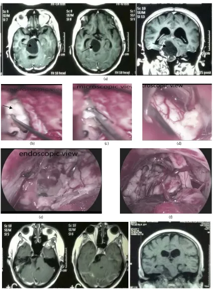
Case (1): Female patient, 38 years old, complained of headache, tinnitus, Rt facial pain and gradual progressive weakness of the Lt side of the body. MRI brain with contrast showed Rt CPA epidermoid tumor (Figure 1).
DOI: 10.4236/ojmn.2018.82016 192 Open Journal of Modern Neurosurgery
(a)
(b) (c) (d)
(e) (f)
[image:6.595.85.513.65.648.2](g)
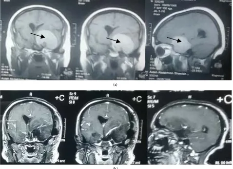
DOI: 10.4236/ojmn.2018.82016 193 Open Journal of Modern Neurosurgery of vision of left eye, her visual Acuity: 6/60 on lt side, MRI brain with contrast showed lt medial sphenoid wing meningioma (Figure 2).
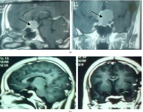
Case (3): Female patient, 60 years old, complained of headache and visual de-terioration of both eyes, her visual acuity was 6/18 on Rt side and hand motion on Lt side. MRI brain with contrast showed suprasellar meningioma (Figure 3).
4. Results
The study included 30 patients with a median age of 45.36 years (range: 23 - 66 years). They were 11 males (37%) and 19 females (63%). Medial Sphenoidal wing meningiomas were diagnosed in 8 females (80% of Group 1 patients) and in only 2 males (20% of Group 1 patients). They occurred in patients with a me-dian age of 49.5 years (range: 25 - 59 years). Suprasellar meningiomas occurred in 6 females (60% of Group 2 patients) and in 4 males (40% of Group 2 patients). The median age of Group 2 patients was 50.6 years (range: 25 - 66 years). CPA epidermoids were diagnosed in 5 females and 5 males (50% of Group 3 patients for each). The median age of Group 3 patients was 36 years (range: 23 - 46 years). CPA epidermoids were noticed at more young age groups than other two
(a)
[image:7.595.61.539.342.688.2](b)
DOI: 10.4236/ojmn.2018.82016 194 Open Journal of Modern Neurosurgery
(a)
[image:8.595.62.541.72.448.2](b)
Figure 3. MRI brain with contrast (sagittal and coronal views) showed suprasellar meningioma: (a) Pre-operative (The arrow-heads); (b) Post-operative excision through endoscope assisted microscopic surgery.
groups. Similarly, medial sphenoid wing meningiomas occurred predominantly at female patients than males.
4.1. Intra-Operative Findings
DOI: 10.4236/ojmn.2018.82016 195 Open Journal of Modern Neurosurgery
4.2. Gross Tumor Resection Rate Using Endoscope-Assisted
Microsurgery (EAM)
For Group 1, gross total resection was obtained in 7 patients (70%), and was not possible in 3 patients (30%). For Group 2, gross total resection was possible in all 10 patients (100%). For Group 3, gross total resection of the tumor was possible in all 10 patients (100%). In all study patients, the incidence of gross total resec-tion of the tumors was 90% (Table 4).
4.3. Radiological Resection Rate
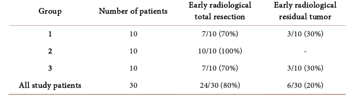
Total resection of the tumor in early postoperative radiology was obtained in7 patients of 10 (70%) of Group 1 patients, 10 patients of 10 (100%) of Group 2 patients, in 7 patients of 10 (70%) of Group 3 patients. This represents an overall percentage of 80% radiological resection rate of all study groups (Table 5).
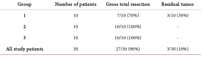
[image:9.595.208.539.377.469.2]Total resection of the tumor in 3-month follow up post operative radiology was obtained in 7 patients of 10 (70%) of Group 1 patients, 10 patients of 10 (100%) of Group 2 patients, in 10 patients of 10 (100%) of Group 3 patients with overall percentage of 90% of all study patients. The disappearance of the tumor residual in 3 patients with CPA epidermoid may be due to filling of the space
Table 3. Intra-operative findings.
Group Number of cases using microscope Total resection alone
Total resection after introduction
of the endoscope
Residual tumor after endoscope assisted microsurgery
1 10 3 4 3
2 10 7 3 -
[image:9.595.208.540.502.592.2]3 10 - 10 -
Table 4. Gross tumor resection rate using endoscope-assisted microsurgery.
Group Number of patients Gross total resection Residual tumor
1 10 7/10 (70%) 3/10 (30%)
2 10 10/10 (100%) -
3 10 10/10 (100%) -
All study patients 30 27/30 (90%) 3/30 (10%)
Table 5. Early radiological tumor resection rate using endoscope-assisted microsurgery.
Group Number of patients Early radiological total resection Early radiological residual tumor
1 10 7/10 (70%) 3/10 (30%)
2 10 10/10 (100%) -
3 10 7/10 (70%) 3/10 (30%)
[image:9.595.198.540.629.734.2]DOI: 10.4236/ojmn.2018.82016 196 Open Journal of Modern Neurosurgery with CSF not with epidermoid residual because it disappeared at 3 months fol-low up. Otherwise, if residual, it would not disappear. So, diffusion MRI for epi-dermoid cases is mandatory to exclude residual (Table 6).
4.4. Peri-Operative Complications
Peri-operative complications occurred in 5 patients (17%) of all study patients. Vascular injury occurred in 2 patients with recurrent medial sphenoid wing me-ningioma, control of bleeding occurred, surgery aborted due to severe brain edema in one patient and due to anesthetic causes in the other one, 2nd sitting
was done with near total resection of the tumor using endoscope-assisted mi-crosurgery. Transient Facial nerve palsy had occurred in 2 patients with CPA epidermoid that improved within 2 weeks after surgery. 6th nerve palsy occurred in one patient with CPA epidermoid that improved within one month after sur-gery.
5. Discussion
[image:10.595.208.542.631.727.2]Endoscope-assisted microsurgery is a practical indication for application of en-doscopic technology. Enen-doscopic approach minimizes potential morbidity of approaching skull base tumors. This technique is usually associated with im-proved clinical outcome, shorter operating times, and decreased need for exten-sive surgical manipulation that may lead to greater morbidity [12]. The current study involved a heterogenous group of skull base tumors divided into 3 groups. Group 1 patients were 10 cases with medial sphenoid wing meningiomas, Group 2 patients were 10 cases with suprasellar meningiomas, and Group 3 patients were 10 cases with CPA epidermoids. In this study, endoscope-assisted micro-surgery was performed. This technique allowed good microsurgical access as-sisted with excellent lighting and visualization through the endoscope. With a total gross resection in 90% of patients, the results of the current series are con-sidered. Gross tumor resection rate was 70% in group 1 patients and was 100% in both group 2 and group 3 patients. Total resection of the tumor in early post operative radiology was obtained in 70% of Group 1 patients, 100% of Group 2 patients, in 70% of Group 3 patients and in 80% of all study patients. Total re-section of the tumor in the 3-month follow up post operative radiology was ob-tained in 70% of Group 1 patients, 100% of Group 2 patients, in 100% of Group
Table 6. 3-month post-operative radiological tumor resection rate using endoscope-assisted microsurgery.
Group Number of patients Gross total resection Residual tumor
1 10 7/10 (70%) 3/10 (30%)
2 10 10/10 (100%) -
3 10 10/10 (100%) -
DOI: 10.4236/ojmn.2018.82016 198 Open Journal of Modern Neurosurgery a study on eight patients harboring an epidermoid tumor of the CPA that treated using an endoscope-assisted microsurgical technique. A retrosigmoid subocci-pital approach was used in five patients and a pterional transsylvian approach was chosen in the remaining three. In four patients the lesion was resected mi-crosurgically and the endoscope was used repeatedly to verify complete tumor removal, whereas most of the tumor mass was removed with the aid of an oper-ating microscope in the other four. Tumor parts extending into other cranial compartments that were not visible through the microscope were removed un-der endoscopic view by using rigid rod-lens scopes with 30 and 70 degrees angles of view. All epidermoids were completely evacuated and the membranes were widely resected. Large tumors occupying both the middle and posterior cranial fossa were removed through a single small opening without enlarging the cra-niotomy. Permanent hearing loss and permanent hypacusis were observed in one patient each. One patient with facial and one with abducent nerve palsy re-covered within 6 and 4 months, respectively. In the present study, transient fa-cial nerve palsy occurred in 2 patients and tansient 6th nerve palsy occurred in one patient. A transient weakness of the chewing muscles was encountered in one patient in Schroeder et al. study. Postoperative magnetic resonance imaging revealed no residual tumor in any patient in Schroeder et al. study and in 3 pa-tients in the early post operative radiological study and after 3 months there was no residual tumor in.
6. Limitations
There were some limitations in this study including dependency only on the type and site of the lesion to differentiate the groups of the study patients. Inside each group, there were no differentiating criteria like the size of the lesion, its extensions, presence of recurrence, adherence to surrounding vessels. The follow up period of 3 months post-surgical procedure is another limitation in this study. Follow up for longer periods after surgical excision may be indicated to obtain more accurate results.
7. Conclusion
Endoscopic-assisted microsurgical approach is a reliable, safe and effective op-tion for adequate surgical resecop-tion of skull base tumors. The technique allowed proper inspection of the tumor relations and vascularity, detection of any resi-dual portions, providing better chance for gross total resection with minimal tissue damage or vascular injury as well as convenient clinical outcome. It proved very useful for tumors that can insinuate themselves around corners be-tween neurovascular structures and going to hidden pockets like epidermoids as concluded from our study and from others as Schroeder et al.
References
Sur-DOI: 10.4236/ojmn.2018.82016 199 Open Journal of Modern Neurosurgery gery. Youmans Neuroslogical Surgery, 116, 1267-1284.
https://doi.org/10.1016/B978-1-4160-5316-3.00119-2
[2] Tatagiba, M., Matthies, C. and Samii, M. (1996) Microendoscopy of the Internal Auditory Canal in Vestibular Schwannoma Surgery. Neurosurgery, 38, 737-740. https://doi.org/10.1227/00006123-199604000-00021
[3] Maio, S.D. and Sekhar, L.N. (2012) Skull Base Approaches. In: Principles of Neuro-logical Surgery. 3rd Edition, 667-679.
https://doi.org/10.1016/B978-1-4377-0701-4.00043-9
[4] Landriel, F. and Black, P. (2012) Meningiomas. In: Principles of Neurological Sur-gery. 3rd Edition, 541-564.
https://doi.org/10.1016/B978-1-4377-0701-4.00036-1
[5] Rhoton, Jr. A.L. (2000) The Cerebellopontine Angle and Posterior Fossa Cranial Nerves by the Retrosigmoid Approach. Neurosurgery, 47, S93-129.
https://doi.org/10.1093/neurosurgery/47.3.S93
[6] De Monte, F. (1993) Neoplasms and Cranial Nerves of the Posterior Fossa. In: Bar-row, D.L., Ed., Surgery of the Cranial Nerves of the Posterior Fossa. American As-sociation of Neurological Surgeons, Park Ridge, 253-254.
[7] Bonneville, F., Savatovsky, J. and Chiras, J. (2007) Imaging of Cerebellopontine An-gle Lesions: An Update. European Radiology, 17, 2908-2920.
https://doi.org/10.1007/s00330-007-0680-4
[8] Samii, M. and Gerganov, V.M. (2008) Surgery of Extraaxial Tumors of the Skull Base. Neurosurgery, 62, 1153-1168.
https://doi.org/10.1227/01.neu.0000333782.19682.76
[9] Cushing, H. and Eisenhardt, C. (1962) Meningiomas. Their Classification, Regional Behavior, Life History and Surgical End Results. Hafner, New York, 225-241. [10] Gregorius, F.K., Hepler, R.S. and Stern, W.E. (1975) Loss and Recovery of Vision
with Suprasellar Meningiomas. Journal of Neurosurgery, 42, 69-75. https://doi.org/10.3171/jns.1975.42.1.0069
[11] Galzio, R.J. and Tschabitscher, M. (2010) Endoscope-Assisted Microneurosurgery. Principles, Methodology and Applications. Endo: Press™, Tuttlingen.
[12] Choudhri, O., Feroze, A.H., Nathan, J., Cheshier, S. and Guzman, R. (2014) Ven-tricular Endoscopy in the Pediatric Population: Review of Indications. Child’s Nervous System, 30, 1625-1643. https://doi.org/10.1007/s00381-014-2502-8
[13] Abolfotoh, M., Bi, W.L., Hong, C.K., Almefty, K.K., Boskovitz, A., Dunn, I.F. and Al-Mefty, O. (2015) The Combined Microscopic-Endoscopic Technique for Radical Resection of Cerebellopontine Angle Tumors. Journal of Neurosurgery, 123, 1301-1311. https://doi.org/10.3171/2014.10.JNS141465
[14] Presutti, L., Alicandri-Ciufelli, M., Rubini, A., Gioacchini, F.M. and Marchioni, D. (2014) Combined Lateral Microscopic/Endoscopic Approaches to Petrous Apex Le-sions: Pilot Clinical Experiences. The Annals of Otology, Rhinology, and Laryngol-ogy, 123, 550-559. https://doi.org/10.1177/0003489414525342
[15] Zhang, J., Shrestha, R., Cai, B.W., Zhou, P.Z., Li, Y.P. and Jiang, S. (2014) Manage-ment of Large Medial Sphenoid Wing Meningiomas: A Series of 178 Cases. Turkish Neurosurgery, 24, 664-671.
DOI: 10.4236/ojmn.2018.82016 200 Open Journal of Modern Neurosurgery [17] Bander, E.D., Singh, H., Ogilvie, C.B., Cusic, R.C., Pisapia, D.J., Tsiouris, A.J., Anand, V.K. and Schwartz, T.H. (2017) Endoscopic Endonasal versus Transcranial Approach to Tuberculum Sellae and Planum Sphenoidale Meningiomas in a Similar Cohort of Patients. Journal of Neurosurgery, 128, 40-48.
https://doi.org/10.3171/2016.9.JNS16823
[18] Koutourousiou, M., Fernandez-Miranda, J.C., Stefko, S.T., Wang, E.W., Snyderman, C.H. and Gardner, P.A. (2014) Endoscopic Endonasal Surgery for Suprasellar Me-ningiomas: Experience with 75 Patients. Journal of Neurosurgery, 120, 1326-1339. https://doi.org/10.3171/2014.2.JNS13767
[19] Lu, Z.F., Cheng, X.B., Zhao, Y.G. and Shi, B.Z. (2013) Twenty-Nine Cases of Resec-tion of Suprasellar Meningioma through Small Bone Window: An Interhemispheric Approach. Contemporary Oncology, 17, 525-529.