http://www.scirp.org/journal/ojgas ISSN Online: 2163-9469
ISSN Print: 2163-9450
DOI: 10.4236/ojgas.2018.83010 Mar. 29, 2018 94 Open Journal of Gastroenterology
A Case of Simultaneous Triple Primary Cancers
of the Hypopharynx, Esophagus, and Stomach
Which Were Dissected by Endoscopic
Laryngo-Pharyngeal Surgery Combined with
Endoscopic Submucosal Dissection
Kenro Kawada
1*, Taro Sugimoto
2, Ryuhei Okada
2, Kazuya Yamaguchi
1, Yuudai Kawamura
1,
Masafumi Okuda
1, Yuuichiro Kume
1, Andres Mora
1, Tairo Ryotokuji
1, Takuya Okada
1,
Akihiro Hoshino
1, Yutaka Tokairin
1, Yasuaki Nakajima
1, Yusuke Kiyokawa
2, Fuminori Nomura
2,
Yoshuke Ariizumi
2, Shohei Tomii
3, Takashi Ito
3, Takahiro Asakage
2, Yusuke Kinugasa
1,
Tatsuyuki Kawano
11Department of Gastrointestinal Surgery, Tokyo Medical and Dental University, Tokyo, Japan 2Department of Head and Neck Surgery, Tokyo Medical and Dental University, Tokyo, Japan 3Department of Human Pathology, Tokyo Medical and Dental University, Tokyo, Japan
Abstract
A 65-year-old man was admitted to our hospital following 6 months of dys-phagia. At first, conventional endoscopy showed a reddish and depressed le-sion in the stomach and an elevated lele-sion in the posterior wall of the hypo-pharynx. An endoscopic biopsy showed adenocarcinoma in the stomach, and squamous cell carcinoma in the hypopharynx. On the further examination, trans-nasal endoscopy with narrow band imaging (NBI) was performed. During the trumpet maneuver, a huge protruded lesion was observed and it reached to the orifice of the esophagus. Other superficial lesion located at left pyriform sinus was detected by NBI system as brownish area with brown dots. Furthermore, superficial esophageal cancer in the cervical esophagus was de-tected. Finally, 4 carcinomas in upper gastrointestinal tract were dede-tected. Among them, the hypopharyngeal cancer was the most advanced (T3N0). The patient hoped to preserve his voice and swallowing function, endoscopic la-ryngo-pharyngeal surgery (ELPS) was performed for the hypopharyngeal cancer. Endoscopic mucosal resection (EMR) was performed for the esopha-geal cancer, and Endoscopic submucosal dissection (ESD) was performed for the gastric cancer. Under collaboration between a head and neck surgeon and an endoscopist, the tumor was resected en-bloc. The histopathological find-How to cite this paper: Kawada, K.,
Su-gimoto, T., Okada, R., Yamaguchi, K., Kawamura, Y., Okuda, M., Kume, Y., Mo-ra, A., Ryotokuji, T., Okada, T., Hoshino, A., Tokairin, Y., Nakajima, Y., Kiyokawa, Y., Nomura, F., Ariizumi, Y., Tomii, S., Ito, T., Asakage, T., Kinugasa, Y. and Kawano, T. (2018) A Case of Simultaneous Triple Primary Cancers of the Hypopharynx, Eso-phagus, and Stomach Which Were Dissected by Endoscopic Laryngo-Pharyngeal Surgery Combined with Endoscopic Submucosal Dissection. Open Journal of Gastroenter-ology, 8, 94-102.
https://doi.org/10.4236/ojgas.2018.83010
DOI: 10.4236/ojgas.2018.83010 95 Open Journal of Gastroenterology ings of hypopharyngeal cancer were squamous cell carcinoma, subeipthelial invasion, 29 × 28 × 4.2 mm. The others were diagnosed as mucosal cancers. The patient is currently alive with no recurrence at 28 months after the sur-gery; there is no stricture at the cervical esophagus. Endoscopic laryngopha-ryngeal surgery for the tumor of pharyngo-esophageal junction can provide a less invasive treatment.
Keywords
Endoscopic Laryngopharyngeal Surgery, Hypopharyngeal Cancer, Multiple Primary Cancer, Trumpet Maneuver, Endoscopic Submucosal Dissection
1. Background
Recently, multiple primary malignant cancers in the same patient frequently have been found because of the advancing age of patients and improvements in the diagnostic tool in identifying early stage mucosal cancer of the head and neck. Synchronous and metachronous occurrence of squamous cell carcinomas within the head and neck region is well known as field cancerization [1] [2] [3]. Most cases of hypopharyngeal cancer are detected at an advanced stage, and re-quire extensive treatment and are usually associated with a loss of function of swallowing or speaking. Several reports [4] [5] [6] have indicated the use fullness of narrow band imaging (NBI) improving the detection of superficial hypopha-ryngeal cancer. However, circumferential observation of the hypophahypopha-ryngeal- hypopharyngeal-mucosa is difficult during conventional endoscopy due to its anatomically closed nature. We previously reported that the utility of trans-nasal endoscopy using the trumpet maneuver for precise inspection before treatment [7] [8]. We here in report a case of simultaneous triple primary cancers of the hypo-pharynx, esophagus, and stomach which were dissected by Endoscopic laryn-go-pharyngeal surgery (ELPS) combined with Endoscopic submucosal dissec-tion (ESD).
2. Case Presentation
The patient was a 65-year-old man with dysphasia as the chief complaint. He had a heavy drinking career and also had some history of smoking. He had nothing remarkable in his family history. He had an anamnesis of the enlarge-ment type cardiomyopathy From around July 2015, he visited at the clinic near his home, but he was diagnosed as having no abnormality. From September 2015, the dysphagia progressed to the extent that he was unable to tolerate solid foods. He was admitted to our hospital. At first, conventional endoscopy showed a reddish and depressed lesion in the stomach (Figure 1) and an elevated lesion in the posterior wall of the hypopharynx (Figure 2). An endoscopic biopsy showed adenocarcinoma in the stomach, and squamous cell carcinoma in the hypopharynx. On the further examination, trans-nasal endoscopy with narrow Copyright © 2018 by authors and
Scientific Research Publishing Inc. This work is licensed under the Creative Commons Attribution International License (CC BY 4.0).
DOI: 10.4236/ojgas.2018.83010 96 Open Journal of Gastroenterology Figure 1. The early gastric cancer detected by conventional
endoscopy with indigo carmine.
Figure 2. The protrude lesion was observed by conventional endoscopy. The distal side of the lesion was invisible.
band imaging (NBI) was performed. We have routinely performed the trumpet maneuver using trans-nasal endoscopy for patients with esophageal cancer since 2009, using the following procedure.
[image:3.595.261.486.281.451.2]DOI: 10.4236/ojgas.2018.83010 97 Open Journal of Gastroenterology (a) (b)
[image:4.595.208.536.67.246.2]Figure 3. (a) The huge and protruded lesion was observed by trans-nasal endoscopy with NBI system using trumpet maneuver. The distal side of the tumor was invaded to the cer-vical esophagus; (b) The Brownish area was observed at the left piriform sinus with NBI system.
Figure 4. The cervical esophageal cancer detected by transnasal endoscopy with NBI system.
Computed tomography revealed hypertrophy of the wall of the hypopharynx, and no lymph nodal metastasis, or distant metastasis. The prognosis was based on advanced hypopharyngeal cancer (T3N0M0, Stage III). The patient’s treatment method was decided after the discussion with head and neck surgeon and gas-troenterologist. He was recommended to undergo chemo-radiotherapy. Howev-er, his performance status was not so good, he refused invasive treatments and hoped to preserve his voice and swallowing function. At first, we decided to perform endoscopic resection for hypopharynegeal cancers and superficial eso-phageal cancer cooperated with head and neck surgeon.
3. Therapeutic Procedure
[image:4.595.280.467.313.500.2]DOI: 10.4236/ojgas.2018.83010 98 Open Journal of Gastroenterology treatment and head and neck surgery [9]. The endoscope is passed through the nasal route, and the surgeon inserts the curved forceps and the electric knife through the mouth. Endotracheal intubation is administered under general anesthesia, and the larynx is lifted using a Sato’s curved laryngoscope (Nagashi-ma Medical instruments Company, Ltd., Toyo, Japan) developed by Sato et al., so we can get a good , wide working space. At First iodine staining was per-formed, we could recognize the lateral margin of the tumor. Then the margin was marked using electric knife in coagulation mode.
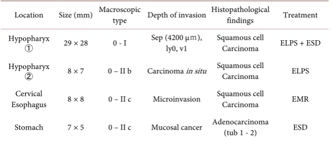
[image:5.595.247.499.505.693.2]After the marking, inject a solution of epinephrine (0.02 mg/ml), indigo car-mine, and saline into the subepithelial layer beneath the lesion. After the injec-tion, make the cutting 2 mm outside of the marking. At the distal side of the hypopharyngeal cancer and the esophageal cancer, the electric knife couldn’t reach, so it was difficult for the head and neck surgeon to perform the operation alone. Endoscopic mucosal resection (EMR) for the superficial cervical esopha-geal cancer was performed. Then an endoscopist dissected the distal margin us-ing the electric knife inserted through a flexible endoscope (Endoscopic submu-cosal dissection technique). At the proximal side of the hypopharyngeal tumor, the cutting and dissection procedure was performed combination of the trans-orally inserted curved forceps in one hand and the electric knife in the other hand (Figure 5). Finally, the tumor was resected en bloc. The superficial lesion located at left piriform sinus was dissected by ELPS procedure. All the tumors were resected one piece (Figure 6). The total operation time was 80 min. The patient was extubated immediately after surgery. The fasting period was 5 days after surgery, and the postoperative hospital stay was ten days. 2 months later, the gastric cancer was dissected by endoscopic submucosal dissection. All specimens were microscopically evaluated (Table 1). The histopathological finding of the main tumor was squamous cell carcinoma, 29 × 28 mm, subepi-thelial invasion (the tumor thickness was 4200 μm), and a negative lateral and
DOI: 10.4236/ojgas.2018.83010 99 Open Journal of Gastroenterology Figure 6. 3 resected specimens (two hypopharyngeal cancers, one
esophageal cancer).
Table 1. Histopathological findings.
Location Size (mm) Macroscopic type Depth of invasion Histopathological findings Treatment
Hypopharyx
① 29 × 28 0 - I
Sep (4200 μm),
ly0, v1 Squamous cell Carcinoma ELPS + ESD Hypopharyx
② 8 × 7 0 – II b Carcinoma in situ Squamous cell Carcinoma ELPS
Cervical
Esophagus 8 × 8 0 – II c Microinvasion Squamous cell Carcinoma EMR
Stomach 7 × 5 0 – II c Mucosal cancer Adenocarcinoma (tub 1 - 2) ESD
vertical margin (Figure 7). During the follow-up period of 28 months, there is no stricture at the cervical esophagus (Figure 8). The phonation and swallowing functions were preserved. Close collaboration between head and neck surgeons and endoscopistscan provide good results in treating tumors of the pharyn-go-esophageal junction.
4. Discussion
Regarding the theory of the development of multiple primary cancers, Slaughter et al. [1] proposed the field cancerization theory that explained the concept of multicentricorigin, and this theory is still accepted. It has been reported that the application of magnifying endoscopy with the NBI system drastically changes the diagnostic strategy for the early detection of early head and neckcancers [4] [5] [6]. However,some areas are difficult to observe with conventional endos-copy. We previously reported the utility of trans-nasal endoscopy using the trumpet maneuver for precise inspection before the treatment [7].
[image:6.595.209.542.298.440.2]DOI: 10.4236/ojgas.2018.83010 100 Open Journal of Gastroenterology Figure 7. A histopathological examination revealed a
diag-nosis of invasive squamous cell carcinoma with negative lat-eral and vertical margin. The tumor thickness was 4200 μm.
Figure 8. At 28 months after treatment, no local recurrence was observed.
Trans-oral surgery is becoming a major strategy in the treatment of
la-ryngo-phageygealcancer [10]. If the lesion was diagnosed as carcinoma in
situ or carcinoma with invasion to the subepithelial layer, Endoscopic laryn-go-pharyngeal surgery was indicated as a minimally invasive treatment. It has been reported that the ratio of subepithelial invasion or muscular invasion in type 0 - I were 100% [11]. In determining the treatment strategy, the stage and location of the tumor and the general status of the patient should be considered. The 0 - I located at hypopharyngeal lesion of the present case seems to have a deep subepithelial invasion. The indication for ELPS was thought to be difficult.
Chemo-radiotherapy is widely performed at present but is associated with a high frequency of early and late toxicities [12]. Total pharyngo-laryngo-esophagectomy is considered the most complicated and most invasive surgery for the ga-stro-intestinal tract [13].
[image:7.595.259.488.304.471.2]DOI: 10.4236/ojgas.2018.83010 101 Open Journal of Gastroenterology junction dissected by ELPS with ESD before [14]. In Japan, most cases of super-ficial squamous cell carcinoma located at the cervical esophagus are treated by endoscopically, so we selected the endoscopic treatment for both hypopharyn-geal and cervical esophahypopharyn-geal cancer. In this case, we could easily perform surgery without any complications. After endoscopic resection for superficial pharyngeal cancer, additional treatments such as radiation therapy or radical resection may be needed. We use a “resect and watch starategy [15]”, it means that observe un-til development of secondary diseases, including local recurrence, neck lymph node metastasis, and metachronous pharyngeal cancer.
The follow-up examinations after treatment, included cervical ultrasound and the measurement of his tumor marker levels every three months, and computed tomography every six months. Balloon dilation was not required. The patient is currently alive with no recurrence at 28 months after the surgery. More cases and longer follow-up periods will be required to obtain conclusive findings, and future studies will need to determine the indications of this treatment.
5. Conclusion
We have been experienced a case of simultaneous multiple cancers located at hypopharynx, cervical esophagus, stomach cured by endoscopic treatment. It is a less invasive treatment than conventional surgery and is becoming a major strategy in the treatment of superficial pharyngeal cancer.
References
[1] Slaughter, D.P., Southwick, H.W. and Smejkal, W. (1953) Field Cancerization in Oral Stratified Squamous Epithelium; Clinical Implications of Multicentric Origin. Cancer, 6, 963-968.
https://doi.org/10.1002/1097-0142(195309)6:5<963::AID-CNCR2820060515>3.0.C O;2-Q
[2] Panosetti, E., Luboinski, B.,Mamelle, G., et al. (1989) Multiple Synchronous and Metachronous Cancers of the Upper Aerodigestive Tract: 9-Year Study. Laryngos-cope, 99, 1267-1273. https://doi.org/10.1288/00005537-198912000-00011
[3] Jones, A.S., Morar, P., Phillips, D.E., et al. (1995) Second Primary Tumors in Pa-tients with Head and Neck Squamous Cell Carcinoma. Cancer, 75, 1343-1353. https://doi.org/10.1002/1097-0142(19950315)75:6<1343::AID-CNCR2820750617>3. 0.CO;2-T
[4] Muto, M., Nakane, M., Katada, C., et al. (2004) Squamous Cell Carcinoma in Situ at Oropharyngeal and Hypopharyngeal Mucosal Sites. Cancer, 101, 1375-1381. https://doi.org/10.1002/cncr.20482
[5] Watanabe, A., Tsujie, H., Taniguchi, M., et al. (2006) Laryngoscopic Detection of Pharyngeal Carcinoma in Situ with Narrowband Imaging. The Laryngoscope, 116, 650-654. https://doi.org/10.1097/01.mlg.0000204304.38797.34
[6] Muto, M., Minashi, K., Yano, T., et al. (2010) Early Detection of Superficial Squamous Cell Carcinoma in the Head and Neck Region and Esophagus by Narrow Band Imaging: A Multicenter Randomized Controlled Trial. Journal of Clinical Oncology, 28, 1566-1572. https://doi.org/10.1200/JCO.2009.25.4680
Hypopha-DOI: 10.4236/ojgas.2018.83010 102 Open Journal of Gastroenterology ryngeal Cancer at the Pharyngoesophageal Junction Which Is Detected by Trans-nasal Endoscopy Using Trumpet Maneuver. Open Journal of Gastroenterology, 5, 1-6.
[8] Kawada, K., Kawano, T., Sugimoto, T., et al. (2015) Observation of the Pharynx to the Cervical Esophagus Using Transnasal Endoscopy with Blue Laser Imaging. In: Amornyotin, S., Ed., Endoscopy-Innovative Uses and Emerging Technologies, 2172-2175.
[9] Sato, Y., Omori, T. and Tagawa, M. (2006) Surgical Treatment for Hypopharyn-gealsuperficial Cancer: Endoscopic Laryngo-Pharyngeal Surgery. Nihon Jibiinkoka Gakkai Kaiho, 109, 581-586.
[10] Tateya, I., Shiotani, A., Satou, Y., et al. (2016) Transoral Surgery for Laryn-go-Pharyngeal Cancer—The Paradigm Shift of the Head and Neck Cancer Treat-ment. Auris Nasus Larynx, 43, 21-32. https://doi.org/10.1016/j.anl.2015.06.013 [11] Tateya, I., Morita, S., Muto, M., et al. (2015) Magnifying Endoscope with NBI to
Predict the Depth of Invasion in Laryngo-Pharyngeal Cancer. The Laryngoscope, 125, 1124-1129.https://doi.org/10.1002/lary.25035
[12] Nakajima, A., Nishiyama, K., Morimoto, M., et al. (2012) Definitive Radiotherapy for T1-2 Hypopharyngeal Cancer: A Single-Institution Experience. International Journal of Radiation Oncology, Biology, Physics, 82, e129-e135.
https://doi.org/10.1016/j.ijrobp.2011.03.040
[13] Sagawa, N., Okushiba, S., Ono, K., et al. (2000) Reconstruction after Pharyngo La-ryngo Esophagectomy: Comparison of Elongated Stomach Roll with Microvascular Anastomosis with Gastric Pull up Reconstruction or Something like That. Langen-beck’s Archives of Surgery, 385, 34-38. https://doi.org/10.1007/s004230050008 [14] Kawada, K., Kawano, T., Sugimoto, T., et al. (2017) Case of Superficial Cancer
Lo-cated at the Pharyngoesophageal Junction Which Was Dissected by Endoscopic La-ryngopharyngeal Surgery Combined with Endoscopic Submucosal Dissection. Case Reports in Otolaryngology, 2017, Article ID: 1341059.
https://doi.org/10.1155/2017/1341059