BACTERIOLOGICAL PROFILE OF VENTILATOR ASSOCIATED
PNEUMONIA IN INTENSIVE CARE UNIT
*Dr. A. Ravishankar Reddy1, Dr.A. Swathi2, Dr.V. Ramadevi3, Dr. K. Kamal Chand4.
1
Associate Professor. Department of microbiology, Kamineni Academy of Medical Sciences & Research Center, Kamineni Hospitals, L. B. Nagar, Hyderabad 500068. A.P.
2
Assistant professor, KAMS & RC. Department of microbiology, Kamineni Academy of Medical Sciences & Research Center, Kamineni Hospitals, L. B. Nagar, Hyderabad 500068.
A.P.
3
Associate professor, Department of microbiology, Govt. Medical College. Nizamabad.
4
Professor, KAMS & RC. Kamineni Academy of Medical Sciences & Research Center, Kamineni Hospitals, L. B. Nagar, Hyderabad 500068. A.P.
ABSTRACT
Interventions to prevent pneumonia in the ICU should combine
multiple measures targeting the invasive devices, microorganisms, and
protection of the patient. VAP is particularly common in patients with
ARDS, after tracheotomy, in patients with COPD, and in injured and
burned patients. It is the most common cause of hospital acquired
infection and death among patients admitted in ICU. So we aimed to
study the incidence of VAP, their microbiological profile in the
intensive care unit of Kamineni Hospitals. A Prospective study
conducted on 300 randomly selected Patients after Institutional Ethics
Committee clearance has been taken. The diagnosis of VAP was
established on the basis of Clinical Pulmonary Infection Score. A
MiniBAL sample was collected Culture was done on blood agar and
Mac Conkey agar 97 patients developed VAP. Results were tabulated
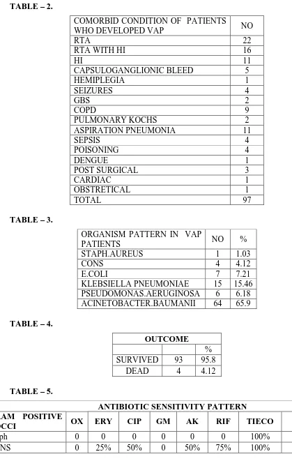
Incidence of VAP was found to be 32.3 %, the organisms isolated in VAP patients are
Acinetobacter- 65.9%, Klebsiella pneumoniae - 15.46%, E.coli - 7.21%, Pseudomonas -
6.18%. Conclusion: Clinicians must focus on eliminating or minimizing the incidence of
VAP through preventive techniques. The causes of VAP and the likelihood of infection by an
antibiotic-resistant strain can be predicted based on the patient characteristics, the duration of
hospitalization, the duration of mechanical ventilation, prior exposure to antibiotic therapy,
Volume 3, Issue 6, 943-954. Research Article ISSN 2277 – 7105
Article Received on 14 June 2014,
Revised on 09 July 2014, Accepted on 04 August 2014
*Correspondence for
Author
Dr. A. Ravishankar Reddy
Associate Professor.
Department of microbiology,
Kamineni Academy of Medical
Sciences & Research Center,
Kamineni Hospitals, L. B.
Nagar, Hyderabad 500068.
and prior colonization patterns. Local microbiology and antibiotic susceptibility data are
essential for making informed antibiotic treatment choices.
KEYWORDS: Ventilator-associated pneumonia (VAP), Aerobic Gram negative bacteria (AGNB), Bronchoalveolar lavage (BAL), Clinical Pulmonary Infection Score (CPIS).
INTRODUCTION
Use of Mechanical ventilation has increased many folds since its first usage in polio
epidemics in 1950s. Ventilator-associated pneumonia (VAP) refers to pneumonia that occurs
more than 48 hours after endotracheal intubation. It is the most common cause of
hospital-acquired infections among patients admitted in ICU 1. The incidence of VAP ranges from 6.8% to 44% and its occurrence is associated with increased length of hospital stay, mortality,
and financial burden2. VAP does seem to be associated with a significantly higher risk of death 3-9.
PATHOPHYSIOLOGY
Pneumonia represents the host’s inflammatory response to the microbial invasion of the
normally sterile lung parenchyma. The magnitude of this response depends on the size and
type of the inoculum, the virulence of the organisms involved, and the competence of the
host’s immune system. There are only four routes through which bacteria can reach the lower
respiratory tract to cause VAP: contiguous spread, hematogenous spread, inhalation, and
aspiration. Hematogenous or contiguous routes of invasion are very rare. Contamination of
the ventilator circuits is universal and has no clinical implications. Therefore, the ventilator
circuit change interval does not affect the incidence of VAP 10.
VAP are caused by the aspiration of infected secretions from the oropharynx 11, 12 Critical illness leads to the rapid colonisation of the oropharynx with potentially pathogenic bacteria
caused by changes in host defences, previous antibiotic exposure, and changes in either the
bacterial adhesins or host surface receptors 13. Aerobic Gram negative bacteria (AGNB) and Staphylococcus aureus rapidly replace normal flora. It remains contentious whether the
aspiration of infected material from the stomach plays an important role 14, 15, 16. However, alkalinisation of the normally acid environment in the stomach leads to overgrowth with
commonly used in ICU, and gain access to the trachea along folds in the cuff. These
organisms can then gain access to and colonise the biofilm that rapidly coats the inner surface
of the tracheal tube 19. This is commonly followed by colonisation of the trachea with pathogenic organisms. The infected material is then propelled into the distal airways by the
inspiratory flow provided by the mechanical ventilator. Occasionally, contaminated
nebulisers, ventilation circuits or humidifiers may be the source of the infected material 20.
MATERIALS AND METHODS
A Prospective study was conducted after Institutional Ethics Committee clearance has been
taken in Kamineni Hospitals. 300 Patients admitted in the emergency / ICU, requiring
intubation and mechanical ventilation for more than 72 hours was considered eligible for
inclusion. Written informed consent was obtained from nearest relative of the patients.The
lack of specificity of a clinical diagnosis of VAP has led to efforts to improve the diagnostic
criteria. A simplified strategy for the management of suspected VAP Adapted from Torres et
al 21. The Clinical Pulmonary Infection Score (CPIS) 22 was developed by weighting of the various clinical criteria usually used for the diagnosis of VAP was used by this study. A
Mini-BAL was performed on all ventilated patients for identification of VAP pathogens. The
microorganisms isolated were identified based on standard bacteriological procedures
including Gram’s stain, colony morphology on blood agar and Mac Conkey agar, and
biochemical reactions 23.
RESULTS
97 patients developed VAP. The organisms identified as Acinetobacter, Klebsiella
Pneumoniae, Klebsiella Pneumoniae, Pseudomonas, Cons, Staph. Aureus. The results are
tabulated.
DISCUSSION
VAP is the most common complication after mechanical ventilation with the incidence
estimated to be 3% per day during first 5 days of ventilation, 2% per day between days 5 and
10 of ventilation and 1% per day thereafter 24 Akcaet al. in their study have discovered following factors to be responsible for multi resistant bacterial infection of early onset –
emergency intubation, aspiration, and Glasgow Coma Scale (GCS) less than 9 25, Bronchard
Our study included maximum number of patients needing intubation and mechanical
ventilation with the diagnosis of head injury (28%), poisoning (4%), central nervous system
disease (12%), respiratory failure (22%), RTA (22%),and others (12%). Association of
emergency intubation, micro aspiration and low GCS were all associated in most of our
patients and may have been responsible for a high incidence of early onset VAP compared to
late onset VAP. After this study, VAP prevention bundles have been instituted to decrease the
incidence of high early onset VAP. Risk factors for pneumonia include use of nasogastric
tube, continuous enteral feeding, prolonged mechanical ventilation (>1 day), use of
H2-receptor antagonist, sucralfate, muscle relaxants, corticosteroids, barbiturates, and inotropic
agents, positive end-expiratory pressure, intense sedation, re-intubation, and tracheotomy.
Multidrug-resistant pathogens such as A. baumannii, Klebsiella pneumoniae, P. aeruginosa,
and E. coli were found to be the common organisms causing VAP. This highlights the need
for treatment of the VAP cases with second-line antibiotics effective against these MDR
pathogens. This finding also emphazises the need for stringent preventive measures against
VAP, as the treatment of an established VAP becomes very expensive, with case fatality rate
27
. Emergence of A. baumannii as a causative organism for VAP many of whom were
carbapenemase producing (31.25%) is a new finding in our study. A. baumannii are aerobic
Gram-negative bacilli and is known for being an opportunistic pathogen responsible for a
number of significant opportunistic infections and possession of various intrinsic drug
resistance gene. Multidrug resistant and carbapenemase nonfermenters were chiefly
responsible for late onset VAP. In our study the rate of carbapenemase-producing bacteria
among all GNB was 29.39% which is higher than other studies published in recent past 28. Various other studies from India have shown a rate between 18.75 and 26% 29, 30. Relatively high rate of carbapenemase may be due to increase prevalence of these bacteria as cross
colonizer in hospitals, especially in developing countries with poor maintenance of infection
control practice. The increase incidence of carbapenemase production might be as a result of
rampant use of carbapenem group of antibiotics and natural selection tool of bacteria like
plasmid and chromosomal-mediated gene transfer among species of
carbapenemase-producing Enterobacteriaceae. It is fast becoming a major health threat among ICU of
developing countries 31. The study also indicated that there was less correlation between the initial prophylactic antibiotic and the bacterial sensitivity. The cause maybe multifactorial,
common causes being change in microbial flora causing infection from time to time, lack of
awareness of causative organism, and their sensitivity pattern, continuation of initial
antibiotic policy is warranted to decrease the misuse of these drugs. Following this study a
stringent antibiotic policy was instituted with the collaboration of intensivist, physicians,
microbiologists, and hospital infection control team. We had observed that Cephalosporins
were the most favored drug as first-line treatment but its effectivity was found to be poor 32. A high sensitivity was seen for Tigecycline and Polymyxin B against Gram-negative isolates
and Vancomycin and Linezolid for Gram-positive isolates. It may be so because these drugs
were reserved as second-line of antibiotic therapy 33. A limitation of our study was it being conducted in a resource-limited setting, with small number of patients with VAP and in a
single center, few patients being lost as they left against medical advice due to financial
constraint and increased cost of treatment. We suggest further multi-centric study with larger
patient population to confirm our findings, in particular the high incidence of carbapenemase
along with other MDR pathogen in Indian ICU.
Diagnosis of VAP; Adapted from Torres et al Clinical suspicion of VAP
Infiltration on chest radiograph + one or more of the following • Purulent tracheal secretions
• Fever • Leucocytosis
Immediate retrieval of respiratory secretions for quantitive culture (either bronchoscopic samples or tracheal aspirates)
Start empirical broad spectrum antibiotic(s) guided by microbiologist
VAP confirmed as highly probable either
clinically or microbiologically
Re-evaluation at 48–72 hours
Yes No
Continue antibiotics for eight days Narrow spectrum
depending on microbiological data
Stop antibiotics if no other source of sepsis
The Clinical Pulmonary Infection Score (CPIS)
Clinical Pulmonary Infection Score (CPIS)
Criterion Score
Fever (°C)
38.5 but 38.9 1
>39 or <36 2
Leukocytosis
<4000 or >11,000/L 1
Bands >50% 1 (additional)
Oxygenation (mmHg)
PaO2/FIO2 <250 and no ARDS 2
Chest radiograph
Localized infiltrate 2
Patchy or diffuse infiltrate 1
Progression of infiltrate (no ARDS or CHF) 2
Tracheal aspirate
Moderate or heavy growth 1
Same morphology on Gram's stain 1 (additional)
Maximal scorea 12
a
At the time of the original diagnosis, the progression of the infiltrate is not known and tracheal aspirate culture results are often unavailable; thus, the maximal score is initially 8–10.
Abbreviations: ARDS, acute respiratory distress syndrome; CHF, congestive heart failure.
Table – 1. Age distribution of patients requiring Mechanical ventilation
Age Frequency
2012 2013
0-9 yrs 0 1
10-19 yrs 3 2
20-29 yrs 11 4
30-39 yrs 8 4
40-49 yrs 10 4
50-59 yrs 21 2
60-69 yrs 16 3
>70 yrs 4 4
TABLE – 2.
COMORBID CONDITION OF PATIENTS
WHO DEVELOPED VAP NO
RTA 22
RTA WITH HI 16
HI 11
CAPSULOGANGLIONIC BLEED 5
HEMIPLEGIA 1
SEIZURES 4
GBS 2
COPD 9
PULMONARY KOCHS 2
ASPIRATION PNEUMONIA 11
SEPSIS 4
POISONING 4
DENGUE 1
POST SURGICAL 3
CARDIAC 1
OBSTRETICAL 1
TOTAL 97
TABLE – 3.
ORGANISM PATTERN IN VAP
PATIENTS NO %
STAPH.AUREUS 1 1.03
CONS 4 4.12
E.COLI 7 7.21
KLEBSIELLA PNEUMONIAE 15 15.46
PSEUDOMONAS.AERUGINOSA 6 6.18
ACINETOBACTER.BAUMANII 64 65.9
TABLE – 4.
OUTCOME
%
SURVIVED 93 95.8
DEAD 4 4.12
TABLE – 5.
ANTIBIOTIC SENSITIVITY PATTERN GRAM POSITIVE
COCCI OX ERY CIP GM AK RIF TIECO VAN LZ
Staph 0 0 0 0 0 0 100% 100% 100%
TABLE – 6.
ANTIBIOTIC SENSITIVITY PATTERN
GRAM NEGATIVE
BACILLI CIP GM AK NETIL CEF+SUL PIP+TAZ IM MM P-B COL
ECOLI 0 0 28.50% 14.20% 57.10% 57.10% 71.40% 85.40% 57.10% 57.10%
KLEBSIELLA 6.60% 0 26.60% 20% 26.60% 26.60% 78% 80% 80% 80%
PSEUDOMONAS 33.30% 33.30% 83.30% 83.30% 83.30% 83.30% 83.30% 83.30% 50% 50%
CONCLUSION
Clinicians must focus on eliminating or minimizing the incidence of VAP through preventive
techniques. Interventions to prevent pneumonia in the ICU should combine multiple
measures targeting the invasive devices, microorganisms, and protection of the patient. VAP
is particularly common in patients with ARDS, after tracheotomy, in patients with COPD,
and in injured and burned patients. Careful monitoring, MiniBAL sample surveillance and
implementation of VAP bundles are important in preventing and for early diagnosis of
complications of mechanical ventilators. The microbial causes of VAP are many and varied.
Most cases are caused by routine bacterial pathogens that reach the lung after aspiration of
oropharyngeal secretions or direct inoculation into the airways. The causes of VAP and the
likelihood of infection by an antibiotic-resistant strain can be predicted based on the patient
characteristics, the duration of hospitalization, the duration of mechanical ventilation, prior
exposure to antibiotic therapy, and prior colonization patterns. Local microbiology and
antibiotic susceptibility data are essential for making informed antibiotic treatment choices.
Simple and effective preventive measures can be instituted easily and at minimal costs. Such
measures might include NIV, diligent respiratory care, hand hygiene, elevation of head, oral
and not nasal cannulation, minimization of sedation, chest physiotherapy, prone positioning,
the timing of tracheostomy, institution of weaning protocols, judicious use of antibiotics,
de-escalation, and leveraging PK/PD characteristics for antibiotics administered. More costly
interventions should be reserved for appropriate situations.
BIBLIOGRAPHY
1. Wagh H, Acharaya D. Ventilator Associated Pneumonia- an Overview. Br J Med Pract
2009; 2:16-9.
2. Peter JV, Chacko B, Moran JL. Comparison of closed endotracheal suction versus open
endotracheal suction in the development of ventilator associated pneumonia in intensive
care patients: An evaluation using meta analytic techniques. Indian J Med Sci 2007;
61:201-11.
3. Heyland DK, Cook DJ, Griffith L, et al. The attributable morbidity and mortality of
ventilator-associated pneumonia in the critically ill patient. The Canadian Critical Trials
Group. Am J Respir Crit Care Med 1999; 159:1249–56.
4. Fagon JY, Chastre AJ, Hance AJ, et al. Nosocomial pneumonia in ventilated patients: a
cohort study evaluating attributable mortality and hospital stay. Am J Med 1993; 94:281–
5. Bercault N, BoulainT. Mortality rate attributable to ventilator-associated nosocomial
pneumonia in an adult intensive care unit: a prospective casecontrol study. Crit Care Med
2001; 29:2303–9.
6. Craig CP, Connelly S. Effect of intensive care unit nosocomial pneumonia on duration of
stay and mortality. Am J Infect Control 1984; 12:233–8.
7. Baker AM, Meredith JW, Haponik EF. Pneumonia in intubated trauma patients.
Microbiology and outcome. Am J Respir Crit Care Med 1996; 153:343–9.
8. Cunnion KM, Weber DJ, Broadhead WE, et al. Risk factors for nosocomial pneumonia:
comparing adult critical care populations. Am J Respir Crit Care Med 1996; 153: 158–62.
9. Papazian L, Bregeon F, Thirion X, et al. Effect of ventilator-associated pneumonia on
mortality and morbidity. Am J Respir Crit Care Med 1996; 154:91–7.
10.Dreyfuss D, Djedaini K, Gros I, et al: Mechanical ventilation with heated humidifiers and
head moisture exchangers: Effects on patient colonization and incidence of nosocomial
pneumonia. Am J Respir Crit Care Med 1995; 151:986–992
11.Estes RJ, Meduri GU. The pathogenesis of ventilator-associated pneumonia: I.
Mechanisms of bacterial transcolonization and airway inoculation. Intensive Care Med
1995; 21:365–83.
12.Kollef MH. The prevention of ventilator-associated pneumonia. N Engl J Med 1999;
340:627–34.
13.Garrouste-Orgeas M, Chevret S, Arlet G, et al. Oropharyngeal or gastric colonization and
nosocomial pneumonia in adult intensive care unit patients. A prospective study based on
genomic DNA analysis. Am J Respir Crit Care Med 1997; 156:1647–55.
14.Bonten MJ, Gaillard CA, van Tiel FH, et al. The stomach is not a source for colonization
of the upper respiratory tract and pneumonia in ITU patients. Chest 1994; 105 : 878–84.
15.Craven DE, Steger KA, Barber TW. Preventing nosocomial pneumonia: state of the art
and perspectives for the 1990’s. Am J Med 1991; 91:44–53.
16.Du Moulin GC, Paterson DG, Hedley-Whyte J, et al. Aspiration of gastric bacteria in
antacid-treated patients: a frequent cause of postoperative colonisation of the airway.
Lancet 1982; i: 242–5.
17.Donowitz LG, Page MC, Mileur BL, et al. Alterations of normal gastric flora in critical
care patients receiving antacid and ciimetidine therapy. Infect Control 1986; 7:23–6.
18.Seegobin RD, van Hasselt GL. Aspiration beyond endotracheal cuffs. Can Anaesth Soc J
19.Sottile FD, Marrie TJ, Prough DS, et al. Nosocomial pulmonary infection: possible
etiological significance of bacterial adhesion to endotracheal tubes. Crit Care Med 1986;
14: 265–70.
20.Craven DE, Lichtenberg DA, Goularte TA, et al. Contaminated medication nebulizers in
mechanical ventilator circuits. Source of bacterial aerosols. Am J Med 1984;77: 834–8.
21.Kollef MH. Inadequate antimicrobial treatment: an important determinant of outcome for
hospitalized patients. Clin Infect Dis 2000; 31:S131–8.
22.Harrison's Principles of Internal Medicine (18e)
23.Mackie TJ and McCartney JE (1996) Practical medical microbiology, 14th edition. New
York: Churchill Livingstone 978p.
24.Rello J, Torres A, Ricart M, Valles J, Gonzalez J, Artigas A, et al. Ventilator-associated
pneumonia by Staphylococcus aureus: Comparison ofmethicillin-resistant and
methicillin-sensitive episodes. Am J Respir Crit Care Med 1994;150:1545-9.
25.Akca O, Koltka K, Uzel S, Cakar N, Pembeci K, Sayan MA, et al. risk factors for early
onset ventilator associated pneumoniain critical care patients. Anesthesiology
2000;93:638-45.
26.Bronchard R, Albaladejo P, Brezac G, Geffory A, Seince PF, Morris W, et al. Early onset
pneumonia risk factors and consequences in head trauma patients. Anesthesiology
2004;100:234-9.
27.Erbay RH, Yalcin AN, Zencir M, Serin S, Atalay H. Costs and risk factors for
ventilator-associated pneumonia in a Turkish university hospital’s intensive care unit: A
case-control study. BMC Pulm Med 2004;4:3.
28.Dey A and Bairy I. Incidence of multidrug-resistant organisms causing
ventilator-associated pneumoniain a tertiary care hospital: A nine months’ prospective study. Ann
Thorac Med 2007;2: 52-7.
How to cite this article: Thakuria B, Singh P, Agrawal S, Asthana V. Profile of infective
microorganisms causing ventilator-associated pneumonia: A clinical study from resource
limited intensive care unit. J Anaesthesiol Clin Pharmacol 2013;29:361-6.
29.Joseph NM, Dutta SS, Badhe AS, Rsistha D, Parija SC. Ventilator associated pneumonia
in a tertiary care hospital in India: Role of multidrug resistant pathogen. J Infect Dev
Ctries 2010;4:218-25.
30.Solanke V, Pai C, Urhekar AD. Comparative bacteriological study of community
31.Miriagou V, Cornaglia G, Edelstein M, Galani I, Giske CG, Gniadkowski M, et al.
Acquired carbapenemases in Gram-negative bacterial pathogens: Detection and
surveillance issues. Clin Microbiol Infect 2010;16:112-22.
32.Dancer SJ. The Problem with cephalosporins. J Antimicrob Chemother 2001;48:463-78.
33.Tsering D, Das S, Adhiakari L, Pal R, Singh T. Extended spectrum beta-lactamase