INTERNAL FIXATION OF TYPE C FRACTURE OF DISTAL
HUMERUS
Dr. S. Santhosh*
Final year M.S (ORTHO) P.G, Dept. of Orthopaedics Sree Balaji Medical College and
Hospitals Biher, No.7, Clc Works Road, Chromepet, Chennai-600044.
ABSTRACT
Background: Type C fracture of distal humerus is a relatively
uncommon fracture. Internal fixation is difficult but anatomical
reduction is needed to prevent poor functional outcome and
degenerative changes. Methods: Twenty two cases with type C fracture
of distal humerus including 6 having grade I compound fracture were
treated with open reduction and internal fixation. Dual 3.5mm
reconstruction plates in two planes were used in 17 patients and single
plate was used in 5 patients. Patients were followed up for a mean period
of 45 months (24 to 60 months). Results: All the fractures united at a
mean duration of 13 weeks (8 to 20 weeks). Mean loss of extension
(flexion deformity) was 280 (50 to 600). Mean range of movement achieved was 1060. Complications were few, except restriction of movement. Conclusion: Internal fixation is a good method of
treatment for this type of fracture to get restoration of the articular surface anatomy, stable
fixation and early mobilization.
KEYWORDS: Internal fixation; Distal humerus fracture.
INTRODUCTION
Intra-articular bicondylar fractures of distal humerus (Type C, AO classification) are difficult to
manage. Malunion, stiffness and osteoarthrosis are common. Many methods like close
reduction, hanging arm cast, traction, limited internal fixation, open reduction with rigid
fixation and elbow replacement have been described. In the last few decades, the popularity
of internal fixation of this fracture is growing fast.[1-3] Being intra-articular fracture the importance of anatomical reduction is vital. Surgical treatment gives a chance for accurate
Volume 7, Issue 18, 1086-1091. Research Article ISSN 2277– 7105
Article Received on 04 Sept. 2018,
Revised on 24 Sept. 2018, Accepted on 15 Oct. 2018
DOI: 10.20959/wjpr201818-13590
*Corresponding Author
Dr. S. Santhosh
Final year M.S (ORTHO)
P.G, Dept. of Orthopaedics
Sree Balaji Medical College
and Hospitals Biher, No.7,
Clc Works Road,
Chromepet,
Patient was kept in lateral position, arm resting on a bolster which was kept in front of the
chest. Tourniquet was used, but released as soon as the exposure was completed.
Transolecranon approach was used in all the cases. A transverse osteotomy was used but the
subchondral bone was fractured by levering the osteotome instead of cutting it. The ragged
edge created in this manner helped in accurate reduction at the time of olecranon fixation.
Fracture haematoma was cleaned. Assessment of the fracture anatomy was done. It may be
different from what was seen on X-ray. No fragment was discarded except the very small
one. Reduction was done and fixed temporarily with 1.5mm K-wires. Reduction forceps with
points was useful to hold the condyles. Anatomical reduction was the aim. Reconstruction of
the trochlea is the most important part. Stenosis of the olecranon fossa was avoided at all
cost. Valgus and varus position were checked. Normal anterior tilt of the condyle or humero
-capitulum angle was checked. Defect in the inter condylar area, if any, was filled with
cancellous bone graft. Inter condylar fracture was fixed with a 4 mm cancellous screw as lag
screw. But compression was avoided in presence of comminution. If medial or lateral column
was broken as a butterfly fragment or a wedge, it was fixed to the proximal fragment with lag
screw to make the fracture anatomy simpler before applying plate. Two 3.5 reconstruction
plates in two perpendicular planes were used in 17 cases. One 3.5 mm reconstruction plate
was kept on the posterior surface of the lateral column and one on the medial side of the
medial column. Main problem was to achieve a good purchase of the distal screw in low
fractures. In such cases, the lag screw which was fixing the intercondylar component of the
Fig 1. Pre-operative X-ray of a 30 years
old male with type C fracture.
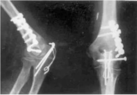
reconstruction
Fig 2. Post-operative X-ray. Two 3.5 plates were used. The lag screw fixing the intercondylar fracture was through the last
This increased the fixation to the distal fragment. Single plate was used in 5 patients. If the
plate was encroaching the ulnar groove, the nerve was transposed anteriorly and noted
carefully for future reference. Olecranon was fixed with tension band wiring. A below elbow
slab was applied at 70º to 80º flexion. Exercise was started as soon as pain subsided, usually in
one week. Only active exercise was given. No passive mobilization was done. Muscle
strengthening exercises was given after the union of the fracture. Result was assessed with
criteria of Riseborough and Radin (1969) and criteria of Jupiter et al and Mayo elbow
performance score (Table I, II).
Table I. Criteria of Riseborough and Radin. (1969).
Flexion contracture flexion Subjective symptom
good <30 >115 minor fair 30-60 >115 Minor poor >60 <115 major
Table II: Criteria of Jupiter etal (1985) Range of movement (degrees).
Loss of extension flexion pain disability
excellent <15 >130 none none good <30 >120 slight minimal fair <40 >90 With activity moderate poor <40 >90 varible severe
RESULT
Mean age of the patient was 34 years (20 to 45 years). Patients were followed up for a mean
duration of 45 month (24 to 60 months). All the fracture united in a mean duration of 13
[image:3.595.79.521.74.287.2]Table III: Functional results with three assessment criteria (expressed in number of
patients and its percentage in bracket.).
Riseborough and radin criteria
Jupiter et al criteria
Mayo elbow performance score
Excellent - 0 6(27.2) Good 15(68.2) 15(68.2) 12(54.5) Fair 5(22.7) 3(13.6) 4(18.2) Poor 2(9.1) 4(18.2) 0
DISCUSSION
Type C fractures of distal humerus are difficult to manage inspite of the advancement in
fixation technique. Though the range of movement is better in surgically treated patients,
stiffness is the most important complication. It is due to intra- articular adhesion, periarticular
fibrosis, myositis ossificans and malunion. Accurate restoration of articular surface prevents
osteoarthrosis. Reported[8] mean range of movement in conservatively treated patients is 470. Mean range of movement obtained in this series (1060), is less than 1150 and 1080 reported.[4,9] Result appears better with Mayo elbow performance score as 18 patients (81.8%) were rated
as excellent or good. The reason is that this score provide only 20 points to motion and 80
points to pain, instability and function. These patients rarely complained of pain and
[image:4.595.179.416.614.744.2]Fig 4. X-ray after the union of the fracture. Medial wedge fragment was fixed with
interfragmentary screw from lateral side and single plate for lateral column was used.
Perfect bony anatomy was restored.
Transolecranon approach is must for these fractures. We did not find any difficulty in
reduction and fixation of olecranon and none of the cases developed nonunion of olecranon.
Anatomical reduction of articular surface was achieved in all cases without comminution at
articular surface, but in cases with comminution some amount of step at the articular surface
was common. Fixation of the distal fragment in low fracture is a problem as there is hardly any
space to accommodate two screws for each plate. Preoperatively placement of the screws
should be meticulously planned. Passing the lag screw which fixes the intercondylar fracture
through the plate is one way to increase the fixation (fig.2). If the purchase of the screw is
poor, double tension band wiring is one option.[9] If there is wedge fragment from the medial or lateral column it should be fixed to the shaft of humerus with lag screw first to make the
fracture anatomy simple.
This fracture in young patients should be treated with internal fixation through a
transolecranon approach. Single method of fixation could not be applied to every case.
Meticulous planning should be done for every case. Functional results are better than
radiological results.
REFERENCES
1. Gabel GT, Hanson G, Bennett JB, Noble PC, Tullos HS. Intra- articular fracture of
the distal humerus in the adult. Clin Orthop, 1987; 216: 99-108.
2. Holdsworth BJ, Mossad MM. Fractures of the adult distal humerus: elbow function after
[image:5.595.179.413.71.233.2]Shoulder Elbow Surg, 2002; 11(1): 48- 52.
6. Riseborough EJ, Radin EL. Intercondylar T fractures of the humerus in the adults (a
comparison of operative and non-operative treatment in twenty nine cases J Bone Joint
Surg (Am), 1969; 51: 130.
7. Wallace E, Miller MD. Comminuted fracture of the distal end of the humerus in adult.
J Bone Joint Surg (Am), 1964; 46: 644-57.
8. Zhao J, Wang X, Zhang Q. Surgical treatment of comminuted intra- articular fractures
of the distal humerus with double tension band Osteo- synthesis. Orthopaedics, 2000;