warwick.ac.uk/lib-publications
A Thesis Submitted for the Degree of PhD at the University of Warwick
Permanent WRAP URL:
http://wrap.warwick.ac.uk/102838/
Copyright and reuse:
This thesis is made available online and is protected by original copyright. Please scroll down to view the document itself.
Please refer to the repository record for this item for information to help you to cite it. Our policy information is available from the repository home page.
1
Pathways
that Regulate Renal Development, Fibrosis, and
Metabolic Disease in Mouse Models
Abdulsalam Soofi, MS, BS
Research Lab Specialist Senior
Department of Pathology-Medical School
University of Michigan, Ann Arbor
Michigan, USA
Thesis submitted for the degree of Doctor of Philosophy
Division of Metabolic & Vascular health
Warwick Medical School
University of Warwick
United Kingdom
2017
4 Contents: Page
- Acknowledgment. . . 6
- Declaration. . . 7
- Acknowledgments of contributions. . . 11
- Abstract . . . 12
- Chapter I. . . 14
General Background I.a- Pax2 Roles and Regulation in Kidney Development, Disease, and Repair/ Regeneration I.a.1- Mammalian Pax Gene Family I.a.2- Pax2 Role in Kidney Development I.a.3- Pax2 Role in Kidney Repair and Regeneration I.a.4- Pax2 & TGFb Superfamily Interaction During Kidney Development I.b- Kidney Biology and Function. . . . . . . . . . . . . . . . 21
I.c- Chronic Kidney Disease and Fibrosis. . . 24
I.d- Obesity; Origin and Characterization of Adipose Tissue. . . 26
I.e- Obesity and Liver . . . 29
I.f- The Transforming Growth Factor β (TGFβ) Signaling Pathway . . . 30
I.f.1- TGFb Signaling Transduction/ Diagram I.f.2- TGFb Prologues (TGFb-1, 2, 3) in Kidney I.f.3- Regulation of Receptor Activation I.g.- Kielin/Chordin-like Protein (KCP) Characterization . . . 37
I.g.1- KCP Functions and Attributions
I.g.2- Original Description of KCP
5
- Chapter II . . . 42
Papers Resulting from Research for this Thesis
Paper 1- Soofi, A., Levitan, I., and Dressler, G.R. (2012). Two novel EGFP insertion alleles reveal unique aspects of Pax2 function in embryonic and adult kidneys. Developmental Biology 365, 241-250.
Paper 2- Soofi, A., Zhang, P., and Dressler, G.R. (2013). Kielin/chordin-like protein attenuates both acute and chronic renal injury. Journal of the American Society of Nephrology 24, 897-905
Paper 3- Soofi, A., Wolf, K.I., Ranghini, E.J., Amin, M.A., and Dressler, G.R. (2016). The kielin/chordin-like protein KCP attenuates nonalcoholic fatty liver disease in mice. American Journal of Physiology Gastrointestinal and Liver Physiology 311, G587-G598.
Paper 4- Soofi, A., Wolf, K.I., Emont, M.P., Qi, N., Martinez-Santibanez, G., Grimley, E., Ostwani, W., and Dressler, G.R. (2017). The kielin/chordin-like protein (KCP) attenuates high-fat diet-induced obesity and metabolic syndrome in mice. Journal
Biological Chemistry 292, 9051-9062.
- Chapter III . . . 132
Thesis Conclusions, Reflections and Future Directions
III.a- Thesis Conclusions
III.b- Thesis Reflections and Future Directions
III.b.1- The ‘How I would do the experiments differently now’
question?
III.b.2- Further experiments based on this thesis
- List of references . . . . . . 141
- Candidate List of Publications . . . 170
6
Acknowledgment
I would like to express my sincere gratitude to my advisor Prof. Victor Zammit,
medical school, University of Warwick, UK. I admire him for his patience, motivation,
and the continuous support he gave me during the process of my Ph.D application and
thesis. His immense knowledge and clear guidance helped me write this thesis in best
way possible.
My sincere thanks also goes to Prof. Greg Dressler, Department of Pathology,
medical school, University of Michigan, for providing me the opportunity to join his lab.
He gave me the freedom and trust to do my own work independently. Without his strong
support, it would not be possible to conduct this research and obtain my degree.
I would like to thank my friend Dr. Adam Stein and Dr. Patel, medical school,
University of Michigan for their support and encouragement.
I thank my fellow lab mates and dear friends Dr. Egon Ranghini and Dr. Edward
Grimley for the insightful discussions and support.
Last but not the least; I would like to thank my family; my wife for supporting me
spiritually for the last 20 years and my children, Aisha and Hamzah, who inspired me to
11
Acknowledgment of Contributions
My extended acknowledgments to the core faclities at the University of Michigan for their contributions.
- Thanks to the Transgenic Animal Model Core Facility:
Tom Saunders and E. Hughes for the generation of ES cells carrying the Egfp-neo cassette. M. Van Keuren for helping to generate the KCP transgenic mice,
- Thanks to the Mouse Genomic Core Facility:
Jante Hoff for help with the UUO surgery
- Thanks to the Images Analysis, Microscope & Image Analysis laboratory Core:
Chris Edward for his help with image analysis of the embryonic kidney life images. Judy Poore and Jeff Harrison for tissue processing and sectioning.
- Thanks to the DNA Sequencing Core:
Craig Johnson for affymetrix analyses
- Thanks to the Animal Phenotyping Core:
Elisabeth Limback and Melanie Schmitt for help with the metabolic analysis.
12
ABSTRACT
The kidney is an essential organ that maintains homeostasis, maintains water and mineral balance, and removes metabolic waste products from the body. In mammals, the kidney derives from the intermediate mesoderm (IM) and develops through a multistep process where undifferentiated mesenchyme is converted into a highly complex organ. Several transcriptional regulators, including the Pax2 gene, have been identified in the specification and maintenance of this multistep process. The Pax2 gene marks the IM shortly after gastrulation, when the mesoderm becomes compartmentalized into paraxial, intermediate, and lateral plate. Pax2 expression in the IM distinguishes all of the cells fated to become epithelia in the urogenital tract and is necessary to establish and maintain this phenotype.Pax2null mutants do develop a nephric duct (Brophy et al., 2001; Soofi et al., 2012), but the duct is completely absent in a Pax2/8double mutant, suggesting that these Pax genes function redundantly in this early IM domain; however, in Pax2 homozygous mutant mice, the metanephric mesenchyme neither responds to inductive signals nor does the mutant mesenchyme aggregate into early renal vesicles resulting in a lack of kidneys, ureters, and genital track. We describe two new alleles of Pax2 created by inserting the Enhanced Green Fluorescent Protein coding region into the 5' untranslated leader sequence. One allele is a hypomorph that generates less protein and exhibits structural defects in kidneys and ureters upon homozygosity. A second allele is a true null that can be used to image Pax2 expressing cells in a mutant background. Organ culture and embryo analyses point to a loss of epithelial cell polarity and increased mobility in cells that have deleted Pax2 function. These experiments provide new insight into the role of Pax2 protein levels in determining correct renal architecture and cell fate.
13 proteins in enhancing or suppressing renal interstitial fibrosis, respectively, the results of this thesis will show how the expression of this secreted protein KCP could diminished renal fibrosis in mouse models of chronic and acute kidney disease.
In vivo, KCP-KO mice are viable and fertile but are more sensitive to tubular injury and exhibit significant pathology after recovery. Also, deletion of KCP sensitized mice to developing obesity and associated complications such as liver steatosis and glucose intolerance. In contrast, transgenic mice that expressed KCP in the kidney, liver, and brown adipose tissues were resistant to developing high fat diet induced obesity and had significantly reduced white adipose tissue. This data demonstrates that modulation of the TGFβ signaling with secreted inhibitors or enhancers can alter the profile of adipose tissue, which reduces obesity and impaired the progression of metabolic disease.
The Metabolic Syndrome is reaching epidemic proportions in the developed world, primarily due to the increased availability of high caloric foods and the decrease in daily physical activity. Energy balance is critical for maintaining normal body weight and homeostasis. When caloric intake chronically exceeds energy expenditure, white adipose tissue stores excess energy in the form of triglycerides, leading to obesity and related complications such as type-2 diabetes, a condition also referred to as metabolic syndrome which is a condition of chronic sub-clinical inflammation.
In mice, the TGFβ superfamily has been implicated not only in the development and differentiation of white and brown adipose tissues, but also in the induction of the pro-inflammatory state that accompanies (Tseng et al., 2008). The work outlined in this thesis suggests that altering the TGFβ superfamily signaling pathway by a secreted protein (KCP) can attenuate renal fibrosis and the negative effects of obesity-associated metabolic syndrome. Providing a conceptual basis for the use of small molecule analogues of KCP to attenuate profibrotic pathways that depend on continued TGFβ
14
Chapter I. General Background
Ia. Pax2 Roles and regulation in Kidney development, disease, and
Repair/Regeneration
I.a.1- Mammalian Pax Gene Family
The PAX family is classified into four groups according to their structural similarity, sequence homology, the presence or absence of an octapeptide motif and also according to its homeodomain or partial homeodomain (Dahl et al., 1997) (Table 1). Pax proteins are characterized by the presence of a 128 amino acid sequence in their structure, which constitutes a DNA-binding domain, the paired domain (PD) (Chi and Epstein, 2002). Each Pax protein has a c-terminal region, rich in serine and threonine, that is responsible for transcriptional activation of target genes (Chi and Epstein, 2002; Ward et al., 1994).
15
I.a.2- Pax2 Role in Kidney Development
In mouse embryos, Pax2 is expressed around the 9-somite stage in the nephric duct primordium (Bouchard et al., 2000; Bouchard et al., 2002; Torres et al., 1995). Pax2 mutant embryos initially form a nephric duct, which degenerates by apoptosis during the elongation process, and fail to form normal mesonephric tubules (Dressler et al., 1990). As a result, Pax2-deficient embryos completely lack metanephric kidneys (Bouchard et al., 2002; Torres et al., 1995). Surprisingly, Pax8 null embryos show normal nephric duct and kidney development but die postnatally due to developmental defects in the thyroid gland (Mansouri et al., 1998). However, in the context of Pax2 gene deficiency, Pax8 inactivation exacerbates urogenital defects such that the pro/mesonephros is completely absent and the prospective renal tissue undergoes massive apoptosis (Bouchard et al., 2002). This finding demonstrates the functional redundancy between Pax2 and Pax8 as pro/mesonephros development is initiated normally with either Pax2 or Pax8 present (Bouchard et al., 2002). The fact that only Pax2 is required for later renal development
17 these findings underline the complexity of the Pax2 regulatory network in the ductal epithelium and further suggest that some regulatory interactions are maintained but are utilized differently in different systems.
The metanephros is the site of adult kidney development in the vertebrate embryo and responds to the concerted action of several genetic regulators. Metanephric development in mice begins at E10.5 by induction of the nephric duct to form the ureteric bud, which invades the metanephric mesenchyme and initiates branching morphogenesis. Ureteric bud formation is initiated by the action of the mesenchymal signal Gdnf that binds the co-receptor complex Ret/GFR 1 expressed in the nephric duct epithelium (Chi et al., 2009). This crucial interaction induces cell shape changes and proliferation that leads to the invasion of the metanephric mesenchyme by the ureteric bud (Chi et al., 2009; Dressler, 2009). Accordingly, inactivation of Gdnf, Ret or Gfr1 prevents ureteric bud formation, leading to renal agenesis (Skinner et al., 2008). Pax genes act on this system at several distinct levels during kidney development. In the nephric duct, Ret is a direct regulatory target of Gata3 (Boualia et al., 2013; Grote et al., 2008; Marcotte et al., 2014). The Pax2/8-Gata3 cascade is therefore necessary to establish the responsiveness of the nephric duct to kidney induction. Among the transcriptional regulators are Osr1, Pax2, Eya1, Hox11 and Six1/2 (Brophy et al., 2001; Wellik et al., 2002; Xu et al., 1999; Xu et al., 2003). Inactivation of each of these genes leads to a down regulation or loss of Gdnf expression in the metanephric mesenchyme which prevents normal kidney development.
I.a.3- Pax2 Role in Kidney Regeneration and Repair
19 singularly re-expressed among other transcription factors, such as Pax8, WT1, Wnt4 and BF-2, which are also present during development (Maeshima et al., 2002a). These studies emphasize that the presence of Pax2 may potentially influence renal regeneration, conducting key events as it does during development, but that actions of Pax2 in renal recovery are still not fully understood. Thus, Pax2 expression would drive tubular kidney cells to proliferate. In addition, the expression of this gene has also been demonstrated to prevent apoptosis. Torban and coworkers (Torban et al., 2000) used different strategies, in vivo and in vitro, to confirm that the primary function of Pax2 is preventing apoptosis, but demonstrated that Pax2 does not lead to proliferation. Most of the literature is in agreement with the view that Pax2 protects cells from apoptosis; however, further studies are necessary to better clarify other features of Pax2 actions in cell biology. There are very few in vivo approaches to directly associate Pax2 with renal recovery after injury, especially showing the participation of Pax2 in key processes related to tissue repair in vivo. It can be considered a growing field of interest as judged by the increasing number of studies showing that different factors known to influence renal tissue regeneration are now being related to Pax-2 gene expression (Maeshima et al., 2002a; Zhang et al., 2004).
I.a.4- Pax2 &TGF
β
Superfamily Interaction in Kidney Development
20 prologues in tissue development depends on the presence or absence of specific TGFβ
superfamily co-receptors such as the type III TGFβ receptor (TGFBR3), commonly referred to as betaglycan, an accessory receptor (Stenvers et al., 2003), The betaglycan heterozygous kidneys exhibited accelerated ureteric branching with a transient decrease in BMP4 expression at E11.5 and a subsequent cascade of changes in the gene regulatory network that governs metanephric development, including significant increases in Pax2, Eya1, Gdnf, Ret, Wnt4, and WT1 expression. In contrast, betaglycan null kidneys exhibited renal hypoplasia (Walker et al., 2011).
Lindoso R S et al., 2009, reported that activin A and TGFβ1 promote downregulation of Pax2 expression inhibiting cellular proliferation (Lindoso et al., 2009). In kidneys, Activins are expressed during development and re-expressed after injury periods (Maeshima et al., 2001; Tuuri et al., 1994). These proteins act as autocrine factors and play different roles in the kidney, such as activation of renal interstitial fibroblasts (Yamashita et al., 2004).
The putative mechanism of action of activin A is regulation of the expression of transcription factors like Pax2 (Nakamura et al., 1990). Data presented by Maeshima and coworkers showed that Pax2-positive cells present specific activin A receptors (ActR-II) and that administration of activin A leads to a reduction in the number of cells therefore BrdU/Pax2 double positive in vivo (Maeshima et al., 2002a). Activin A leads to reduction of Pax2 expression in the kidney culture system during embryonic development as well as in tubular cell lineages (Maeshima et al., 2002a; Maeshima et al., 2002b; Maeshima et al., 2006). The inhibition of activin A, either by follistatin or by superexpression of a mutant truncated receptor, leads to increases in Pax2 expression and cell growth promotion (Maeshima et al., 2002a). Another member of the TGFβ
21 known for modulating important growth regulatory gene products, affects the stability of Pax2 mRNA and consequently promotes a reduction of the Pax2 protein in the cell.
I.b- Kidney Biology and function
22 The kidneys consist of two essential parts: an outer part “cortex” and an inner part “medulla” (Figure 2). Each adult kidney contains about one million nephrons and each nephron contains a glomerulus surrounded by a thin-walled, bowl-shaped structure named Bowman capsule. The nephron also contains a small tube that drains filtrate from the space in the Bowman capsule and a collecting duct that drains urine from the tubule and regulates urine concentration (Kim et al., 2007). Each tubule has three interconnected parts: the proximal convoluted tubule, the loop of Henle, and the distal convoluted tubule. The distal tubule connects to the collecting duct, a continuous highly arborized epithelial network with a quite distinct origin from the contiguous renal tubule. The collecting duct epithelium displays a distinct cortical-medullary axis of branching, and cellular organization. The medullary collecting ducts are highly water permeable in order to facilitate water retention which is critical for sodium retention (Al-Awqati and Gao, 2011; Pearce et al., 2015).
Figure 1. The intermediate mesoderm: its origin and derivatives. A) Cross section embryo at E8.5B) the Wolffian duct at E9.0. C) Mesonephric tubules at E10. D) Outgrowth of the ureteric bud (UB). E) The UB has bifurcated and induced mesenchyme surrounds the tips.G) Live image of E115.5 eGFP kidney of WT embryos. Images A to E are from Dressler, 2009 and G done by the author.
G
[image:21.612.133.532.150.351.2]23 The primary function of the kidneys is to maintain the proper balance of water and minerals in the body. Mineral balance is maintained by tightly controlled ion fluxes that are external (intestine and kidney) and internal (between bone and other organs), and are regulated and coordinated by many endocrine signals among these organs (Kuro and Moe, 2016). An additional function is filtration and excretion of waste products from the processing of food, drugs, and harmful substances. Blood is filtered through small pores in the glomerulus, leaving behind blood cells and large molecules, such as proteins. The Figure 2. Adult Kidney Anatomy: A) Adult Kidney image obtained from Kidney Cross Section Diagram – Human Anatomy System. B) Scanning Electron Microscope (SEM) of the Cortex. C) SEM of Glomeruli D) a see through the filtration barrier. Images B, C and D done by the author.
A
B
C
[image:22.612.113.547.153.497.2]24 glomerular basement membrane acts as a filtration barrier, reducing entry of larger molecular weight serum solutes into the nephron (> 15 kDa) such as serum albumin (Miner, 2011; Suh and Miner, 2013). In healthy adults, about 180 liters of fluid is filtered into the kidney tubules each day. Nearly all of this fluid, (and the electrolytes contained in it), is reabsorbed by the kidney. The prevalence of chronic kidney disease substantially increases with increasing metabolic syndrome risk factors (Chen et al., 2004). There are a number of pathologic links between metabolic syndrome and chronic kidney disease (Abrass, 2004). Contemporary research highlights the relationship between hyperinsulinemia and modifications within the kidney, including glomerular hypertrophy, mesangial matrix proliferation, and glomerulosclerosis. These changes are thought to be secondary to glomerular hyperfiltration as well as inflammatory mediators from increased adiposity. Additionally, obesity-related kidney damage has been posited to be due to a series of alterations like hyperlipidemia, increased oxidative stress, increased salt intake, and activation of the sympathetic nervous system (Palatini, 2012). Also hyperglycemia, hypertension (Eckel et al., 2005; Wong et al., 2016) and protein damage due to glycation may contribute to kidney damage (Faria and Persaud, 2017) .
I.c- Chronic Kidney Disease and Fibrosis
Chronic kidney diseases (CKD) can be due to structural or functional abnormalities typically characterized by active inflammation and renal fibrosis. While the primary pathology leading to most forms of CKD differs significantly, all forms of progressive renal diseases, including glomerulonephritis, chronic interstitial nephritis, and diabetic nephropathy, exhibit interstitial fibrosis (Eddy, 1996; Fogo, 2000). Despite the strong correlation between tubulointerstitial fibrosis and the loss of renal function, the molecular mechanisms underlying fibrosis have remained elusive. However, evidence pointing to the TGFβ superfamily of proteins as primary regulators of fibrosis is accumulating. Indeed, TGFβ1 is generally regarded as the key mediator in the development of renal fibrosis (Flanders, 2004). Transgenic mice overexpressing TGFβ
25 1996; Ledbetter et al., 2000). Furthermore, inhibition of TGFβ by neutralizing antibodies can improve injury in various models of kidney disease (Ziyadeh et al., 2000). In the normal kidney, the expression of TGFβ is weak; however, many disease states, including diabetes mellitus, increase TGFβ activity (Yamamoto et al., 1996). TGFβ induces resident fibroblasts to produce extracellular matrix components, such as type IV collagen and fibronectin, leading to the formation of tubulointerstitial fibrosis (Marti et al., 1994; Martin et al., 1998). BMPs may play an important role in kidney development and kidney regeneration (Cirio et al., 2014; Tsujimura et al., 2016). Animal studies have shown that systemic administration of BMP7 can reverse damage induced kidney fibrosis (AKI), improve cartilage damage, and inhibit the formation of bone metastases resulting from prostate or breast cancer, and increase energy expenditure by inducing the formation of brown adipocyte tissue. BMP7 thus seems a very promising new therapeutic agent in the treatment of a variety of disease states, including obesity and obesity-related disorders such as type 2 diabetes mellitus and cardiovascular disease. An understanding of the complexities of the interplay between the TGFβ1 signaling pathway and the development of CVD, CKD, and obesity with insulin resistance are important (Figure 3).
[image:24.612.151.453.431.615.2]26
I.d- Obesity; Origin and Characterization of adipose tissue
In humans, two types of adipose tissue can be distinguished both histologically and functionally: white adipose tissue (WAT) and brown adipose tissue (BAT). Whereas WAT is the main tissue for storage of triglycerides in the form of fat, BAT has evolved to generate heat through uncoupled mitochondrial fatty acid oxidation (Cannon and Nedergaard, 2004). Much progress has been made toward understanding the developmental origins of brown and white adipocytes, although all aspects have not been resolved. Lineage- tracing studies of adipose tissue and muscle are both considered to be of mesodermal origin (Gesta et al., 2007). Adipocytes develop from mesenchymal stem/progenitor cells which derive from embryonic stem cells. When triggered by appropriate developmental cues, these cells become committed to adipocyte lineages, i.e. the preadipocytes (Figure 4). More recently, Seale et al., used a myogenic marker, myf5, to perform cell fate mapping in the mouse and found that both skeletal muscle and interscapular brown fat, but not white fat, arise from progenitors expressing myf5 (Seale et al., 2008) . In addition to these discrete interscapular brown fat cells, uncoupling protein1 (UCP-1-positive) brown adipocytes are also found systemically distributed in the body, especially within white fat depots (Cousin et al., 1992) and between muscle bundles (Almind et al., 2007). Interestingly, these “systemic” brown adipocytes, such as those present in white fat and muscle, are not derived from myf5-expressing precursors (Seale et al., 2008), suggesting different developmental origins for these different pools of brown fat (beige adipose cells). We are still early in the process of understanding the similarities and differences between brown and beige adipose cells, and we do not yet have a clear picture of their relative importance in energy homeostasis.
27 confirmation that adult humans have brown adipose tissue (BAT) has transformed our understanding of how adipose tissue regulates metabolism and energy balance once again.
(Cypess et al., 2009; van Marken Lichtenbelt et al., 2009; Virtanen et al., 2009). The relatively new finding that some adult humans have substantial amounts of heat-dissipating brown adipose tissue has raised the prospect that in humans it may be an important contributor to energy balance and a possible therapeutic target for the treatment of metabolic disease. The primary function of BAT is to maintain core body temperature in response to cold stress by generating heat, a process known as non-shivering thermogenesis (Cannon and Nedergaard, 2004). Brown adipocytes are distinct from white adipocytes in that their abundant mitochondria express uncoupling protein 1 (UCP1), which uncouples substrate oxidation from ATP production so that heat is produced (Cannon and Nedergaard, 2004). Consequently, activated BAT has a large
-KCP +
[image:26.612.165.526.181.423.2]28 capacity for glucose and lipid uptake per gram of tissue, and may contribute towards the regulation of glycaemia and lipidaemia in mouse models of diabetes and dyslipidaemia (Arbeeny et al., 1995; Bartelt et al., 2011). In line with its remarkable capacity for substrate oxidation, BAT is activated in rodents in response to excess nutrient consumption, such as eating a high-fat diet, a process known as diet-induced thermogenesis (Rothwell and Stock, 1983).
Obesity is a considerable public health problem that affects a sizeable part of the world population across all age and racial/ethnic groups. Obesity is a worldwide epidemic that predisposes individuals to cardiometabolic complications, such as type 2 diabetes mellitus (T2DM) and nonalcoholic fatty liver disease (NAFLD). The obesity spreading patterns around the world are remarkably predictable, low and middle-income countries are presently going through the same rapid transition from normal weight to overweight to obesity as parts of Europe and the United States already have done. According to the Center for Disease Control (CDC), more than 30% of adults are obese in United States (Ogden et al., 2013). The obesity epidemic is multifactorial, but can be mostly attributed to increased consumption of high calorie foods, decreased physical activity, and an acceptance by individuals that being overweight or obese is simply normal. In obesity, adipocytes undergo hypertrophy, which leads to an imbalanced secretion of adipokines. Adipose tissue secretes polypeptides hormones/factors like Leptin, adiponectin and resistin called “adipokines”. Collectively, adipose tissue-secreted factors are involved in energy homeostasis and regulation of glucose and lipid metabolism, immunity, and neuroendocrine systems (Ahima and Lazar, 2008, 2013). Intriguingly, other studies in humans show a very strong and consistent association between resistin and inflammation and/or inflammatory diseases (Senolt et al., 2007). Several developmental regulators hold crucial roles in adipocyte differentiation. Therefore, improved knowledge on the mechanisms underlying the formation of adipose tissue and its role in energy homeostasis is needed for preventing the growing prevalence of obesity and the inappropriate accumulation of ectopic (non-adipose) lipid.
29 (Keophiphath et al., 2009). Identification of the pathogenic molecular mechanisms involved, and effective therapeutic approaches are required.
I.e- Obesity and liver
The liver is a key metabolic organ which regulates a variety of processes vital for maintaining metabolic homeostasis. These processes include control of glucose production, lipid metabolism, and dysregulation of which are symptomatic of the metabolic syndrome. The liver is a multicellular organ that relies on two highly conserved mechanisms: the ability to store energy to prevent starvation and the ability to fight infection. White Adipose tissue has the potential to store large amounts of triglycerides whereas the liver stores a limited amount of glycogen for use during starvation or to combat stressful situations. During the course of obesity, the adipose tissue’s ability to store excess energy is compromised, leading to ectopic lipid accumulation in non-adipose tissues such as muscle and liver (van Herpen and Schrauwen-Hinderling, 2008). The response of the liver to damage and inflammation is a complex process involving parenchymal and non-parenchymal cells as well as monocyte-derived hepatic macrophages (Gressner and Bachem, 1995; Morinaga et al., 2015). The failure to regulate this inflammation during the progression of obesity causes pathological chronic hepatic inflammation characterized by the advance of fatty liver to steatohepatitis, fibrosis, cirrhosis, and eventually liver failure (Buzzetti et al., 2016; Robinson et al., 2016).
30
I.f- The transforming growth factor
β
(TGF
β
) signaling pathway
I.f.1- TGFβ Signal transduction
Since the purification of its first ligand, TGFβ1, from human platelets in 1983 (Assoian et al., 1983), a considerable body of research has focused on this superfamily and more than 30 ligands have been discovered in humans (Feng and Derynck, 2005; Massague, 2008). According to their sequence similarity and biological effects, the TGFβ superfamily can be divided into two distinct groups, the TGFβ/activin/nodal subfamily and bone morphogenetic proteins (BMPs)/anti-muellerian hormone (AMH)/growth and differentiation factors (GDFs) subfamily. The TGFβ signaling regulates a diverse set of cell processes. For example, TGFβs cause cell cycle arrest in epithelial and hematopoietic cells and control mesenchymal cell proliferation and differentiation, while BMPs are important for the differentiation of osteoblasts and the survival of renal mesenchymal cells (Massague, 1998; Patel and Dressler, 2005; Reddi, 1998). In fact, TGFβ superfamily plays a key role throughout the whole development process and is involved in the formation of nearly all organs.
31 1999). Besides R-Smads and Co-Smads, TGFβ signaling can induce the expression of a third group of Smad proteins, Smad 6 and 7 (Inhibitory Smads, I-Smads), which inhibits TGFβ signaling through competitive receptor binding and blocking the interaction between R-Smads and Co-Smads (Hayashi et al., 1997; Imamura et al., 1997). The TGFβ
superfamily is widely involved in embryogenesis and subsequent organogenesis, as it interacts with other signaling pathways, such as Wnt and Notch signaling. Since TGFβ
32
33 I.f.2- TGFβ Prologues (TGFβ1, 2, 3) in the Kidneys
Studies of human kidney specimens have confirmed that three major prologues TGFβ1, TGFβ2, and TGFβ3 are expressed in the kidney (Ito et al., 2010). While functional redundancy between the TGFβ prologues has been long recognized, there is a growing body of evidence for the existence of nonredundant functions in inflammation and organ development (Ren et al., 2009). TGFβ1 is the predominant and best-characterized member, while TGFβ2 and TGFβ3 are less well known. In the normal adult kidney, glomerular expression of TGFβ2 and TGFβ3 is seen mainly in podocytes, whereas TGFβ1 is primarily detected in the tubules but not in the glomeruli (Ito et al., 2010). Interestingly, glomerular expression of TGFβ1, generally with TGFβ2 and TGFβ3, was detected in podocytes in kidney biopsy specimens from patients with proliferative glomerulonephritis and in mesangial cells in diabetic nephropathy and IgA nephropathy (Ito et al., 2010). Moreover, increased expression of TGFβ1 was associated with development of severe glomerulonephritis and glomerulosclerosis (Ito et al., 2010).
34 TGFβ2 and TGFβ3, which carry important implications for therapeutic targeting strategy. One of the major tasks ahead will be to further delineate the roles and specificity of the TGFβ prologues to concrete targets in normal physiology and to aberrant targets in the altered conditions of disease states.
I.f.3- Regulation of Receptor Activation
Despite the diversity of the ligands for the TGFβ superfamily, they all share similar sequence and structure features (Feng and Derynck, 2005). As for TGFβ
paralogues (TGFβ1, 2, 3), its mature form is cleaved from homodimeric proproteins (pro-TGFβ) and remain associated with its N-terminal peptides, called the latency-associated proteins (LAP), to form the latent TGFβ complex. A family of large secretory glycoproteins known as latent-TGFβ-binding protein (LTBPs) covalently bind to LAP via disulfide linkages to form the TGFβ large latent complex. LTBPs are not required for maintenance of TGFβ latency but may instead facilitate the secretion and storage of the TGFβ–LAP complex, which may be covalently anchored to the extracellular matrix (ECM) from where it can be released in a regulated manner (Figure 5) (Annes et al., 2003; Hyytiainen et al., 2004; Massague, 2012). Whether the ligands from other TGFβ
subfamily undergo the same secreting process is not clear.
35 TGFβ/Activin subfamily and BMP subfamily. TGFβ and Activin showed a high affinity for type II receptors and the type I receptors were recruited only after the ligand-type II receptor complex was formed (Massague, 1998). In contrast, from the analysis of binding affinity, BMPs interacted with the type I receptors first, then the type II receptors (Liu et al., 1995). No matter of this sequential issue, the activation of type I receptors and its interaction with Smad proteins required the phosphorylation of its GS domain by type II receptors (Feng and Derynck, 2005; Massague, 1998; Shi and Massague, 2003).
The regulation of TGFβ receptor activation comprises two aspects: (1) controlling the access of TGFβ ligands to their receptors; (2) controlling the activation of type I receptors. Two classes of molecules with opposing function regulate the access of TGFβ
ligands to their receptors. One class consists of a variety of soluble proteins that sequester TGFβ ligands and prevent their binding to the receptors. A separate class consists of membrane-anchored proteins, including betaglycan and endoglin, which may function as accessory receptors to enhance TGFβ signaling (Massague and Chen, 2000; Shi and Massague, 2003).
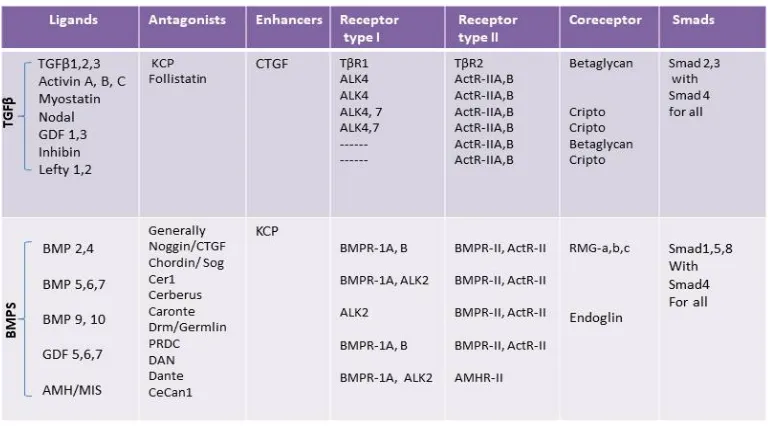
[image:34.612.150.534.391.604.2]
Table 2. Representation of ligands, antagonists, receptors, coreceptors and smads proteins relationships to the TGFβ and BMP branches of the TGFβ superfamily signaling pathway (Lin et al., 2005; Massague, 2008; Soofi et al., 2013).
36 region. For example, Noggin contains a carboxy-terminal cysteine-rich (CR) domain, while Chordin contains four cysteine-rich repeats (Massague and Chen, 2000). The CR domain confers the antagonists to form a homodimer to match the structure of BMP ligand homodimers. The crystal structure of the Noggin-BMP7 complex directly showed that Noggin inhibited BMP7 by blocking the surfaces that were required to interact with the type I and type II BMP receptors (Groppe et al., 2002). Those antagonists are expressed during embryogenesis and are critical for the dosal-ventral patterning and left-right asymmetry. Interestingly, although most of the BMP antagonist shared the CR domain, not all proteins containing CR domain counteract BMP. In this thesis it is shown that instead of blocking BMP signaling, the CR domain protein KCP (Kielin/chordin-like protein) enhanced BMP-receptor interactions and counteract the TGFβ interactions (figure 6) (Lin et al., 2005).
Figure 6. The secreted protein KCP enhances BMP and suppresses TGFβ.
37
I.g.- Kielin/chordin-like protein (KCP) Characterization
I.g.1. KCP Functions and Attributions
The original description of KCP was made in a series of publications from the Dressler laboratory (Lin et al 2005; Lin et al 2006). The newly-described gene, KCP, encodes a protein with homology to the extracellular regulators of the TGFβ superfamily of secreted signaling peptides. KCP is a large secreted protein with 18 repeated cysteine-rich domains. KCP is expressed in the developing kidney at both early and late stages, and its expression is correlated with the formation of early epithelial structures within the intermediate mesoderm, and to the formation of the proximal tubules in the more devolved metanephric kidney. In the mammalian kidney, BMP7 plays an essential role in development and disease. BMP7-null mice show arrested renal development at around E14.5, resulting in severe renal hypoplasia (Dudley et al., 1995; Luo et al., 1995). BMP7 is also an anti-fibrotic agent that can reduce interstitial fibrosis, a common pathology in a broad spectrum of chronic renal diseases. Administration of recombinant BMP7 has shown remarkable efficacy in the reduction of glomerular and interstitial fibrosis in mouse models of chronic renal disease (Zeisberg et al., 2003a; Zeisberg et al., 2003b).
cysteine-38 rich domains that bind BMPs to inhibit signaling (Larrain et al., 2000).
Unlike previously described CR domain proteins, KCP is a potent paracrine enhancer of BMP signaling. KCP increases the affinity of ligand to receptor and/or enhances the stability of the ligand-receptor complex. Given the role of BMP7 in renal disease, we analyzed the phenotypes of KCP KO mice and the KCP transgenic mice in two independent models of renal injury. Both strains of KCP mice were used in the Diet Induced Obesity (DIO) study. The data point to an important role for KCP to enhance BMP signaling, attenuate the initiation, and progression of fibrotic disease after renal injury. KCP also protects mice from the DIO, fatty liver, and metabolic syndrome. These conditions will be discussed in more detail in Chapter II (Soofi et al., 2016; Soofi et al., 2013).
I.g.2- Original Description of KCP
39 The KCP tissue-specific expression patterns were confirmed and expanded upon using whole mount of in situ hybridization using embryos. In mouse embryos at E9.5, the limb bud mesenchyme was positive for KCPRNA (Figure 7B). Expression in the kidney region could be detected as early as E9 in the intermediate mesoderm (Figure 7B, arrow). By E10, the mesonephric tubules and nephric ducts were clearly positive for KCP
mRNA (Figure 7C and D, arrow). At later stages, high levels of KCP mRNA localized to the developing tubules (Figure 7G, arrows and H).
A
CR domain
Signal peptide
Signal peptide [image:38.612.119.422.255.531.2]CR domain vWF-D domain
Figure 7. The Structure and Expression pattern of the endogenous KCP protein.
A) KCP protein structure representation. B) Whole mount in situ hybridization of E9 embryo showing staining in limb bud mesenchyme (arrow) and in the nephric duct (arrowhead). C, D) E10 whole mount embryo indicating kcp mRNA expression in the mesonephric tubules (arrow). E) A section taken through an E9 embryo showing kcp mRNA in the forelimb bud mesenchyme (arrowheads) and in the intermediate mesoderm (arrow). F) E10 whole-mount embryo section indicating Kcp
mRNA expression in the mesonephric tubules (arrow). G) A bisected E16 kidney with kcp
40
I.g.3- Generation of KCP KO mice
Generation of The KCP-KO mice was done by homologues recombination in the mouse germline by replacing exons 2-21 of KCP with lacZ and a Geneticin cassete (Figure 8a). By inserting the lacZ gene in frame, its expression was expected to reflect the endogenous pattern of KCP in the embryo. Thus, lacZ was expected to serve as a useful marker to detect KCP expression cells (Lin J., 2005). The targeting vector deletes amino acids 67-774, which includes most of the CR domains. Also, as expected the LacZ/neo cassette inhibits the expression of coding sequences downstream of 774 aa.
[image:39.612.136.521.382.479.2]Germline chimera’s mice were obtained from the injected ES cells clones. The chimeras were then backcrossed with C57Bl/6J to generate the offspring containing the KCP heterozygote mice. Further homozygous KCP–KO mice were generated by crossing KCP heterozygous mice and genotyped by Southern blotting followed by PCR. KCP-KO mice were viable and fertile without obvious abnormalities.
Figure 8. Generation KCP-KO (a) Schematic diagram of the Kcp targeting vector that was designed to delete exons 2–21, spanning amino acids 67–774 of the coding region. The lacZ gene was inserted in frame after amino acid 67 in exon 2 (Lin et al., 2005).
41 The TGFβ pathway can directly transduce extracellular cues from the cell-surface transmembrane receptors to the nucleus through intracellular mediators, known as Smads. The Smad family is well conserved (Feng et al., 1998; Moustakas and Heldin, 2009; Patterson and Padgett, 2000). In most vertebrates, there are eight Smads, compared to six in the Caenorhabditis genus and four in Drosophila species (Huminiecki et al., 2009). Smads proteins can be divided into three functional groups: (1) Receptor-regulated Smads (R-Smads, 1/2/3/5/8); (2) Common Smad (Co-Smad, 4); (3) Inhibitory Smad (I-Smad, 6/7) illustrated in figure 5, page 22.
132
CHAPTER III
III. a. Thesis Conclusions, Reflections, and Future Directions
III. a. 1- Thesis Conclusions
In this thesis, evidence has been presented through the published articles, showing a that kidney development in mammals is the final product of three successive embryonic steps that are characterized by the transformation of intermediate mesoderm cells. The development of the first kidney, the transient pronephros, is initiated by signals from the somite and surface ectoderm that induce cells in the intermediate mesoderm to undergo the transition to epithelial cells forming the nephric duct (Mari and Winyard, 2015; Mauch et al., 2000; Obara-Ishihara et al., 1999). The caudal migration of the nephric duct subsequently induces the adjacent nephrogenic mesoderm to aggregate and form the tubules of the mesonephros, the second embryonic kidney. On further extension, the nephric duct reaches the metanephrogenic mesenchyme at the level of the developing hindlimb, where the ureteric bud evaginates from the nephric duct and invades the surrounding mesenchyme. Both the ureter and mesenchyme subsequently undergo reciprocal inductive interactions to form the nephrons and collecting ducts of the metanephros, the third and adult kidney. In humans, new nephron formation, or nephrogenesis, starts during the 5th week of gestation, the first glomeruli appear at the 9th week, and the last new nephron is formed by the 36th week of gestation. In mice, nephrogenesis starts at embryonic day 10.5, with the first glomeruli appearing at embryonic day 14 and the last new nephron approximately appearing 1 week to 10 days after birth (Mari and Winyard, 2015; Saxen and Sariola, 1987; Soofi et al., 2012).
133 gene Pax8 (Bouchard et al., 2002) and the homeodomain protein Lhx1 (Tsang et al., 2000). Kidney development starts when the ureteric bud (UB) invades the metanephric mesenchyme (MM) and transmits inductive signals, such as Wnt9b (Carroll et al., 2005), to promote condensation of the MM around the UB tips. These UB tip associated Cap mesenchyme cells (CM) continue to express Pax2 and are the stem cells of the nephron that generate all of the epithelial derivatives, including distal, proximal, and glomerular epithelium (Kobayashi et al., 2008; Mugford et al., 2008a). The CM undergoes a mesenchymal-to-epithelial transition to generate all the epithelial cells of the developing nephron. However, the Pax2 expression is down-regulated in the podocyte precursor cells and the mature epithelial cells of the nephron as development comes to an end (Ryan et al., 1995). This Pax2 positive intermediate mesoderm generates the nephric, or Wolffian, duct, an outgrowth of the duct called the UB, and the surrounding MM. Pax2 null mutant do develop a nephric duct, but the duct is completely absent in a Pax2; Pax8 double mutants. Pax2 and Pax8 have redundant function in kidney development. Pax8 mutant embryos develop a normal urogenital system, but mice die shortly after birth due to defect in the thyroid gland development. In contrast, a Pax2 mutant results in complete renal agenesis because the nephric duct is abnormal and the metanephric mesenchyme cannot respond to inductive signals.
134 urogenital track (CAKUT). Among the causes of CAKUT are heterozygous mutations in the Pax2 gene, which lead to Papillorenal Syndrome, and result in hypoplastic kidneys, vesicoureteral reflux, progressive renal failure, and optic nerve coloboma. Losing of Pax2 function results in complete renal and reproductive tract agenesis in mice (Soofi et al., 2012; Torres et al., 1995). In the absence of Pax2, the IM cells assume a pattern of gene expression more consistent with paraxial mesoderm and its derivatives (Ranghini and Dressler, 2015). Despite its central role in kidney development and renal disease, the biochemistry of Pax2 and its effects on gene regulation are not well characterized in a developing tissue. Few target genes have been identified, including many known kidney developmental regulators, such as Gdnf, c-Ret, Six2, Sal1, and Lhx1, but also affected are genes and proteins associated with glycosylation, cell membranes, cell-cell signaling, and cell adhesion (Ranghini and Dressler, 2015). Furthermore, ectopic or deregulated expression of Pax2 is also seen in Wilms' tumor (Dressler and Douglass, 1992), renal cell carcinoma (Gnarra and Dressler, 1995), and polycystic kidney disease (Ostrom et al., 2000), where it is thought to promote proliferation and/or survival. The reactivation of Pax2 expression is also observed in adult kidneys after acute injury, suggesting a critical role for Pax2 in regenerating the epithelia (Humphreys et al., 2008; Imgrund et al., 1999; Kusaba et al., 2014). We also discussed in Chapter I that Pax2 re-expression is regulated by the TGFβ superfamily singling pathway.
135 kidney has the ability to repair itself. With a mild injury, this repair can result in the return to a structural and functional state that is indistinguishable from normal. However, when the repair is more severe or superimposed on baseline kidney abnormalities, the repair process can lead to fibrosis, which can facilitate progression to chronic kidney disease. Acute kidney injury (AKI) can thus result in incomplete repair and persistent tubulointerstitial inflammation, with a proliferation of fibroblasts and excessive deposition of extracellular matrix, a common feature of many different kinds of kidney diseases and a primary determinant of progression to end-stage renal failure (Forbes et al., 2000). Whether AKI is associated with ischemia reperfusion injury, sepsis or toxins, there is a rapid loss of proximal tubular cell cytoskeletal integrity and cell polarity. There is shedding of the proximal tubule brush border, loss of polarity with mislocalization of adhesion molecules, and other membrane proteins such as the Na+K+ATPase and β- integrins (Thadhani et al., 1996; Zuk et al., 1998). Normal cell-cell interactions are disrupted with an injury. When the injury is severe, there is apoptosis and necrosis (Thadhani et al., 1996). Viable and nonviable cells are desquamated leaving regions where the basement membrane remains the only barrier between the filtrate and the peritubular interstitium.
136 Mice homozygous for a KCP null allele are hypersensitive to developing renal interstitial fibrosis, a disease stimulated by TGFβ but inhibited by BMP7. Transgenic mice that express KCP in adult kidneys showed significantly less expression of collagen IV, α-smooth muscle actin, and other markers of disease progression in the unilateral ureteral obstruction model of renal interstitial fibrosis. In an acute tubular necrosis model, mice expressing KCP were more resistant to high doses of folic acid and showed better recovery at lower doses. The data demonstrates that extracellular regulation of the TGFβ/BMP signaling axis by cysteine-rich domain proteins can reduce disease severity in animal models of renal injury (Soofi et al., 2013).
Recently, we examined the effects of KCP loss or gain of function in mice that were maintained on either a regular or a high-fat diet. Loss of KCP sensitized mice to obesity and associated complications such as hepatic steatosis and glucose intolerance. In contrast, transgenic mice that expressed KCP in the kidney, liver, and brown adipose tissues were resistant to developing high-fat diet induced obesity and had significantly reduced white adipose tissue. The data demonstrate that shifting the TGFβ superfamily signaling with a secreted inhibitor or enhancer can alter the profile of adipose tissue to reduce obesity, and can inhibit the initiation, and progression of hepatic steatosis to significantly reduce the effects of high-fat diet induced metabolic disease (Soofi et al., 2016).
137
III. b. Thesis Reflection and Future Direction
III.b.1. The ‘how I would do the experiments differently now’ question
All work in this thesis has been peer-reviewed and published in quality Journals. That said, any published work is far from being 100% perfect, and we learn something new on a daily basis. Every day, we do and should become better at what we do. Today I will attempt to discuss some experiments and techniques, and how we may perform them differently. Some of these observations were also made by the examiners, Dr. Paul Winyard and Dr. Peter Hohenstein.
138 Paper 1- Soofi, A., Levitan, I., and Dressler, G.R. (2012). Two novel EGFP insertion alleles reveal unique aspects of Pax2 function in embryonic and adult kidneys. Developmental Biology365, 241-250.
Figure 1. Section B, we decided not to show a blot of a negative egfp/egfp (Pax2 mutant), even though there is no Pax2 expression. The internal control (Tubulin) could indicate that they are in the blot.
Figure 3. We should have specified if we were comparing littermates to each other or gene expression independent of the phenotype. It may have been better to compare the diferent gene expression in the similar phenotype of the Pax2 Eneo/Eneo E11-11,5 embryonic kidneys.
Figure 6. Sections E-F, if we were going to talk about cell planar polarity we should have used better polarity markers.
Paper 2- Soofi, A., Zhang, P., and Dressler, G.R. (2013). Kielin/chordin-like protein attenuates both acute and chronic renal injury. Journal of the American Society of Nephrology 24, 897-905
Figure 2. Section A, to confirm that indeed Myc-KCP is localized to the ER compartment in the epithelial cells; we should have co-stained cells with ER markers and markers of other cell compartments as control. For example, we could use Golgi or mitochondrial markers as the negative compartments for the Myc-KCP expression.
Figure 6. Section D, we should have blotted for the total protein Smad3 and Smad1 to normalized the p-Proteins and determine the ratio of the relative unit for each protein. Furthermore, we could also measure Creatinine and the blood urea nitrogen (BUN) as a secondary indication of kidney damage.
Paper 3- Soofi, A., Wolf, K.I., Ranghini, E.J., Amin, M.A., and Dressler, G.R. (2016). The kielin/chordin-like protein KCP attenuates nonalcoholic fatty liver disease in mice. American journal of Physiology Gastrointestinal and Liver Physiology311, G587-G598.
139 Paper 4- Soofi, A., Wolf, K.I., Emont, M.P., Qi, N., Martinez-Santibanez, G., Grimley, E., Ostwani, W., and Dressler, G.R. (2017). The kielin/chordin-like protein (KCP) attenuates high-fat diet-induced obesity and metabolic syndrome in mice. Journal of Biological Chemistry292, 9051-9062.
Even though intensive work was done in this project addressing the effect of normal, gain and loss of KCP in white and brown adipocyte tissue, very little was done about other fat depots, such as the subcutaneous fat tissue (Soofi et al., 2017). Experiments will be seriously considered for future projects in this field, yet it is difficult to cover all in one study.
III. b. 2. Further experiments based on this thesis
140 interested in continuing these studies using the combination of the Pax2-egfp alleles and the new made Pax2 conditoinal KO mice in kidney development as well as in adult kidney regeneration after injury. Another recent publication from our lab by Gerimely E etal., 2017 used the Pax2-egfp mice as an important model to report that Pax proteins are re-expressed or ectopically expressed in cancer and other diseases of abnormal proliferation, making them attractive targets (Grimley et al., 2017). This new data confirms that small molecules targeting the DNA binding paired domain can be identified and may be good lead compounds for developing tissue and cell-type specific anticancer therapies. We will be focusing on future experiments in pursuing the role of Pax2 in Kidney development, kidney disease, and regeneration, as well as the role Pax2 plays in other organs like the female and male reproductive system.
141
List of References
Including
text of the thesis and all publications
Abrass, C.K. (2004). Overview: obesity: what does it have to do with kidney disease? Journal of the American Society of Nephrology 15, 2768-2772.
Abreu, J.G., Ketpura, N.I., Reversade, B., and De Robertis, E.M. (2002). Connective-tissue growth factor (CTGF) modulates cell signalling by BMP and TGF-beta. Nature Cell Biology 4, 599-604.
Ahima, R.S., and Lazar, M.A. (2008). Adipokines and the peripheral and neural control of energy balance. Molecular Endocrinology 22, 1023-1031.
Ahima, R.S., and Lazar, M.A. (2013). Physiology. The health risk of obesity--better metrics imperative. Science 341, 856-858.
Al-Awqati, Q., and Gao, X.B. (2011). Differentiation of intercalated cells in the kidney. Physiology (Bethesda) 26, 266-272.
Almind, K., Manieri, M., Sivitz, W.I., Cinti, S., and Kahn, C.R. (2007). Ectopic brown adipose tissue in muscle provides a mechanism for differences in risk of metabolic syndrome in mice. Proceedings of the National Academy of Sciences of the United States of America 104, 2366-2371.
Amiel, J., Audollent, S., Joly, D., Dureau, P., Salomon, R., Tellier, A.L., Auge, J., Bouissou, F., Antignac, C., Gubler, M.C., et al. (2000). PAX2 mutations in renal-coloboma syndrome: mutational hotspot and germline mosaicism. European Journal of Human Genetics 8, 820-826.
Annes, J.P., Munger, J.S., and Rifkin, D.B. (2003). Making sense of latent TGFbeta activation. Journal Cell Scince 116, 217-224.
142 Asrih, M., and Jornayvaz, F.R. (2015). Metabolic syndrome and nonalcoholic fatty liver disease: Is insulin resistance the link? Molecular and Cellular Endocrinology 15;418 Pt 1:55-65.
Assoian, R.K., Komoriya, A., Meyers, C.A., Miller, D.M., and Sporn, M.B. (1983). Transforming growth factor-beta in human platelets. Identification of a major storage site, purification, and characterization. Journal of Biological Chemistry 258, 7155-7160.
Banks, A.S., McAllister, F.E., Camporez, J.P., Zushin, P.J., Jurczak, M.J., Laznik-Bogoslavski, D., Shulman, G.I., Gygi, S.P., and Spiegelman, B.M. (2015). An ERK/Cdk5 axis controls the diabetogenic actions of PPARgamma. Nature 517, 391-395.
Bartelt, A., Bruns, O.T., Reimer, R., Hohenberg, H., Ittrich, H., Peldschus, K., Kaul, M.G., Tromsdorf, U.I., Weller, H., Waurisch, C., et al. (2011). Brown adipose tissue activity controls triglyceride clearance. Nature Medicine 17, 200-205.
Basson, M.A., Akbulut, S., Watson-Johnson, J., Simon, R., Carroll, T.J., Shakya, R., Gross, I., Martin, G.R., Lufkin, T., McMahon, A.P., et al. (2005). Sprouty1 is a critical regulator of GDNF/RET-mediated kidney induction. Developemntal Cell 8, 229-239.
Basson, M.A., Watson-Johnson, J., Shakya, R., Akbulut, S., Hyink, D., Costantini, F.D., Wilson, P.D., Mason, I.J., and Licht, J.D. (2006). Branching morphogenesis of the ureteric epithelium during kidney development is coordinated by the opposing functions of GDNF and Sprouty1. Developmental Biology 299, 466-477.
Berry, R., Jeffery, E., and Rodeheffer, M.S. (2014). Weighing in on adipocyte precursors. Cell Metabolism 19, 8-20.
Berry, R., and Rodeheffer, M.S. (2013). Characterization of the adipocyte cellular lineage in vivo. Nature Cell Biology 15, 302-308.
143 Bonventre, J.V., and Yang, L. (2010). Kidney injury molecule-1. Current Opinion Critical Care 16 (6) 556-61.
Border, W.A., Okuda, S., Languino, L.R., Sporn, M.B., and Ruoslahti, E. (1990). Suppression of experimental glomerulonephritis by antiserum against transforming growth factor beta 1. Nature 346, 371-374.
Bottinger, E.P., and Bitzer, M. (2002). TGF-beta Signaling in Renal Disease. Journal of the American Society of Nephrology 13, 2600-2610.
Boualia, S.K., Gaitan, Y., Tremblay, M., Sharma, R., Cardin, J., Kania, A., and Bouchard, M. (2013). A core transcriptional network composed of Pax2/8, Gata3 and Lim1 regulates key players of pro/mesonephros morphogenesis. Developmental Biology 382, 555-566.
Bouchard, M., Pfeffer, P., and Busslinger, M. (2000). Functional equivalence of the transcription factors Pax2 and Pax5 in mouse development. Development 127, 3703-3713.
Bouchard, M., Souabni, A., Mandler, M., Neubuser, A., and Busslinger, M. (2002). Nephric lineage specification by Pax2 and Pax8. Genes & Development 16, 2958-2970.
Boutet, A., De Frutos, C.A., Maxwell, P.H., Mayol, M.J., Romero, J., and Nieto, M.A. (2006). Snail activation disrupts tissue homeostasis and induces fibrosis in the adult kidney. EMBO Journal 25, 5603-5613.
Brophy, P.D., Lang, K.M., and Dressler, G.R. (2003). The secreted frizzled related protein 2 (SFRP2) gene is a target of the Pax2 transcription factor. Journal of Biological Chemistry 278, 52401-52405.
Brophy, P.D., Ostrom, L., Lang, K.M., and Dressler, G.R. (2001). Regulation of ureteric bud outgrowth by Pax2-dependent activation of the glial derived neurotrophic factor gene. Development 128, 4747-4756.
144 Bush, K.T., Sakurai, H., Steer, D.L., Leonard, M.O., Sampogna, R.V., Meyer, T.N., Schwesinger, C., Qiao, J., and Nigam, S.K. (2004). TGF-beta superfamily members modulate growth, branching, shaping, and patterning of the ureteric bud. Developmental Biology 266, 285-298.
Buzzetti, E., Pinzani, M., and Tsochatzis, E.A. (2016). The multiple-hit pathogenesis of non-alcoholic fatty liver disease (NAFLD). Metabolism 65, 1038-1048.
Cai, Q., Dmitrieva, N.I., Ferraris, J.D., Brooks, H.L., van Balkom, B.W., and Burg, M. (2005). Pax2 expression occurs in renal medullary epithelial cells in vivo and in cell culture, is osmoregulated, and promotes osmotic tolerance. Proceedings of the National Academy of Sciences of the United States of America 102, 503-508.
Cai, Y., Lechner, M.S., Nihalani, D., Prindle, M.J., Holzman, L.B., and Dressler, G.R. (2002). Phosphorylation of Pax2 by the c-Jun N-terminal kinase and enhanced Pax2-dependent transcription activation. Journal of Biological Chemistry 277, 1217-1222.
Cannon, B., and Nedergaard, J. (2004). Brown adipose tissue: function and physiological significance. Physiological Reviews 84, 277-359.
Capdevila, J., and Belmonte, J.C. (1999). Extracellular modulation of the Hedgehog, Wnt and TGF-beta signalling pathways during embryonic development. Current Opinion in Genetics & Development 9, 427-433.
Carroll, T.J., Park, J.S., Hayashi, S., Majumdar, A., and McMahon, A.P. (2005). Wnt9b plays a central role in the regulation of mesenchymal to epithelial transitions underlying organogenesis of the mammalian urogenital system. Developmental Cell
9, 283-292.
Carroll, T.J., and Vize, P.D. (1999). Synergism between Pax-8 and lim-1 in embryonic kidney development. Developmental Biology 214, 46-59.
Cassuto, H., Olswang, Y., Heinemann, S., Sabbagh, K., Hanson, R.W., and Reshef, L. (2003). The transcriptional regulation of phosphoenolpyruvate carboxykinase gene in the kidney requires the HNF-1 binding site of the gene. Gene 318, 177-184.
145 Chen, J., Bardes, E.E., Aronow, B.J., and Jegga, A.G. (2009b). ToppGene Suite for gene list enrichment analysis and candidate gene prioritization. Nucleic Acids Research 37, W305-311.
Chen, J., Muntner, P., Hamm, L.L., Jones, D.W., Batuman, V., Fonseca, V., Whelton, P.K., and He, J. (2004). The metabolic syndrome and chronic kidney disease in U.S. adults. Annals of internal medicine 140, 167-174.
Chi, N., and Epstein, J.A. (2002). Getting your Pax straight: Pax proteins in development and disease. Trends Genetics 18, 41-47.
Chi, X., Michos, O., Shakya, R., Riccio, P., Enomoto, H., Licht, J.D., Asai, N., Takahashi, M., Ohgami, N., Kato, M., et al. (2009). Ret-dependent cell rearrangements in the Wolffian duct epithelium initiate ureteric bud morphogenesis. Development Cell 17, 199-209.
Chia, I., Grote, D., Marcotte, M., Batourina, E., Mendelsohn, C., and Bouchard, M. (2011). Nephric duct insertion is a crucial step in urinary tract maturation that is regulated by a Gata3-Raldh2-Ret molecular network in mice. Development 138, 2089-2097.
Christian, J.L. (2000). BMP, Wnt and Hedgehog signals: how far can they go? Current Opinions Cell Biolology 12, 244-249.
Cirio, M.C., de Groh, E.D., de Caestecker, M.P., Davidson, A.J., and Hukriede, N.A. (2014). Kidney regeneration: common themes from the embryo to the adult. Pediatric Nephrololgy 29, 553-564.
Clarke, J.C., Patel, S.R., Raymond, R.M., Jr., Andrew, S., Robinson, B.G., Dressler, G.R., and Brophy, P.D. (2006). Regulation of c-Ret in the developing kidney is responsive to Pax2 gene dosage. Human Molecular Genetics.
Cousin, B., Cinti, S., Morroni, M., Raimbault, S., Ricquier, D., Penicaud, L., and Casteilla, L. (1992). Occurrence of brown adipocytes in rat white adipose tissue: molecular and morphological characterization. Journal of Cell Science 103 ( Pt 4), 931-942.
146 Curthoys, N.P., and Gstraunthaler, G. (2001). Mechanism of increased renal gene expression during metabolic acidosis. American Journal Physiology Renal-Physiololgy 281, F381-390.
Cypess, A.M., Lehman, S., Williams, G., Tal, I., Rodman, D., Goldfine, A.B., Kuo, F.C., Palmer, E.L., Tseng, Y.H., Doria, A., et al. (2009). Identification and importance of brown adipose tissue in adult humans. New England Journal of Medicine 360, 1509-1517.
Dahl, E., Koseki, H., and Balling, R. (1997). Pax genes and organogenesis. Bioessays 19, 755-765.
de Miranda, D.M., Dos Santos Junior, A.C., Dos Reis, G.S., Freitas, I.S., Carvalho, T.G., de Marco, L.A., Oliveira, E.A., and Simoes, E.S.A.C. (2014). PAX2 polymorphisms and congenital abnormalities of the kidney and urinary tract in a Brazilian pediatric population: evidence for a role in vesicoureteral reflux. Molecular Diagnosis & Therapy 18, 451-457.
Dehbi, M., Ghahremani, M., Lechner, M., Dressler, G., and Pelletier, J. (1996). The paired-box transcription factor, PAX2, positively modulates expression of the Wilms' tumor suppressor gene (WT1). Oncogene 13, 447-453.
Derynck, R., and Zhang, Y.E. (2003). Smad-dependent and Smad-independent pathways in TGF-beta family signalling. Nature 425, 577-584.
Divoux, A., and Clement, K. (2011). Architecture and the extracellular matrix: the still unappreciated components of the adipose tissue. Obesity reviews : an Official Journal of the International Association for the Study of Obesity 12, e494-503.
Donath, M.Y., and Shoelson, S.E. (2011). Type 2 diabetes as an inflammatory disease. Nature reviews Immunology 11, 98-107.
Dooley, S., and ten Dijke, P. (2012). TGF-beta in progression of liver disease. Cell and Tissue Research 347, 245-256.
147 Dressler, G.R. (2006). The cellular basis of kidney development. Annual Review Cell and Developmental Biology 22, 509-529.
Dressler, G.R. (2009). Advances in early kidney specification, development and patterning. Development 136, 3863-3874.
Dressler, G.R. (2011). Patterning and early cell lineage decisions in the developing kidney: the role of Pax genes. Pediatric Nephrology 26, 1387-1394.
Dressler, G.R., Deutsch, U., Chowdhury, K., Nornes, H.O., and Gruss, P. (1990). Pax2, a new murine paired-box-containing gene and its expression in the developing excretory system. Development 109, 787-795.
Dressler, G.R., and Douglass, E.C. (1992). Pax-2 is a DNA-binding protein expressed in embryonic kidney and Wilms tumor. Proceedings of the National Academy of Sciences of the United States of America 89, 1179-1183.
Dressler, G.R., Wilkinson, J.E., Rothenpieler, U.W., Patterson, L.T., Williams-Simons, L., and Westphal, H. (1993). Deregulation of Pax-2 expression in transgenic mice generates severe kidney abnormalities. Nature 362, 65-67.
Dudley, A.T., Lyons, K.M., and Robertson, E.J. (1995). A requirement for bone morphogenetic protein-7 during development of the mammalian kidney and eye. Genes Development 9, 2795-2807.
Dupont, S., Mamidi, A., Cordenonsi, M., Montagner, M., Zacchigna, L., Adorno, M., Martello, G., Stinchfield, M.J., Soligo, S., Morsut, L., et al. (2009). FAM/USP9x, a deubiquitinating enzyme essential for TGFbeta signaling, controls Smad4 monoubiquitination. Cell 136, 123-135.
Eccles, M.R., and Schimmenti, L.A. (1999). Renal-coloboma syndrome: a multi-system developmental disorder caused by PAX2 mutations. Clinical Genetics 56, 1-9.
Eckel, R.H., Grundy, S.M., and Zimmet, P.Z. (2005). The metabolic syndrome. Lancet 365, 1415-1428.