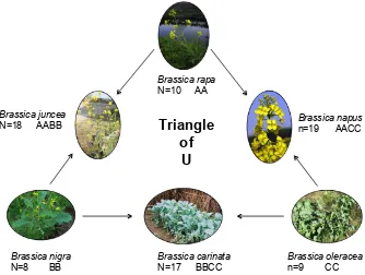
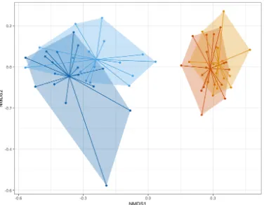
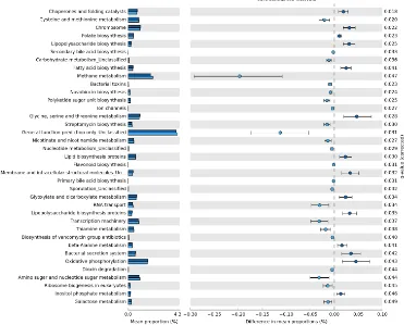
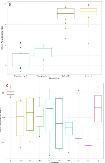
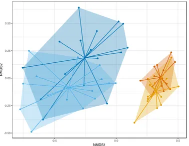
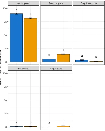
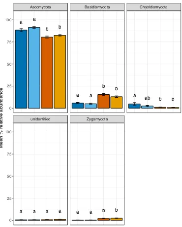
Characterisation of plant (Brassica spp ) and microbial rhizosphere functions
Full text
Figure
Related documents
An additional measureable plastic strain recovery is observed after each unloading step of interrupted (i.e. loading-unloading) tensile tests. This is contributing toward the
The paper is discussed for various techniques for sensor localization and various interpolation methods for variety of prediction methods used by various applications
carpentry, cooperatives, and some pesantren provide santri with experience in agriculture, fisheries, besides the center of community development. With the hope
Figure 5(a) also shows that if the base station re- broadcast sufficient CDM messages so that on aver- age, at least one copy of such authentic CDM mes- sage can reach sensor node
the firm’s customers concerning the attributes of products or processes by which products are made are shared by employees as well: for moral reasons, some
/ L, and a different adsorbent mass, is shown in Figure 5. The results obtained show that the removal rate increases quickly at first, until a time of 60 min, and
That observation is in accordance with the median time to response in our trial (only 2 cycles). However, even though combination therapy may achieve earlier responses in a
5.15: The cellular uptake and distribution of AgNPs and AgNWs (50 µg/ml) together with CBS, CSE and MPST enzymes in microglia cells after 24 h exposure : confocal analysis on
