warwick.ac.uk/lib-publications
Original citation:
Fullam, Elizabeth, Prokes, Ivan, Fütterer, Klaus and Besra, Gurdyal S.. (2016) Structural and
functional analysis of the solute-binding protein UspC from Mycobacterium tuberculosis that
is specific for amino sugars. Open Biology (6).
Permanent WRAP URL:
http://wrap.warwick.ac.uk/80035
Copyright and reuse:
The Warwick Research Archive Portal (WRAP) makes this work of researchers of the
University of Warwick available open access under the following conditions.
This article is made available under the Creative Commons Attribution 4.0 International
license (CC BY 4.0) and may be reused according to the conditions of the license. For more
details see:
http://creativecommons.org/licenses/by/4.0/
A note on versions:
The version presented in WRAP is the published version, or, version of record, and may be
cited as it appears here.
rsob.royalsocietypublishing.org
Research
Cite this article:
Fullam E, Prokes I, Fu¨tterer
K, Besra GS. 2016 Structural and functional
analysis of the solute-binding protein UspC
from
Mycobacterium tuberculosis
that is specific
for amino sugars.
Open Biol.
6
: 160105.
http://dx.doi.org/10.1098/rsob.160105
Received: 12 April 2016
Accepted: 26 May 2016
Subject Area:
biochemistry/structural biology/microbiology
Keywords:
amino sugars, X-ray crystallography,
Mycobacterium tuberculosis, ATP-binding
cassette transporters, nutrient acquisition,
peptidoglycan
Authors for correspondence:
Elizabeth Fullam
e-mail: [email protected]
Gurdyal S. Besra
e-mail: [email protected]
Electronic supplementary material is available
at http://dx.doi.org/10.1098/rsob.160105.
Structural and functional analysis of the
solute-binding protein UspC from
Mycobacterium tuberculosis
that is specific
for amino sugars
Elizabeth Fullam
1,2, Ivan Prokes
3, Klaus Fu¨tterer
2and Gurdyal S. Besra
21School of Life Sciences, University of Warwick, Coventry CV4 7AL, UK
2School of Biosciences, University of Birmingham, Edgbaston, Birmingham B15 2TT, UK 3Department of Chemistry, University of Warwick, Coventry CV4 7AL, UK
EF, 0000-0001-6245-1614; KF, 0000-0001-7445-5372
Mycobacterium tuberculosis (Mtb), the aetiological agent of tuberculosis, has evolved to scavenge nutrients from the confined environment of host macro-phages with mycobacterial ATP-binding cassette (ABC) transporters playing
a key role in nutrient acquisition.Mtb-UspC (Rv2318) is the solute-binding
protein of the essential transporter UspABC, one of fourMtbABC transporters
implicated by homology in sugar acquisition. Herein, we report the structural
and functional characterization ofMtb-UspC. The 1.5 A˚ resolution structure
of UspC reveals a two subdomain architecture that forms a highly acidic carbohydrate-substrate binding cleft. This has allowed a distinct preference ofMtb-UspC for amino sugars as determined by thermal shift analysis and sol-ution saturation transfer difference-NMR. Taken together our data support the functional assignment of UspABC as an amino-sugar transporter. Given the limited availability of carbohydrates within the phagosomal
envi-ronmental niche duringMtbintracellular infection, our studies suggest that
UspABC enablesMtbto optimize the use of scarce nutrients during
intracellu-lar infection, linking essentiality of this protein to a potential role in recycling components of cell-wall peptidoglycan.
1. Introduction
Mycobacterium tuberculosis(Mtb) is a major human pathogen and is the causa-tive agent of tuberculosis (TB). TB remains a major global health threat and is the leading cause of mortality worldwide from a single infectious agent, with an excess of nine million new cases of TB each year claiming the lives of 1.5 million people annually [1]. While TB can be treated, the regimen extends over six to nine months. Premature termination of therapy in combination with a static pool of anti-tubercular drugs are among the major factors in caus-ing the emergence of drug-resistant strains, which now includes extensively drug-resistant and untreatable forms of the disease [2,3]. Clearly, there is an urgent need to address this global health problem.
Mtbis a facultative intracellular organism able to evade the host immune
response and survive within phagosomes for decades. Within this environment,
Mtb has restricted access to nutrients, and mechanisms of nutrient supply
during intracellular infection are poorly understood [4]. A growing body of
evi-dence suggests thatMtbuses host lipids as the main carbon and energy source,
reflected by, firstly, an over-representation of genes in the Mtb genome that
encode enzymes of fatty acid metabolism [5] and, secondly, upregulation of such genes during macrophage infection [6]. Recent studies point to host
lipid cholesterol as a major carbon source used byMtbduring infection.
How-ever, blocking cholesterol uptake and metabolism only partially attenuatesMtb
virulence, suggesting that other yet to be identified carbon sources also have an important role to play [7,8].
It is generally assumed that access ofMtbto host sugars is
particularly limiting. Bioinformatics analysis of the genome
sequence ofMtbhas led to the identification of a number of
transporter systems, four of which have been annotated as carbohydrate importers of the ATP-binding cassette (ABC) superfamily [9–11]. Genome-wide saturation transposon
mutagenesis studies byHimar1 suggest a role for these
sys-tems in the virulence of Mtb [6,12]. Up until recently the
substrates for these Mtb carbohydrate importers have
remained elusive. However, it has since been demonstrated that the LpqY-SugABC transporter system is specific for the uptake of trehalose, which is recycled from the cell-wall glycolipid trehalose monomycolate [13,14]. Importantly, the LpqY-SugABC importer has been demonstrated to be
essential for the virulence of Mtb in vivo[13]. Similarly, the
solute-binding protein UgpB of the UgpAEBC transport
system has been implicated in the recognition of
sn-glycero-3-phosphocholine, a glycolipid that is upregulated during
Mtbinfection of guinea pigs [15,16]. Given the small number
of carbohydrate import systems in Mtb (five) compared to,
for instance, the soil-dwelling Mycobacterium smegmatis
(28 ABC transporters) [10] it is plausible that this discrete set
of transporters inMtbis the result of adaptation to a very
lim-ited set of carbohydrates available in the host environment. Functional roles and substrate specificities of the remaining putative carbohydrate transporters, which include the SugI permease, Rv2038-Rv2041 ABC transporter and the UgpABCE ABC transporter, are not yet known.
UspC fromMtbis a 441-amino acid protein that has been
previously identified in bioinformatics analyses as a putative active importer of carbohydrates across the inner-membrane
of the Mtb cell wall [9,17]. The uspC gene forms part of a
putative three-gene operon,uspABC, of whichuspAanduspB
encode the membrane-spanning subunits of the transporter,
whileuspCis a homologue of ABC transporter-linked
solute-binding proteins. The operon lacks an obvious candidate for encoding the nucleotide-binding domain (NBD), which remains to be identified. It is probable that the UspABC transporter shares the NBD with another mycobacterial ABC transporter [9], which is not unusual among bacterial
ABC transporters [18]. The Mtb UspABC ABC transporter
has been demonstrated to be essential for growthin vitro[19]
and is conserved in Mycobacterium leprae, an obligate
pathogen that has undergone massive gene decay [20], result-ing in a set of genes that are considered core for facilitatresult-ing
intracellular survival in humans. Conservation ofuspABCin
the M. leprae genome underscores the notion that it carries an indispensable function and is highly conserved across mycobacterial genomes (electronic supplementary material, figure S1 and table S1).
Similar to other substrate binding domains of Gram-positive ABC transporters, UspC is predicted to have an N-terminal membrane-associated anchor, comprising resi-dues 7–29 (THMM server [21]), which does not appear to include a known signal peptidase cleavage site (SignalP [22]). Very little is known about the function and substrate(s) of UspC and the associated transporter system. Here, we report structural and biochemical evidence which demon-strates that UspC is able to selectively bind amino sugars,
suggesting that in Mtb the UspABC ABC transporter may
have a key function in the assimilation of amino sugars and
hence have a role in optimizing the use of scarce nutrients available during intracellular infection.
2. Results
2.1. Production of N-terminally truncated UspC from
Mycobacterium tuberculosis
The amino acid sequence of UspC includes an N-terminal 31-residue segment, of which residues 7–31 are predicted to form a trans-membrane anchor-helix, which negatively affected solubility of the full-length recombinant protein.
Therefore, we generated an N-terminally truncatedMtb-uspC
mutant, encoding residues 31–441, by PCR amplification and cloning this gene fragment into a pET-family plasmid contain-ing either N-terminal or C-terminal hexa-histidine affinity tags.
The expression of N-terminally truncated UspC (UspCNt)
in Escherichia coli resulted in 20 mg l21
of soluble protein
that could be purified to apparent homogeneity using Ni2þ
-affinity and anion exchange chromatography (electronic supplementary material, figure S2).
2.2. Crystal structure of
Mycobacterium tuberculosis
UspC
NtUspCNt readily formed crystals in vapour diffusion
exper-iments using a commercial sparse matrix screen (see Material and methods). Phases were determined by
single-wavelength anomalous diffraction data to 2.6 A˚ (electronic
supplementary material, figure S3), exploiting the anomalous signal from bound iodine ions. The structural model was
refined against a native dataset (apotetragonal, table 1) to a
resolution of 1.5 A˚ (figure 1). The UspCNt structure
deter-mined represents the ligand-free form and the model comprises residues 34 –441, plus six additional residues of the partially ordered C-terminal affinity tag (figure 1a). The
fold of UspCNtfollows the architecture of periplasmic
bind-ing proteins for bacterial ABC transporters, consistbind-ing of two subdomains or lobes that enclose the putative carbo-hydrate-binding cleft in the centre of the molecule. Both subdomains consist of two sequence segments, residues 34– 146 and 321– 379 for the N-terminal lobe, and resi-dues 147–320 and 380–440 for the C-terminal lobe, respectively. The subdomains are joined by a central flexible hinge-linker that is localized around residues Asp145, Thr321 and Gly379 (figure 1a). The fold of the N-terminal
subdomain is characterized by a central, mixedb-sheet (b1,
b2, b6 and b15), flanked bya-helices on either face of the
sheet. The C-terminal lobe is predominantlya-helical, with
a small three-stranded b-sheet (b7, b12 and b13) that is
surrounded by a cluster of helices (figure 1a).
UspCNtcrystallized in two different crystal lattices (table 1).
The tetragonal crystal form (space groupP41) contained one
copy of UspCNt in the crystallographic asymmetric unit
(ASU), whereas the monoclinic crystal form (space group
P21) contained two copies. Molecule A of the monoclinic
crys-tal form matches almost exactly the overall structure of the
tetragonal crystal form (figure 1b, r.m.s.d. 0.4 A˚ for 400 aligned
Capositions), while molecule B shows a re-orientation of the
N-terminal domain (figure 1b, r.m.s.d. 0.89 A˚, 300 aligned Ca
positions). The conformational flexibility of UspCNtrevealed
rsob.r
oy
alsocietypublishing.org
Open
Biol.
6
:
160105
by the comparison of the monoclinic to the tetragonal crystal form highlights the potential for structural plasticity between the two domains, which may be functionally significant in ligand binding and is comparable to previously reported carbohydrate-binding domains of ABC transporters, which undergo an opening/closing motion upon ligand binding, exemplified by the structures of GacH (electronic supplemen-tary material, figure S4) [18]. Comparison of molecule B of
the monoclinic crystal form of UspCNt with the tetragonal
structure demonstrates that UspC possesses the capacity to undergo a similar closing motion (figure 1b). Analysis of the
packing interfaces of the monoclinic crystal form of UspCNt,
using the PISA server (http://www.ebi.ac.uk/msd-srv/prot_ int/pistart.html [25]), does not suggest self-assembly of
UspCNtinto dimers or higher oligomers, in line with a gel
filtration experiment where UspCNt (44 kDa) eluted
bet-ween the 29 and 66 kDa calibration markers (electronic supplementary material, figure S5).
2.3. Comparison with other sugar solute-binding
proteins of ATP-binding cassette transporters
The closest structural neighbour of UspC according to secon-dary structure matching (PDBeFold [24]) is the extracellular
solute-binding protein from Alicylclobacillus acidocaldarius
subsp. acidocaldarius DSM446 (PDB entry 4ovj, listed as
[image:4.595.43.553.84.569.2]‘to be published’, no function assigned), aligning with an
Table 1.
X-ray diffraction data and refinement statistics.
X-ray diffraction data
crystal
apo
tetragonal
apo
monoclinic
iodine derivative
beamline
Diamond I04-1
Diamond I04
rotating anode
wavelength (A
˚
)
0.9173
0.9795
1.5418
space group
P4
1P2
1P4
1cell parameters
a,b,c
(A
˚
)
87.85, 87.85, 52.94
87.0, 52.4, 87.9,
b
¼
90.98
87.9, 87.9, 52.95
molecules in ASU
1
2
1
resolution (A
˚
)
45.3 – 1.50
87.9 – 2.41
27.8 – 2.6
high-resolution shell (A
˚
)
1.54 – 1.50
2.47 – 2.41
2.74 – 2.60
R
merge(%)
a10.6 (80.8)
11.3 (66.4)
9.2 (20.9)
total, unique reflections
437 432, 64 354
224 325, 30 838
1147489, 12662
I/s(I
)
a13.8 (3.2)
14.3 (2.3)
80.1 (33.7)
completeness (%)
a99.4 (99.0)
99.6 (95.2)
99.8 (99.3)
multiplicity
a6.8 (6.9)
7.3 (6.0)
90.6 (72.1)
anomalous completeness
a99.8 (99.0)
anomalous multiplicity
a43.4 (33.6)
ShelxC-,d
00/sig.
3.8 – 1.1
FOM
b(27.8 – 2.6 A
˚
)
0.359
refinement
resolution range (A
˚
)
45.3 – 1.50
87.9 – 2.41
unique reflections
61 120
29 276
R
cryst,
R
free(%)
16.2, 18.9
20.3, 26.1
no. of non-hydrogen atoms
3620
5930
protein, solvent
3131, 489
5878, 52
r.m.s.d. bonds, angles (A
˚
,
8)
0.014, 1.59
0.015, 1.7
Wilson B-factor (A
˚
b)
12.3
38.9
average—all atoms (A
˚
b)
14.4
34.7
protein, solvent (A
˚
b)
12.4, 27.0
34.8, 24.5
r.m.s.d. B-factors (A
˚
b)
0.7
0.9
Ramachandran plot
cfavoured region (%)
99
96.5
allowed regions (%)
1
3.25
disallowed (%)
0
0.25
a
Numbers in parentheses refer to the last resolution shell.
b
FOM calculated after phasing, prior to density modification.
c
Ramachandran plot statistics were calculated using M
OLPROBITY
[23].
rsob.r
oy
alsocietypublishing.org
Open
Biol.
6
:
160105
r.m.s.d. of 2.91 A˚ for 352 aligned residues (sequence identity 18%, figure 1c). The functional relationship of UspC with solute ABC transporters is further underscored by the
alignment with the solute binder of theE. colimaltose
trans-porter complex (PDB entry 3puw [26]), which appears as the second highest hit in the search of structural
neigh-bours (r.m.s.d. 2.78 A˚, 326 aligned Caatoms, 18% sequence
identity, electronic supplementary material, figure S6a).
Furthermore, the recently determined structure of the Mtb
solute-binding protein UgpB (PDB entry 4MFI [15]) also
aligns closely with UspC (r.m.s.d. 2.95 A˚, 326 aligned Ca
atoms, sequence identity 17.8%, electronic supplementary material, figure S6b). UgpB is part of the UgpABCE trans-porter system, which has been implicated in the uptake of sn-glycero-3-phosphochline [15]. Thus, the structural com-parison with functionally characterized ABC transporters supports the assignment of UspC as a component of an ABC transporter system.
2.4. The putative ligand-binding cleft of UspC
NtThe molecular surface of UspCNt shows a prominent cleft
between the two subdomains (figure 2a), which is character-istic for periplasmic substrate binding proteins of ABC transporters. Apart from the structural similarity to function-ally characterized solute-binding proteins, several structural
features of the UspCNtinter-lobe cleft suggest a functional
carbohydrate substrate binding unit of the UspABC transpor-ter system. The cleft is lined by several aromatic side chains
(figure 2b), which affords the potential to form p-stacking
interactions with carbohydrate moieties. The most solvent-exposed aromatic residues lining the binding cleft are
Trp46, Tyr77, Phe81 and Tyr103 on the N-terminal lobe, and Tyr292 and Phe402 on the C-terminal lobe (figure 2b). In addition, the electrostatic surface shows a very prominent negatively charged area in and around the ligand-binding cleft (figure 2c), which is similarly characteristic for carbo-hydrate-binding proteins. In UspC, this negative surface patch reflects a cluster of five acidic residues (Asp216, Asp270, Asp273, Glu410 and Asp414), while the acidic patch in the centre of the pocket is linked to Asp145. At the left rim of the pocket, Asp47 and Glu48 form a third promi-nent acidic patch (figure 2c). Promipromi-nent negative surface patches, although less extensive, are also seen in the substrate binding cleft of UgpB and of GacH, a UspC homologue identified by structural similarity (electronic supplementary material, figure S7). Superposition of the structures of UspC with maltotetraose-bound GacH (PDB entry 4K00 [18]) suggests that residues Asp145, Tyr292 and Gln218, which cluster in the centre of the substrate binding cleft (figure 2b), may play a critical role in ligand binding. These residues were subsequently subjected to a mutational analysis (see §2.5).
2.5. Identification of carbohydrate ligands for UspC
NtIn order to identify ligands that bind to Mtb-UspCNt, we
tested a series of carbohydrates for their ability to stabilize
the structure of UspCNtin a thermal shift assay, monitoring
the shift of the melting temperature Tm in response to the
addition of a diverse set of carbohydrate ligands. In total, 31 different carbohydrates were probed, ranging from mono-, to tetra-saccharides, and comprising pentose and hexose carbohydrates, amino-carbohydrates, phosphorylated
(a) (b)
b1
a1
b2 b15
a2
a3
a17
a16
a10
a11
a5
a8
a14
a6
a12 b7
b12
a7
b6
a4
a15
b13
Thr321 Ala146
Gly379
N-terminal lobe
C-terminal lobe
Lys441 (C-terminus)
Ala34
partially ordered affinity tag
a17
a16
b1 b2
a2
a1
mcl: A
b1
b2
a2
a1
mcl: B
4OVJ
[image:5.595.94.500.40.308.2](c)
Figure 1.
Overview of the structure of UspC
Ntand comparison with structural neighbour. (a) Ribbon representation of the structure of UspC
Nt(tetragonal crystal
form) and identification of secondary structure elements. The partially ordered His6-affinity tag forms an extension to the C-terminal helix
a17. (b) Superposition of
the tetragonal structure with molecules A and B of the monoclinic crystal form. (c) Superposition of UspCNt with the closest structural neighbour according to
secondary structure matching using PDBeFold [24]. The structural homologue is solute-binding protein family 1 from
Alicyclobacillus acidocaldarius
subsp.
acido-caldarius
DSM 446 (PDB entry 4OVJ).
rsob.r
oy
alsocietypublishing.org
Open
Biol.
6
:
160105
carbohydrates as well as C2-modified carbohydrates (figures 3a and 4; electronic supplementary material, figure S7). The carbo-hydrates were selected on the basis that they were readily available and would provide a rational basis for a fragment-led approach to identifying important structure–function relationships of key structural components that affect binding toMtb-UspCNt. In figure 3a, we show theTmshift of UspCNt,
in the presence of the respective carbohydrate (100 mM) relative to the protein alone. Strikingly, several amino-monosaccharides
resulted in an increase of Tm of up to 38C relative to the
apo protein, including D-glucosamine, D-galactosamine and
D-mannosamine (figure 3a). This led us to probe the importance
of the amino moiety in recognition and binding to UspCNt.
The amino group at C2 can be tolerated in either the equatorial
or axial stereoisomer (comparison of D-glucosamine with
D-mannosamine, respectively (figures 3a and 4)). Similarly,
the stereo-specificity of the hydroxyl group at C4 can be toler-ated in either axial or equatorial configuration (comparison
ofD-glucosamine withD-galactosamine, respectively). The
pres-ence of an amino group at C1, in the case ofb-D-glucopyranosyl
amine, or C6, in the case of 6-amino-6-deoxy-D-glucopyranose,
did not result in a significant change in the Tm of UspCNt,
whereas an amino group at C3, in the case of kanosamine,
resulted in a shift in theTmof UspCNtof 38C, comparable to
the C2 amino sugars D-glucosamine, D-galactosamine and
D-mannosamine. Together these results indicate that an amino
group at C2 or C3 is able to stabilize the structure of UspCNt,
suggesting that these sugars are themselves ligands or form a fragment of a ligand recognized by UspC.
Modifying the amino moiety at C2 revealed that a free amino group is an essential requirement for binding to
UspCNt, as theTmof UspCNtremained unchanged or increased
only moderately relative to theapoprotein for
2-azido-2-deoxy-D-glucose,N-acetyl-D-glucosamine, 2-deoxy-2-fluoro-D-glucose
and D-glucosamine-2-N-sulfate (figure 3a and electronic
supplementary material, figure S7). Additional moieties decorat-ing the glucosamine unit can also be tolerated, as exemplified by
muramic acid (MurNAc), a lactic acid derivative ofD
-glucosa-mine, and D-glucosamine-6-phosphate, which both show
positiveTmmelting points shifts of 3.58C and 7.98C, respectively.
The increased stability of D-glucosamine-6-phosphate over
D-glucosamine suggests that the 6-phosphoryl group has a
positive additive effect upon binding, which nonetheless remains dependent on the amino group at C2, as no increase
ofTmrelative toapoUspCNtis observed for glucose-6-phosphate.
Given that the cell wall ofMtbcomprises peptidoglycan (PG),
consisting ofb(1,4)-linked disaccharide subunits ofN-acetylated
MurNAc and N-acetylated glucosamine (GlcNAc), we were
interested to examine the commercial sugar chitobiose, a
disac-charide ofb-1,4-linkedD-glucosamine units that, apart from
N-acetylation, mimics the carbohydrate backbone of PG
(figure 4). Addition of chitobiose to UspCNtdid indeed result
in an increase ofTmby 6.78C, a shift greater than that afforded
by the monosaccharides MurNAc or glucosamine alone. By
contrast, the addition ofD-lactosamine, a disaccharide
compris-ingD-galactose inb(1,4)-linkage withD-glucosamine, resulted
in a shift that is comparable to that of the mono-saccharide
D-glucosamine. These results indicate that both of the C2
amino groups of chitobiose have a positive role in binding
and substrate recognition to UspCNt. One hallmark feature
identified from these binding studies is that increasing
the length of theD-glucosamine oligosaccharide to tri- and
tetra-b-1,4-linkedD-glucosamine units in the case of chitotriose
and chitotetraose significantly reduced the binding of these MTT
Glu410 Asp216
Asp270
Asp414 Asp273
Asp145
Glu48
Asp47
Asp216
Asp4 p
p47
Tyr292
Phe402 Tyr77
Trp46 Tyr221 Trp98
Tyr103
Gln218 Asp145 p T
(a) (c)
[image:6.595.92.507.47.326.2](b)
Figure 2.
Putative substrate binding cleft of UspC. (a) Surface representation of UspC
Ntillustrating the putative ligand-binding cleft with N- and C-terminal lobes shown in
blue and orange, respectively. Maltotetraose (MTT) was placed according to the secondary structure-matched superposition of UspC
Ntwith MTT-bound GacH (PDB entry
3K00 [18]). (b) Close-up view of the putative substrate binding cleft, highlighting aromatic side chains with significant exposure to solvent and the residue cluster of
Asp145, Gln218 and Tyr292 in the centre of the cleft, which were subjected to site-directed mutagenesis. Sticks in grey show the position of MTT as derived from
superposition with MTT-bound GacH. (c) Electrostatic surface diagram of UspC
Ntgenerated in CCP4MG [27] shown in an orientation identical to panel (a).
rsob.r
oy
alsocietypublishing.org
Open
Biol.
6
:
160105
carbohydrates to UspCNt, in comparison to chitobiose,
suggesting that binding and recognition is dependent upon the length of the carbohydrate.
Overall, our thermal shift assay identified chitobiose
andD-glucosamine-6-phosphate as ligands with the greatest
effect on the stability of UspCNt. Therefore, these ligands
were used to examine the dose-dependence of stabilization.
We found thatDTmshowed saturation binding behaviour in
response to the addition of these amino sugars, allowing us
to determine an apparent binding affinity Kd,app of 27 mM
and 38 mM for D-glucosamine-6-phosphate and chitobiose,
respectively (figure 3b).
We were not successful in co-crystallizing UspCNt with
chitobiose, D-glucosamine-6-phosphate or D-glucosamine.
However, the superposition of UspCNt with
carbohydrate-bound GacH had suggested a potential role for Asp145, Gln218 and Tyr292 in ligand binding. We therefore generated
point mutants of UspCNt where these side chains were
substituted by alanine. Monitoring the shift in Tm of these
point mutants against our panel of carbohydrates shows a
similar profile of changes in Tm as wild-type UspCNt
(figure 3a). However, a reduction in shift inTmof UspCNtof
theD-glucosamine-6-phosphate and chitobiose was observed
in the cases of the Asp145Ala, Gln218Ala and Tyr292Ala, supporting the notion that these residues do play a role in substrate selectivity.
2.6. Saturation transfer difference-NMR of UspC
Ntwith
D-glucosamine and chitobiose
To gain insight into the molecular basis of carbohydrate recognition saturation transfer difference (STD)-NMR was
employed with UspCNtand the identifiedD-glucosamine and
chitobiose ligands from the thermal shift assays to characterize the epitope of the carbohydrate that is involved in binding to
b-D-glucop
yranosyl amine
6-amino-6-deoxy-D-glucop
yranose
sn-glycero-3-phosphocholine
D-glucose
a,a-D -trehalose
D-glucosamine
D-galactosamineD-mannosamine
kanosamine
D-lactosamine
D
-glucosamine-2-N-sulf ate
N
-acetyl-D-glucosaminemuramic acid
D-glucosamine-6-phosphate D-glucose-6-phosphate
chitobiosechitotriose chitotetraose 0
1 2 3 4 5 6 7 8
D
Tm
(°C)
UspCNT 145 218 292 (a)
0 50 100 150 200 250
[image:7.595.89.497.45.501.2]0 2 4 6 8 10 12
[chitobiose] (mM)
0 50 100 150 200 250
0
[GlcN-6-P] (mM) (b)
2 4 6 8 10 12
D
Tm
(°C)
Figure 3.
Thermal shift assay probing a panel of potential UspC ligands. (a) Bar graph illustrating shifts of
T
mfor a series of carbohydrates. Data shown are from
three independent repeats. UspC
Ntmutants carrying a single alanine substitution at Asp145, Gln218 or Tyr292 are labelled as 145, 218 or 292, respectively.
(b) Saturation binding curve derived from the thermal shift data varying the ligand concentration (0 – 200 mM). The apparent dissociation equilibrium constants
K
d,appderived from these data by fitting a single-site saturation binding model are 27.7
+
6.4 mM (
D-glucosamine-6-phosphate) and 38.1
+
3.5 mM (chitobiose).
rsob.r
oy
alsocietypublishing.org
Open
Biol.
6
:
160105
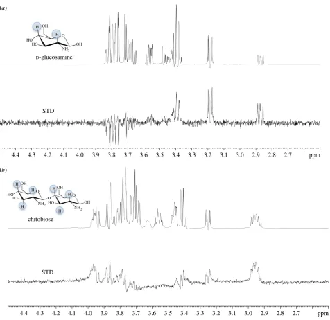
UspCNt. STD effects were observed in the STD-NMR difference
spectrum for both D-glucosamine (50 mM) and chitobiose
(15 mM) and hence provide further support that both of
these amino sugars do bind to the UspCNtprotein (figure 5).
From these experiments, we have determined from the
STD-NMR binding pattern that the H2 (a and b) and H4
(a and b) of D-glucosamine interact non-covalently with
Mtb-UspCNt(figure 5a) and that in the case of chitobiose the
H2 (aandb), H3 (aandb) and H4 (aandb) interact with
Mtb-UspCNt(figure 5b). It has not been possible to determine
which subunit of the b-1,4-linked D-glucosamine units of
chitobiose is involved without higher resolution NMR. Given the additional interaction observed with chitobiose in the
STD-NMR experiment, this goes some way to explaining the
increasedTmshift observed from the thermal shift assays.
3. Discussion
To date, the nutrient requirements ofMtb during infection
inside the human host remain to be fully elucidated [11]. Remarkably, there is little known regarding the identity and properties and mechanisms of the proteins that are involved in the import of essential nutrients. The structural and biochemical analysis of carbohydrate transport systems
are an important step towards understanding howMtb can
O HO HO OH OH D-galactosamine O HO HO OH OH D-mannosamine O HO HO OH OH D-glucopyranosyl amine O HO HO
NH2 NH2 NH2
H2N
OH OH
D-glucosamine
NH2
N3
H2N
O HO HO OH OH 6-amino-6-deoxy-glucopyranose kanosamine O HO OH OH OH O HO HO OH OH 2-azido-2-deoxy-glucopyranose O HO HO NH OH OH O N-acetyl glucosamine NH2 O HO HO NHSO3 OH OH D
-glucosamine-2-N-sulfate
O HO HO F OH OH 2-fluoro-2-deoxy-glucose O HO HO OH OH O P O OH OH D -glucose-6-phosphate O HO HO OH O P O OH OH D-glucosamine 6-phosphate NH2 NH2 O HO O OH OH COOH muramic acid O HO OH OH O HO OH O OH HO D-lactosamine O HO OH OH OH O HO OH O OH HO cellobiose NH2
NH2 NH2
[image:8.595.63.537.29.589.2]NH2 NH2 O HO OH OH O HO HO O OH chitobiose O HO OH OH O O HO O OH O HO HO OH chitotriose O HO HN O OH O O O NH O OH O HO O O R n peptidoglycan backbone NH2 NH2 NH2 NH2 O HO OH OH O O HO O OH O O HO OH O HO HO OH chitotetraose
acquire essential nutrients from a carbohydrate-limited host cell environment.
Our X-ray crystallographic structure determination
revealed that UspC has the same overall fold and architecture as other carbohydrate-binding proteins associated with charac-terized ABC transporter systems [15,18], comprising two subdomains joined by a central hinge region that enclose the (putative) substrate binding cleft. Structural similarity to the
solute-binding unit of the E. coli maltose transporter [26],
the capacity for conformational flexibility between N- and C-terminal lobes (figure 1b), distribution of solvent-accessible aromatic side chains in the binding cleft and the characteristic acidic molecular surface (figure 2b) are structural features fully consistent with the proposed role as the substrate binding unit of the carbohydrate UspABC ABC transporter system.
The thermal shift data have provided the first evidence for carbohydrate-binding selectivity of UspC (figure 3; electronic supplementary material, figure S8). Among the panel of carbo-hydrates tested, there is a clear preference for sugars with a free amino group at C2, whereby adding a phosphate at C6
toD-glucosamine or using the amino-disaccharide chitobiose
markedly increased the thermal stability of UspCNt. Although
thein vitrobinding affinities of these two ligands are relatively
weak to Mtb-UspCNt, when compared with the affinity of
sn-glycero-3-phosophocholine for UgpB (Kd27mM) of the
UgpABCE transporter system [15], binding affinities of up to 8 mM have been reported for a PG recognition lectin with mimics of the PG backbone [28]. Nonetheless, the STD-NMR
data have revealed the specific binding epitope forD
-glucos-amine and chitobiose (figure 5); however, this does not preclude the potential for the further identification of higher affinity substrates.
Recycling and import of saccharides and lipids are
emerging as an essential feature of survival ofMtbin
macro-phages. A prime example is the LpqY-SugABC system, which has been implicated in recycling of trehalose [13], the saccharide component of the primary mycobacterial cell-wall lipid cord factor [29]. Similarly, the UgpABCE transporter
system has been implicated in importing the lipid
sn-glycero-3-phosphocholine [15]. Consistent with low sequence identity 2.7
2.8 2.9 3.0 3.1 3.2 3.3 3.4 3.5 3.6 3.7 3.8 3.9 4.0 4.1 4.2 4.3
4.4 ppm
(a)
D-glucosamine
STD
chitobiose
2.7 2.8 2.9 3.0 3.1 3.2 3.3 3.4 3.5 3.6 3.7 3.8 3.9 4.0 4.1 4.2 4.3
4.4 ppm
(b)
STD OH
HO HO
HO OH
OH O
O O
H H
H
H
NH2
NH2
OH OH
H
H O HO
HO
NH2
H
[image:9.595.61.538.36.498.2]H
Figure 5.
STD-NMR analysis of amino-sugar binding to UspC
Nt.
D-Glucosamine (50 mM) (a) and chitobiose (15 mM) (b) were probed using
1H-NMR in the absence
(top) and presence (bottom) of 30
mM UspC
Nt. The top panel in both (a) and (b) represent the reference NMR spectrum, while the bottom panel in both (a) and (b)
represents the STD-NMR spectrum. The STD effects of the protons involved for each amino sugar are highlighted (blue).
rsob.r
oy
alsocietypublishing.org
Open
Biol.
6
:
160105
between UspC and LpqY (17%) or UgpB (22%), neither
treha-lose norsn-glycero-3-phosphocholine increase the stability of
UspCNt (figure 3), reinforcing the notion that carbohydrate
transport permease systems in mycobacteria have defined sub-strate preferences. Such preferences are also manifested in that relatively subtle changes of structural features in the ligand-binding cleft can have a pronounced effect on ligand affinity.
For instance, binding ofMtbUgpB to
sn-glycero-3-phospho-choline depends on the presence of solvent-exposed Leu205 in the active site cleft, in line with the hydrophobic nature of the intact substrate [15]. Even when Leu205 is substituted
with the corresponding tryptophan from the E. coli UgpB
orthologue, glycerol-3-phosphate binding is not restored [15].
When mapped onto the structure of UspCNt, Leu205 ofMtb
UgpB falls close to Gln218, one of the active site cleft residues mutation of which markedly affected the stabilizing effect of chitobiose.
Given the potential lack of availability of diverse
carbo-hydrates during intracellular infection of Mtb within the
environment of the phagosome, our findings that UspC prefer-entially binds amino sugars are significant. While, to our knowledge, chitobiose is not found within the phagosome, the structural relationship of the binder chitobiose to the PG backbone of the mycobacterial cell wall is striking (figure 4). The UspABC transporter is likely to be localized in the inner-membrane of the cell wall, with the solute-binding UspC protein positioned in the periplasmic space between the inner-membrane and the mycolic acid–arabinogalactan– peptidoglycan core of the mycobacterial cell wall, thus positioning UspC in close proximity to cell-wall amino-sugar substrates. Amino sugars are abundant in the cell wall of Mtb, not least as the dominant component of cell-wall PG, for which the UspC-binder chitobiose can be considered a
deacety-lated analogue. Similarly,D-galactosamine is present through
the modification of interior branched arabinosyl residues in the arabinogalactan layer [30]. It could therefore be envisaged that from a physiological stand point, UspC requires relatively high binding affinities for its amino-sugar substrates to pre-vent the organism from depriving the integral cell wall of amino sugars unless required. If PG were the origin of UspABC substrates, transport would probably require
de-acetylation, which could be mediated byMtbRv1096, known
to deacetylate PG [31], or through additional yet to be ident-ified deacetylases [30]. Hydrolysis of PG is known to be mediated through the lytic transglycosylase resuscitation
pro-moting factors (Rpf) that cleave the glycosidicb-(1,4)-linkage
between alternating MurNAc–GlcNAc residues resulting in disaccharide functional units [32]. It is therefore tantalizing to link the essentiality of the UspABC transporter [19] to a poten-tial functional role in recycling of amino-sugar cell-wall components, thus contributing to evolutionary adaptation to the carbohydrate-limited niche of host macrophages and optimizing the use of scarce carbohydrates within this environ-mental niche. Further experiments are now underway to further investigate this hypothesis.
In conclusion, our data strongly indicate thatMtb-UspC is a
carbohydrate-binding unit of the essential UspABC transpor-ter system, with a substrate preference for sugars containing an amino group at the C2 or C3 position. These data indicate
a potential functional role for the Mtb UspABC transport
system in recycling key components of PG from the
mycobac-terial cell wall, affordingMtb the opportunity to use scarce
nutrients during intracellular infection.
4. Material and methods
All chemicals and reagents were purchased from Sigma-Aldrich, with the exception of all of the carbohydrates used in this study, which were purchased from Carbosynth. Restriction enzymes were obtained from New England Biolabs. Double-distilled water was used throughout.
4.1. Plasmid constructs, protein expression and
purification
Full length and an N-terminal truncated form (codons 31–
440) of Mtb uspC were amplified from Mtb genomic DNA
and cloned into either pET28a (þ) or pET23b (þ) vector
using the NdeI and HindIII restriction enzyme sites resulting
in the constructs: uspC_pet28a, uspC_pet23b, uspC_T_pet28a
anduspC_T_pet23b. The primer sequences are listed in the elec-tronic supplementary material, table S2. Targeted single-site
substitutions were introduced into uspC_T_pet28a using the
primers listed in the electronic supplementary material, table
S2 with Phusion Polymerase and the PCR cycle (988C, 30 s;
20 cycles of 988C, 30 s; 608C, 30 s; 728C, 4 min; followed by
5 min at 728C), followed by digestion with 1ml DpnI. Plasmid
sequences were verified and used for protein expression. Escherichia coli BL21(DE3) competent cells were
trans-formed with theuspCexpression plasmid, grown at 278C to
an optical density at 600 nm (OD600) of 0.8 –1.0 in Terrific
Broth medium (Difco) supplemented with either 50mg ml– 1
kanamycin ( pET28a constructs) or 100mg ml21
ampicillin ( pET23b constructs). Protein production was induced with
1 mM isopropyl-b-thiogalactopyranoside (IPTG) and the
cul-tures were grown at 168C overnight with shaking. The cells
were harvested and resuspended in lysis buffer (50 mM
NaH2PO4, 300 mM NaCl, 10% glycerol pH 7.6 (buffer A)
sup-plemented with 0.1% Triton-X 100 and Complete Protease Inhibitor Cocktail (Roche). The cells were freeze –thawed and sonicated on ice (Sonicator Ultrasonic Liquid Processor XL; Misonix). Following centrifugation (27 000g, 40 min,
48C) the supernatant was loaded onto Ni2þ-affinity resin
(Qiagen). Recombinant UspC was eluted from the Ni2þ
-affi-nity column in buffer A with increasing concentrations of imidazole. Fractions containing the protein were dialysed against 20 mM Tris–HCl, 100 mM NaCl, 10% glycerol pH 8.0 (buffer B). After dialysis, the protein was loaded onto a 1 ml QHP ion exchange column (GE Healthcare) and eluted with buffer B with increasing NaCl concentrations (0.1– 1 M). Fractions containing pure UspC were dialysed at
48C against 50 mM HEPES, 100 mM NaCl, 5% glycerol, pH
7.6. The identity of the protein was confirmed by tryptic digest and nanoLC-ESI-MS/MS (WPH Proteomics Facility, University of Warwick).
4.2. Crystallization and structure determination
Purified UspC (truncated at the N-terminus to remove the
first 31 amino acids: UspCNt) was concentrated by
ultrafiltra-tion (30 kDa cutoff; Amicon Ultra) to 10 mg ml21in 50 mM
HEPES, 100 mM NaCl, 5% glycerol, pH 7.6. Crystals were grown by vapour diffusion in 96-well, sitting-drop plates (SwissSci), using an automatic liquid handling system (Mosquito, TTP Labtech) to pipette drops of 150 nl protein solution mixed with 150 nl reservoir solution. Reservoir
rsob.r
oy
alsocietypublishing.org
Open
Biol.
6
:
160105
conditions producing diffracting crystals are listed in the elec-tronic supplementary material, table S3. Crystals appeared
after 1–3 days at 188C. UspC crystals were mounted
into nylon loops directly from the crystallization drop and flash-frozen in liquid nitrogen prior to data collection.
Diffraction data were recorded for two crystal forms (table 1) at the Diamond Light Source, respectively. Initial phases were determined based on an iodine derivative, using single-wave-length anomalous diffraction data recorded on our in-house source (Rigaku MicroMax007HF, VariMax optics, Saturn 944 CCD). All diffraction data were integrated and scaled using XDS, XSCALE [33] and programs of the CCP4 suite [34]. Heavy atom positions were determined in SHELXD [35], and phases calculated in SHARP [36], followed by solvent-flattening in SOLOMON [37]. An initial model of UspC was generated using ARP/wARP [38], extended manually (COOT [39]) and used to determine a molecular replacement solution for the monoclinic crystal form (PHASER [40]). An improved exper-imental density map could be generated by multi-crystal averaging (DMMULTI [34]), which allowed to build and refine (REFMAC5 [41], PHENIX.REFINE [42]) a complete struc-tural model. The final refined model comprises residues 34–441
of the sequence ofMtb-UspC, plus an additional six residues
originating from the C-terminal affinity tag encoded by the expression plasmid. Crystallographic data and refinement
stat-istics are shown in table 1. Figures were prepared using PYMOL
(www.pymol.org) adopting the Corey–Pauling–Koltun (CPK) colouring scheme: O, red; N, blue; S, yellow and C, green, as indicated in the figure legends.
4.3. Deposition of coordinates and structure factors
Coordinates and structure factors have been deposited in the Protein Data Bank under PDB accession codes 5K2X (tetra-gonal crystal form) and 5K2Y (monoclinic crystal form).
4.4. Protein thermal shift assay
The transition unfolding temperatureTmof the UspCNtprotein
(30mM) was determined in the presence or the absence
of ligands. The carbohydrate screen used a constant ligand concentration of 100 mM, while the saturation binding
experiment probedTm over a concentration range from 0 to
200 mM. Reactions were performed in a total volume of 20ml
using Rotor-Gene Q Detection System (Qiagen), setting the exci-tation wavelength to 470 nm and detecting emission at 557 nm
of the SYPRO Orange protein gel stain, 15final concentration
(Invitrogen). The cycle used was a melt ramp from 30 to 958C,
increasing temperature in 18C steps and time intervals of 5 s.
Flu-orescence intensity was plotted as a function of temperature. The
Tmwas determined using the ROTOR-GENEQ software and the
Analysis Melt functionality. All experiments were performed
in triplicate. To obtain saturation binding data, theDTmvalues
of two experiments were averaged and plotted against
concentration of compound.Kd,appwas determined by fitting a
single-site binding model (GraphPad, PRISM5).
4.5. Saturation transfer difference NMR
UspC was buffer exchanged into deuterated phosphate-buffered saline (PBS) and the ligands dissolved in deuterated PBS. All STD-NMR experiments were recorded on a 600 MHz Bruker Avance III instrument equipped with a 5 mm TBI probe. Acquisitions were performed at 298 K using the stan-dard STD pulse sequence with a shaped Q5 pulse train
(50 ms, 908, 4ms delay between pulses) for selective protein
irradiation, and an alteration between on and off resonances. Presaturation of the protein resonances was performed with an on-resonance radiation at 0.98 ppm; off resonance radiation was applied at 50.0 ppm where no NMR resonances of protein or ligand are present. STD spectra were recorded as described previously [43], with water suppression. A total number of 16 scans were collected for each experiment.
Data accessibility. The datasets supporting this article have been uploaded as part of the electronic supplementary material. Authors’ contributions.E.F., K.F. and G.S.B. conceived and designed exper-iments; E.F., I.P. and K.F. performed experexper-iments; E.F., I.P., K.F. and G.S.B. analysed data; E.F., K.F. and G.S.B. wrote the manuscript. All authors provided final approval of the version to be published. Competing interests.We have no competing interests.
Funding. G.S.B. acknowledges support in the form of a Personal Research Chair from Mr James Bardrick, a Royal Society Wolfson Research Merit Award and from the Medical Research Council (MR/K012118/1). E.F. previously held a Leverhulme Trust Early Career Fellowship and is now a Sir Henry Dale Fellow (the Wellcome Trust, Royal Society, grant no. 104193/Z/14/Z). This work used a crystal imaging system obtained through the Birmingham City Translational Medicine Clinical Research and Infrastructure Trials Platform. We also acknowledge the contribution of the Warwick/ Waters Centre for BioMedical Mass Spectrometry and Proteomics in the School of Life Sciences, University of Warwick.
Acknowledgements.The authors would like to thank Prof. Edith Sim for
helpful discussions; Dr Claudia Sala for providing Mtb genomic
DNA and Professor John McCarthy for access to facilities. We would also like to thank staff at Diamond Light Source beamlines I04 and I041 for technical support and assistance.
References
1. WHO. 2015Global Tuberculosis Report2015. Geneva, Switzerland: WHO.
2. Dheda K, Gumbo T, Gandhi NR, Murray M, Theron G, Udwadia Z, Migliori GB, Warren R. 2014 Global control of tuberculosis: from extensively drug-resistant to untreatable tuberculosis.Lancet Respir. Med.2, 321 – 338. (doi:10.1016/S2213-2600(14)70031-1)
3. Kim DHet al.2008 Treatment outcomes and long-term survival in patients with extensively
drug-resistant tuberculosis.Am. J. Respir. Crit. Care Med. 178, 1075– 1082. (doi:10.1164/rccm.200801-132OC) 4. de Chastellier C. 2009 The many niches and
strategies used by pathogenic mycobacteria for survival within host macrophages.Immunobiology 214, 526 – 542. (doi:10.1016/j.imbio.2008.12.005) 5. Cole STet al.1998 Deciphering the biology of
Mycobacterium tuberculosisfrom the complete genome sequence.Nature393, 537 – 544. (doi:10. 1038/31159)
6. Schnappinger Det al.2003 Transcriptional adaptation ofMycobacterium tuberculosiswithin macrophages: insights into the phagosomal environment.J. Exp. Med.198, 693 – 704. (doi:10.1084/jem.20030846)
7. Pandey AK, Sassetti CM. 2008 Mycobacterial persistence requires the utilization of host cholesterol.Proc. Natl Acad. Sci. USA105, 4376 – 4380. (doi:10.1073/pnas. 0711159105)
rsob.r
oy
alsocietypublishing.org
Open
Biol.
6
:
160105
8. Yam KCet al.2009 Studies of a ring-cleaving dioxygenase illuminate the role of cholesterol metabolism in the pathogenesis ofMycobacterium tuberculosis.PLoS Pathog.5, e1000344. (doi:10. 1371/journal.ppat.1000344)
9. Braibant M, Gilot P, Content J. 2000 The ATP binding cassette (ABC) transport systems of Mycobacterium tuberculosis.FEMS Microbiol. Rev. 24, 449 – 467. (doi:10.1111/j.1574-6976.2000. tb00550.x)
10. Titgemeyer Fet al.2007 A genomic view of sugar transport inMycobacterium smegmatisand Mycobacterium tuberculosis.J. Bacteriol.189, 5903 – 5915. (doi:10.1128/JB.00257-07) 11. Niederweis M. 2008 Nutrient acquisition by
mycobacteria.Microbiology154, 679 – 692. (doi:10. 1099/mic.0.2007/012872-0)
12. Sassetti CM, Rubin EJ. 2003 Genetic requirements for mycobacterial survival during infection.Proc. Natl Acad. Sci. USA100, 12 989 – 12 994. (doi:10. 1073/pnas.2134250100)
13. Kalscheuer R, Weinrick B, Veeraraghavan U, Besra GS, Jacobs WRJr. 2010 Trehalose-recycling ABC transporter LpqY-SugA-SugB-SugC is essential for virulence ofMycobacterium tuberculosis.Proc. Natl Acad. Sci. USA107, 21 761 – 21 766. (doi:10.1073/ pnas.1014642108)
14. Rengarajan J, Bloom BR, Rubin EJ. 2005 Genome-wide requirements forMycobacterium tuberculosis adaptation and survival in macrophages.Proc. Natl Acad. Sci. USA102, 8327 – 8332. (doi:10.1073/pnas. 0503272102)
15. Jiang D, Zhang Q, Zheng Q, Zhou H, Jin J, Zhou W, Bartlam M, Rao Z. 2014 Structural analysis of Mycobacterium tuberculosisATP-binding cassette transporter subunit UgpB reveals specificity for glycerophosphocholine.FEBS J.281, 331 – 341. (doi:10.1111/febs.12600)
16. Somashekar BS, Amin AG, Rithner CD, Troudt J, Basaraba R, Izzo A, Crick DC, Chatterjee D. 2011 Metabolic profiling of lung granuloma in Mycobacterium tuberculosisinfected guinea pigs: ex vivo1H magic angle spinning NMR studies. J. Proteome Res.10, 4186 – 4195. (doi:10.1021/ pr2003352)
17. Lew JM, Kapopoulou A, Jones LM, Cole ST. 2011 TubercuList–10 years after.Tuberculosis91, 1 – 7. (doi:10.1016/j.tube.2010.09.008)
18. Vahedi-Faridi A, Licht A, Bulut H, Scheffel F, Keller S, Wehmeier UF, Saenger W, Schneider E. 2010 Crystal structures of the solute receptor GacH of Streptomyces glaucescensin complex with acarbose and an acarbose homolog: comparison with the acarbose-loaded maltose-binding protein of Salmonella typhimurium.J. Mol. Biol.397, 709 – 723. (doi:10.1016/j.jmb.2010.01.054)
19. Griffin JE, Gawronski JD, Dejesus MA, Ioerger TR, Akerley BJ, Sassetti CM. 2011 High-resolution phenotypic profiling defines genes essential for mycobacterial growth and cholesterol catabolism. PLoS Pathog.7, e1002251. (doi:10.1371/journal. ppat.1002251)
20. Cole STet al.2001 Massive gene decay in the leprosy bacillus.Nature409, 1007 – 1011. (doi:10. 1038/35059006)
21. Krogh A, Larsson B, von Heijne G, Sonnhammer EL. 2001 Predicting transmembrane protein topology with a hidden Markov model: application to complete genomes.J. Mol. Biol.305, 567 – 580. (doi:10.1006/jmbi.2000.4315)
22. Bendtsen JD, Nielsen H, von Heijne G, Brunak S. 2004 Improved prediction of signal peptides: SignalP 3.0.J. Mol. Biol.340, 783 – 795. (doi:10. 1016/j.jmb.2004.05.028)
23. Davis IW, Murray LW, Richardson JS, Richardson DC. 2004 MOLPROBITY: structure validation and all-atom contact analysis for nucleic acids and their complexes.Nucleic Acids Res.32, W615 – W619. (doi:10.1093/nar/gkh398)
24. Krissinel E, Henrick K. 2004 Secondary-structure matching (SSM), a new tool for fast protein structure alignment in three dimensions.Acta Crystallogr. D Biol. Crystallogr.60, 2256 – 2268. (doi:10.1107/S0907444904026460)
25. Krissinel E, Henrick K. 2007 Inference of macromolecular assemblies from crystalline state. J. Mol. Biol.372, 774 – 797. (doi:10.1016/j.jmb. 2007.05.022)
26. Oldham ML, Chen J. 2011 Snapshots of the maltose transporter during ATP hydrolysis.Proc. Natl Acad. Sci. USA108, 15 152 – 15 156. (doi:10.1073/pnas. 1108858108)
27. McNicholas S, Potterton E, Wilson KS, Noble ME. 2011 Presenting your structures: the CCP4mg molecular-graphics software.Acta Crystallogr. D Biol. Crystallogr.67, 386 – 394. (doi:10.1107/ S0907444911007281)
28. Lehotzky RE, Partch CL, Mukherjee S, Cash HL, Goldman WE, Gardner KH, Hooper LV. 2010 Molecular basis for peptidoglycan recognition by a bactericidal lectin.Proc. Natl Acad. Sci. USA107, 7722 – 7727. (doi:10.1073/pnas.0909449107) 29. Noll H, Bloch H, Asselineau J, Lederer E. 1956 The
chemical structure of the cord factor of Mycobacterium tuberculosis.Biochim. Biophys. Acta 20, 299 – 309. (doi:10.1016/0006-3002(56)90289-X) 30. Skovierova Het al.2010 Biosynthetic origin of the
galactosamine substituent of arabinogalactan in Mycobacterium tuberculosis.J. Biol. Chem.285, 41 348 – 41 355. (doi:10.1074/jbc.M110.188110) 31. Yang S, Zhang F, Kang J, Zhang W, Deng G, Xin Y,
Ma Y. 2014Mycobacterium tuberculosisRv1096
protein: gene cloning, protein expression, and peptidoglycan deacetylase activity.BMC Microbiol. 14, 174. (doi:10.1186/1471-2180-14-174) 32. Kana BDet al.2008 The resuscitation-promoting
factors ofMycobacterium tuberculosisare required for virulence and resuscitation from dormancy but are collectively dispensable for growthin vitro.Mol. Microbiol.67, 672 – 684. (doi:10.1111/j.1365-2958. 2007.06078.x)
33. Kabsch W. 2010 Integration, scaling, space-group assignment and post-refinement.Acta Crystallogr. D Biol. Crystallogr.66, 133 – 144. (doi:10.1107/ S0907444909047374)
34. Winn MDet al.2011 Overview of theCCP4suite and current developments.Acta Crystallogr. D Biol. Crystallogr.67, 235 – 242. (doi:10.1107/ S0907444910045749)
35. Sheldrick GM. 2008 A short history of SHELX.Acta Crystallogr. A Found Adv.64, 112 – 122. (doi:10. 1107/S0108767307043930)
36. Bricogne G, Vonrhein C, Flensburg C, Schiltz M, Paciorek W. 2003 Generation, representation and flow of phase information in structure
determination: recent developments in and around SHARP 2.0.Acta Crystallogr. D Biol. Crystallogr.59, 2023 – 2030. (doi:10.1107/S0907444903017694) 37. Abrahams JP, Leslie AG. 1996 Methods used in
the structure determination of bovine mitochondrial F1 ATPase.Acta Crystallogr. D Biol. Crystallogr.52, 30 – 42. (doi:10.1107/S0907444995008754) 38. Langer G, Cohen SX, Lamzin VS, Perrakis A. 2008
Automated macromolecular model building for X-ray crystallography using ARP/wARP version 7.Nat. Protoc.3, 1171– 1179. (doi:10.1038/nprot.2008.91) 39. Emsley P, Lohkamp B, Scott WG, Cowtan K. 2010 Features and development of Coot.Acta Crystallogr. D Biol. Crystallogr.66, 486 – 501. (doi:10.1107/ S0907444910007493)
40. McCoy AJ, Grosse-Kunstleve RW, Adams PD, Winn MD, Storoni LC, Read RJ. 2007 Phaser crystallographic software.J. Appl. Crystallogr.40, 658 – 674. (doi:10.1107/S0021889807021206) 41. Murshudov GN, Skuba´k P, Lebedev AA, Pannu NS,
Steiner RA, Nicholls RA, Winn MD, Long F, Vagin AA. 2011REFMAC5for the refinement of macromolecular crystal structures.Acta Crystallogr. D Biol. Crystallogr.67, 355 – 367. (doi:10.1107/ S0907444911001314)
42. Adams PDet al.2010 PHENIX: a comprehensive Python-based system for macromolecular structure solution.Acta Crystallogr. D Biol. Crystallogr.66, 213 – 221. (doi:10.1107/S0907444909052925) 43. Castro S, Duff M, Snyder NL, Morton M, Kumar CV,
Peczuh MW. 2005 Recognition of septanose carbohydrates by concanavalin A.Org. Biomol. Chem.3, 3869 – 3872. (doi:10.1039/b509243d)