ISSN Online: 2165-3410 ISSN Print: 2165-3402
DOI: 10.4236/aim.2019.912066 Dec. 24, 2019 1012 Advances in Microbiology
Antibacterial Activity of Psidium guajava Leaf
Extract against Selected Pathogenic Bacteria
Anayochukwu Chibuike Ngene
1,2*, John C. Aguiyi
1, Chinedu Joshua Chibuike
2, Virginia O. Ifeanyi
2,
Kelechi M. Ukaegbu-Obi
2, Esther Garos Kim
3, Uchechukwu C. Ohaeri
1, Bruno O. Onyemegbulem
11Africa Center of Excellence in Phytomedicine Research and Development, University of Jos, Jos, Plateau State, Nigeria
2Department of Microbiology, College of Natural Sciences, Michael Okpara University of Agriculture, Umudike, Abia State, Nigeria 3Department of Medical Microbiology, Faculty of Clinical Sciences, College of Health Sciences, University of Jos, Jos, Plateau State, Nigeria
Abstract
This study has been embarked upon due to increased resistance of bacteria to many already synthesized drugs and the high price of the orthodox medicines in the market, the antimicrobial effect of Psidium guajava leaf extracts against selected drug-resistant bacteria will help to reduce the overdependence on orthodox or synthetic drugs which greater population of the society cannot afford due to economic/financial restriction. Ethanolic and aqueous extracts of Psidium guajava were assayed for antibacterial activity using the agar dilu-tion method in the determinadilu-tion of the minimum inhibitory concentradilu-tion (MIC) and minimum bactericidal concentration (MBC). The bioactive com-ponents of the extracts were determined using standard techniques and the inhibitory activities of the extracts were compared to gentamicin, ciproflox-acin, erythromycin, azithromycin, ofloxciproflox-acin, and nitrofurantoin. The MIC of the extract on the test organisms ranges from 10 mg/ml - 40 mg/ml. The sen-sitivity test showed that the organisms were susceptible to Psidium guajava leave extract except Pseudomonas aeruginosa. The phytochemical analysis of the extracts revealed the presence of bioactive compounds such as Tannin, Saponin, Balsam, Flavonoids, Alkaloids, Cardiac glycosides, Carbohydrate, Resins, Terpenes, and Sterols. It has been shown that the leaves of Psidium guajava have antibacterial effects and hence can be used to treat the ailments caused by those organisms at a particular dosage and concentration. Howev-er, more research work on its toxicity level, synergistic or antagonistic inte-raction with other plants or drugs is needed to consolidate its usage.
Keywords
Antibiotic Resistance, Psidium guajava, Minimum Inhibitory Concentration, Minimum Bactericidal Concentration, Zone of Inhibition, Phytochemical Analysis How to cite this paper: Ngene, A.C.,
Aguiyi, J.C., Chibuike, C.J., Ifeanyi, V.O., Ukaegbu-Obi, K.M., Kim, E.G., Ohaeri, U.C. and Onyemegbulem, B.O. (2019) Antibacterial Activity of Psidium guajava Leaf Extract against Selected Pathogenic Bacteria. Advances in Microbiology, 9, 1012-1022.
https://doi.org/10.4236/aim.2019.912066
Received: September 30, 2019 Accepted: December 21, 2019 Published: December 24, 2019
Copyright © 2019 by author(s) and Scientific Research Publishing Inc. This work is licensed under the Creative Commons Attribution International License (CC BY 4.0).
DOI: 10.4236/aim.2019.912066 1013 Advances in Microbiology
1. Introduction
Plants are one of the most important sources of medicines for treating illnesses since the beginning of human civilization [1]. The use of plants and herb extract in the treatment of human ailments is a very ancient art, a practice that has been passed on for generations and Scientists in Africa and other developing coun-tries are researching local plants abundant in the continent for their possible use in traditional medicine [2]. The study on the medicinal plants is essential to promote the proper use of herbal medicine to determine their potential as a source for the new drugs [3]. Research into traditional plants and herbs received a further boost due to the increasing resistance to much orthodox medicine and thus a search for new organic molecules of plants with antimicrobial properties [4]. The World Health Organization (WHO) has categorized more than 20,000 plant species with medicinal properties providing treatment for such diseases as pneumonia, ulcers, diarrhea, bronchitis, colds, and diseases of the respiratory tracts. One method among the many ways in which plants are used in popular remedies is to extract and consume essential plant oils. Essential oils are complex chemical natural products extracted from plant species, and are composed of more than a hundred compounds some of which are responsible for plant aro-mas. They are obtained from different parts of the plants. Flowers, leaves, seeds, barks, and tubers of many plants have medicinal properties [5]. The use of plant extracts and phyto-products is gaining attention due to their availability, cost-effectiveness, proven nature of specificity, biodegradability, low toxicity, and minimum residual toxicity in the ecosystem [6]. Increased developments of resistance to current antibiotics have strengthened scientific research for the discovery of new drugs. However, new leads/hits in drug discovery have been developed from natural sources due to growing scientific links between the fol-kloric medicinal use of some of these natural products especially for plant origin, to biological activity. Hence plants continue to provide a good source for new drugs [7] [8]. Guava is a small tropical tree that grows up to 35 feet tall; it is grown for its fruits in tropics. It is a member of the Myrtaceae family, with about 133 genera and more than 3800 species. The leaves and bark of the Psidium gua-java tree have a long history of medicinal uses that are still employed today [9]. The leaves and bark of the guava plant have been used to treat diarrhea, other gastrointestinal disorders, toothaches, colds, and swelling. Guava is used for skin disorders such as astringent for acne, rashes, and ringworm [10]. [11] tested Psi-dium guajava against dermatophytes, Trichophyton tonsurans, Trichophyton rubrum, Microsporum fulvrum, and Candida albicans. The guava tree, Psidium guajava Linnaeus, is distributed from México down to São Paulo State, Brazil, being indigenous to Central América and part of South America between Co-lombia and Peru [12] [13] [14] [15]. Worldwide literature information is availa-ble about the antimicrobial activity by P. guajava on different members of such bacterial families as Enterobacteriaceae, Vibrionaceae, Micrococcaceae, and Pro-pionibacteriaceae.
DOI: 10.4236/aim.2019.912066 1014 Advances in Microbiology To evaluate the antimicrobial activity of Psidium guajava leaf extract against selected pathogenic bacteria.
Objectives:
1) To determine the bioactive components in Psidium guajava leaf. 2) To isolate and identify selected bacteria.
3) To determine the minimum inhibitory concentration and minimum bacte-ricidal concentration of Psidium guajava on the isolates.
2. Materials and Methods
2.1. Plant Material Collection
Psidium guajava leaves were collected from National Root Crop Research Insti-tute Umudike, Abia state.
2.2. Sources of Isolation of Organisms
The organisms were isolated from various sources including the soil, streams, cow dung, and human nose swab. These samples were collected and the organ-isms were isolated, characterized and identified in the Microbiology Laboratory, Michael Okpara University of Agriculture in Umudike (MOUAU), Abia state.
2.3. Isolation, Characterization and Identification of the Organisms
The different samples of stream water, soil, cow dung and human nose swab (from students living in IBB Hostel, MOUAU) were randomly collected from the school environment, and brought to the laboratory. Ten-fold serial dilution was carried on them using the physiological saline to reduce the microbial load. The serially diluted samples were cultured on different culture medium at tem-perature of 37˚C for 24 hours. The isolated organisms were characterized and identified using various biochemical test [16]. The identified isolates were stored in agar slants and subcultured every two weeks.2.4. Preparation of Extracts
The already collected leaves were air-dried for two weeks and later oven-dried for proper drying. The plants were pulverized using the grinder. The method of [17] was used. Fifty gramme (50 g) of the plant was macerated successively in 250 ml of ethanol and distilled water respectively in conical flask at room tem-perature with shaking every 6 hrs for 48 hrs. Then the extracts were filtered us-ing a muslin cloth and then Whatman filter paper. The filtrate was then evaporated to dryness using a water bath at a temperature of 70˚C to remove the solvents used in the extraction. The extract was stored in an airtight bottle until further usage.
2.5. Phytochemical Screening
The phytochemical tests were analyzed after extraction by two solvents (ethanol and aqueous).
DOI: 10.4236/aim.2019.912066 1015 Advances in Microbiology The ethanol and water extracts (5 ml) each was added differently to a concen-trated sulphuric acid (1 ml) and 0.5 g of Mg. A pink or red coloration that dis-appear on standing (3 min) indicates the presence of flavonoids [16].
Tannins:
Two methods were used to test for tannins. First, about 1 ml of the ethanol extract was added in 2 ml of water in a test tube. 2 to 3 drops of diluted ferric chloride solu-tion was added and observed for green to blue-green (catechic tannins) or a blue-black (garlic tannins) coloration. Second, 2 ml of the aqueous extract was added to 2 ml of water, a 1 to 2 drops of diluted ferric chloride solution was added. A dark green or blue-green coloration indicates the presence of tannins [16]. To 1 ml of aqueous extract was added a few volumes of distilled water in a test tube. The solu-tion was shaken vigorously and observed for a stable persistent froth for 20 min [16].
Test for alkaloids:
Three methods were used to test for alkaloids. First, evaporate 10 ml of con-centered etheric solution, the dry residue was added to 1.5 ml HCl (2%) acid so-lution. After that, 1 to 2 drops of Mayer’s reagent and Wagner was added, and the yellow-white precipitate indicates the presence of the alkaloidal base. Second, evaporate 20 ml of ethanol extract, the dry residue dissolved in 5 ml of HCl (2N) and filtered. A few drops of Mayer’s reagent and Wagner were added, the pres-ence of precipitate indicates the alkaloids. Three, to 15 ml of the aqueous extract was added 2 ml of NH4OH à 10% (pH = 7). The alkaloid was extracted 3 times
with 10 ml chloroform. The chloroform layer was washed 3 times with 2 ml of HCL (10%). This was divided into two portions. Mayer’s reagent was added to one portion and Wagner’s reagent to the other. The formation of a brown or white precipitate was regarded as positive for the presence of alkaloids [16].
Test for sterols and steroids:
Sterols and steroids were sought by the reaction of Liebermann. Ten (10 ml) ml of ethanolic extract was evaporated. The residue was dissolved in 0.5 ml of hot acetic anhydride; we added 0.5 ml of the filtrate chloroform. Treated with the reagent of the Liebermann Burchard test. The appearance, at the interphase, a ring of blue-green, showed a positive reaction [16].
Test for the carbohydrate:
Reducing sugars: two methods were used to test for reducing sugars. First, the ethanol extract (1 ml) was added to 1 ml of water and 20 drops of boiling Fehl-ing’s solution (A and B) in a test tube was added too. The formation of a precipi-tate red-brick in the bottom of the tube indicates the presence of reducing sugars. Second, added to 2 ml of the aqueous solution, 5 - 8 drops of boiling Fehling’s solution. A red-brick precipitate showed the presence of reducing sugars [16].
Starch
The aqueous extract 5 ml was treated with the reagent of the starch (iodine). Any shift to blue-violet indicates the presence of starch [16].
2.6. Preparation of the Inoculums
DOI: 10.4236/aim.2019.912066 1016 Advances in Microbiology plated on MacConkey and Nutrient agar and incubated at 37˚C for 24 hrs. After the incubation, the colony of the organisms was taken and each was inoculated into 7 mls of peptone water in a bijou bottle and shook vigorously to obtain homogeneity of the solution. The turbidity produced by these organism were adjusted and used to match the turbidity (opacity) standard prepared as described by [18].
2.7. Bacterial Sensitivity Testing Using
Agar Well Diffusion Method
An 80 mg/ml of both the ethanol and water extract of P. guajava was constituted by dissolving 0.8 g of the extracts in 2 ml each of 20% v/v dimethyl sulfoxide (DMSO) and two-fold serial dilutions made. Inocula measured up to 1 ml each of the test organisms from the peptone waters in the bijou bottles were intro-duced on the surface of a sterile Mueller-Hinton agar. It was evenly distributed by rocking the plates and allowed to dry but covered to prevent atmospheric contami-nation. After that, the plates were bored in two places using a cork borer where 1 ml of the different leave extracts at different concentrations were placed in each hole. Also, a sterile antibiotic disc was carefully placed on different plates containing dif-ferent isolates using a forceps on the center of each plate, which served as controls. These plates were incubated at 37˚C and examined after 24 hrs of growth. Zones of inhibitions were measured in (mm) using transparent meter rule [19].
2.8. Determination of the Minimum Inhibitory
Concentration of the Plant Extracts
The MIC of the plant extract was carried out using a broth dilution method as described in [19]. Two-fold serial dilutions of the extracts were made using Muller-Hinton broth. From the stock concentrations of the extracts containing 40 mg/ml to obtain 20, 10, 5 and 2.5 respectively of the extracts in the broth. Then 0.1 ml of the standardized inoculums from peptone water was then inocu-lated into the solution in the test tubes. These were all incubated at 37˚C for 24 hrs and observed for turbidity of growth. The lowest concentrations which showed no turbidity in the test tubes were recorded as the MIC.
2.9. Determination of the Minimum Bactericidal
Concentration (MBC)
Tubes showing no visible growth from the MIC test were subcultured unto Mueller-Hinton agar. 0.1 ml of the test organisms were inoculated on the plates. These were also incubated at 37˚C for 24 hrs. The plates with the lowest concen-tration of the extracts that showed no colony growth is recorded as the mini-mum inhibitory concentration (MBC).
3. Statistical Analysis
DOI: 10.4236/aim.2019.912066 1017 Advances in Microbiology
4. Results
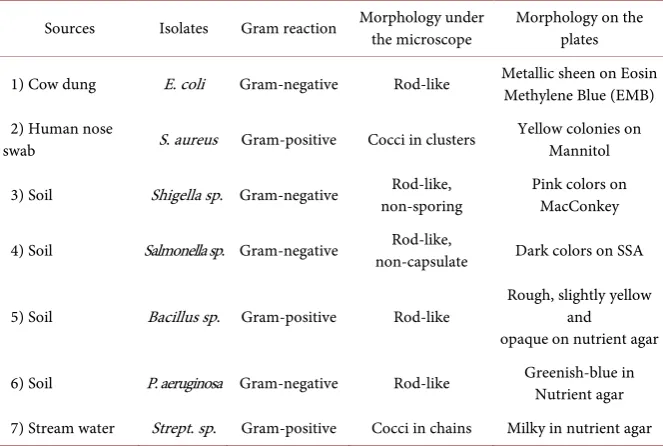
[image:6.595.207.539.316.539.2]The gram staining reaction indicated that four of the organisms were gram- negative while three of them were gram-positive. Out of the three gram-positive identified isolates, one appeared cocci in clusters, the other appeared cocci in chains, while the last was in rod-like form. Out of the four-gram negative iso-lates, all appeared in rod-like form, three were motile while one was non-motile. Also, the morphology of the organisms on different agar plates were investigated and recorded. This is summarized in Table 1. The biochemical test showed that six out of the seven organisms were catalase-positive, four were motile, one was indole positive, one oxidase-positive, and three been citrate positive. Among them, some were negative while some test was not carried on some as they have no relation to such tests. The result is illustrated more in Table 2. From the fol-lowing results above it shows that the folfol-lowing organisms were successfully identified as the isolates. Escherichia coli, Staphylococcus aureus, Shigella sp,
Table 1. Gram staining reaction.
Sources Isolates Gram reaction Morphology under the microscope Morphology on the plates
1) Cow dung E. coli Gram-negative Rod-like Metallic sheen on Eosin Methylene Blue (EMB)
2) Human nose
swab S. aureus Gram-positive Cocci in clusters Yellow colonies on Mannitol
3) Soil Shigella sp. Gram-negative non-sporing Rod-like, Pink colors on MacConkey
4) Soil Salmonella sp. Gram-negative non-capsulate Rod-like, Dark colors on SSA
5) Soil Bacillus sp. Gram-positive Rod-like Rough, slightly yellow and opaque on nutrient agar
6) Soil P. aeruginosa Gram-negative Rod-like Greenish-blue in Nutrient agar
7) Stream water Strept. sp. Gram-positive Cocci in chains Milky in nutrient agar
Table 2. Biochemical test of different isolates.
Isolates Catalase Indole Motility Coagulase Citrate Oxidase
1 + + + − − −
2 + − − + + −
3 + − − − − −
4 + − + − − −
5 + − + − + −
6 + − + − + +
7 − − − − − −
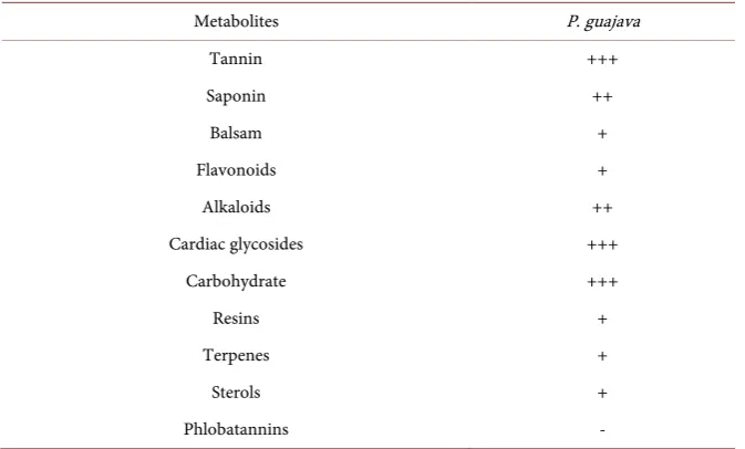
[image:6.595.206.539.570.708.2]DOI: 10.4236/aim.2019.912066 1018 Advances in Microbiology Salmonella sp, Bacillus sp, Pseudomonas aeruginosa, and Streptococcus sp. were isolated and identifted. These were the organisms used to test the strength of the plant extracts. The phytochemical screening of the guava leaf extract shows the presence of metabolites like tannin, saponins, flavonoids, alkaloids cardiac gly-cosides, resins, etc., which occurred in different concentration in the plant ex-tract as shown in Table 3. The sensitivity test shows that the organisms were susceptible to leave extract except P. aeruginosa, which was resistant completely and with few zones. The organisms were also tested on some orthodox standard drugs like gentamicin, ciprofloxacin, erythromycin, etc. which gave a very tangible result. This is shown in Table 4. The leave extract on the different organisms shows that the extracts had their MICs on different concentrations with different organisms ranging from 10 - 40 mg/ml with the gram-positive bacteria having a smaller concentration than the gram-negative bacteria. This is shown in Table 5 and Table 6. The
Table 3. Phytochemical screeningof P. guajava.
Metabolites P. guajava
Tannin +++
Saponin ++
Balsam +
Flavonoids +
Alkaloids ++
Cardiac glycosides +++
Carbohydrate +++
Resins +
Terpenes +
Sterols +
Phlobatannins -
Key; +++, the concentration of the metabolite is high; ++, there is moderate concentration of the metabo-lite; +, there is little amount of the metabolite present; -, there is absence of the metabolite on the plant.
Table 4. Antibacterial sensitivity test
S/N TEST ORGS (mm) ETH (mm) WT (mm) GEN (mm) CPR (mm) ERY (mm) AZ (mm) OFL (mm) NIT
1 E. coli 22 12 20 16 10 - 16 10
2 S. aureus 16 8 20 10 - 15 14 -
3 Shigella sp. 12 8 - 10 6 10 - 14
4 Salmonella sp. 14 6 10 18 10 - 20 10
5 Bacillus sp. 14 - 16 12 - 16 14 6
6 P. aeruginosa - - 8 10 14 - - -
7 Strept. Sp. 18 10 I4 - - 10 12 10
[image:7.595.209.539.552.702.2]DOI: 10.4236/aim.2019.912066 1019 Advances in Microbiology
Table 5. MIC of P. guajava in ethanol.
TEST ORGS./
CONC (mg/ml) Aureus Staph. aeruginosa P. coli E. Strept. Sp Bacillus sp Salmonella sp. Shigella sp.
40 NG G NG NG NG NG NG
20 NG G G NG NG G NG
10 G G G NG G G NG
5 G G G G G G G
2.5 G G G G G G G
[image:8.595.207.542.250.363.2]Key G = growth; NG = no growth.
Table 6. MIC of P. guajava in water.
TEST ORGS/
CONC. (mg/ml) aureus Staph. aeruginosa E. coli Strept. Sp P. Bacillus sp. Salmonella Shigella sp.
40 NG G NG NG NG NG NG
20 G G G G G G G
10 G G G G G G G
5 G G G G G G G
2.5 G G G G G G G
Key G = growth; NG = no growth.
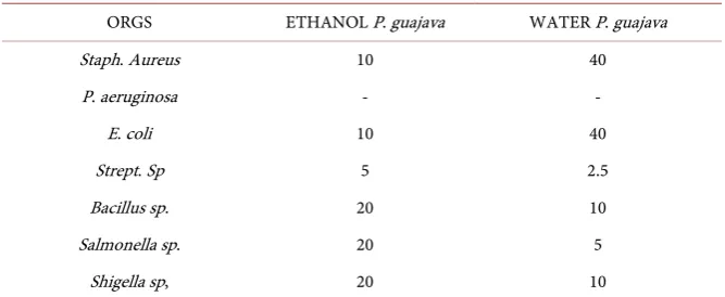
Table 7. MBC of P. guajava in ethanol and water.
ORGS ETHANOL P. guajava WATER P. guajava
Staph. Aureus 10 40
P. aeruginosa - -
E. coli 10 40
Strept. Sp 5 2.5
Bacillus sp. 20 10
Salmonella sp. 20 5
Shigella sp, 20 10
MBCs of the extracts on the different bacteria show Salmonella and Shigella having higher MBC in ethanol extract than in water with E. coli and S. aureus having higher MBC in water than in ethanol. The result also shows no effect on Pseudomonas. This is illustrated in Table 7.
5. Discussions
[image:8.595.206.540.404.541.2]tex-DOI: 10.4236/aim.2019.912066 1020 Advances in Microbiology ture where these plants are grown or probably due to changes in the environ-mental conditions compared to work done by [21]. This may also be that the ac-tive components of the plant extracts are more soluble in ethanol than in water. Also from the result, it was discovered that Pseudomonas aeruginosa has a low susceptibility against the extract from guava with no zone of inhibition seen in water and ethanol extract of the guava, thereby been resistant to it. This result is unique as no one has tested the effect of this plant extracts on Pseudomonas ae-ruginosa. The effectiveness of the P. guajava extracts may be due to the phyto-chemical constituents they contain. Although the constituents are not present in all of them, components like the tannins, saponins, flavonoids, and terpenes have been severally reported to exhibit antibacterial activity and antioxidant ac-tivities [7] [22]. Flavonoids are known to possess good antioxidant properties and guava has been reported to have comparatively high antioxidant activity against 2,2-Diphenyl-1-picryl hydrazyl (DPPH) radicals and the antioxidants are known to exhibit some level of antimicrobial activity due to their ability to mi-nimize the oxidative damage induced by the parasites in the red cells [22].
6. Conclusion
It has been shown that the leaves of Psidium guajava have antibacterial effects and hence can be used to treat the ailments caused by those organisms at a par-ticular dosage and concentration. However, more research work on its toxicity level, possible synergistic or antagonistic interaction with other plants or drugs, is needed to consolidate its usage.
Conflicts of Interest
The authors declare no conflicts of interest regarding the publication of this pa-per.
References
[1] Jyothi, A., Venkatesh, K., Chakrapani, P. and Roja, R. (2011) Phytochemical and Pharmacological Potential of Annonacherimola—A Review. International Journal ofPhytomedicine, 3, 439-447.
[2] Nneamaka, A.I. (1991) Screening of Some Nigerian Medicinal Plants for Bacterial Activity. Amadu Bello University, Zairia, 108 p.
[3] Gajalakshmi, S., Vijayalakshmi, S. and Rajeswari, V. (2012) Phytochemical and Pharmacological Properties of Annona muricata: A Review. International Journal of Pharmacy and Pharmaceutical Science, 4, 3-6.
[4] Sofowora, A. (1993) Medicinal Plants and Traditional Medicine in Africa. 3rd Edi-tion, Spectrum Books Limited Ibadan, Nigeria, 199-204.
[5] Goncalves, F.A., Andrade-Neto, M., Bezerra, J.N.S., Macrae, A., Sousa, O.V., Fon-teles-Filho, A.A. and Vieira, R.H.S.F. (2008) Antibacterial Activity of Guava, Psi-dium guajava Linnaeus, Leaf Extracts on Diarrhea-Causing Enteric Bacteria Isolated from Seabob Shrimp, Xiphopenaeus kroyeri (Heller). Revista do Instituto de Medi-cina Tropical de São Paulo, 50, 11-15.
DOI: 10.4236/aim.2019.912066 1021 Advances in Microbiology
[6] Ogbo, E.M. and Oyibo, A.E. (2008) Effects of Three Plant Extracts (Ocimum gratis-simum, Acalypha wilkesiana and Acalypha macrostachya) on Post-Harvest Patho-gen of Persea Americana. Journal of Medicinal Plants Research, 2, 311-314. [7] Kunle, O.F. and Egharevba, H.O. (2013) Chemical Constituents and Biological
Ac-tivity of Medicinal Plants Used for the Management of Sickle Cell Disease—A Re-view. Journal of Medicinal Plants Research,7, 3452-3476.
[8] Begum, S., Hassan, S.I., Siddiqui, B.S., Shahen, F., Ghayur, M.N. and Gilani, A.H. (2002) Triterpenoids from the Leaves of Psidium guajava. Phytochemistry, 61, 399-403.https://doi.org/10.1016/S0031-9422(02)00190-5
[9] Nwinyi, O.C., Chinedu, N.S., Ajani, O.O., Ikpo, C.O. and Ogunniran, K.O. (2009) Antibacterial Effects of Extracts of Ocimum gratissimum and Piper guineense on
Escherichia coli and Staphylococcus aureus. African Journal of Food Science, 3, 77-81.
[10] Egharevba, H.O., Iliya, I., Ibekwe, N., Abdullahi, M.S., Okwute, S.K. and Okogun, J.I. (2010) Broad Spectrum Antimicrobial Activity of Psidium guajava Linn. Leaf.
NaturalScience, 8, 43-50.
[11] Dutta, B.K., Rahman, I. and Das, T.K. (2000) In-Vitro Study on Antifugal Property of Common Fruit Plants. Journal of Biomedicine, 20, 187-189.
[12] Chomnawang, M.T., Surassmo, S., Nukoolkarn, V.S. and Gritsanapan, W. (2005) Antimicrobial Effects of Thai Medicinal Plants against Acne-Inducing Bacteria.
Journal ofEthnopharmacology, 101, 330-333. https://doi.org/10.1016/j.jep.2005.04.038
[13] Qadan, F., Thewaini, A.J., Ali, D.A., et al. (2005) The Antimicrobial Activities of
Psidium guajava and Juglans regia Leaf Extracts to Acne-Developing Organisms.
American Journal of Clinical Medicine, 33, 197-205.
https://doi.org/10.1142/S0192415X05002783
[14] Robbers, J.E., Speedie, M.K. and Tyler, V.E. (1997) Farmacognosia efarmacobi-otecnologia. São Paulo, Premier, 372.
[15] Vieira, R.H.S.F., Rodrigues, D.P., Gonçalves, F.A., et al. (2001) Microbicidal Effect of Medicinal Plant Extracts (Psidium guajava Linn. and Carica papaya Linn.) upon Bacteria Isolated from Fish Muscle and Known to Induce Diarrhea in Children. Re-vista do Instituto de Medicina Tropical de São Paulo, 43, 145-148.
https://doi.org/10.1590/S0036-46652001000300005
[16] Trease, G.E. and Evans, W.C. (1989) Pharmacology. 11th Edition, Bailliere Tindall Ltd., London, 60-75.
[17] Idris, S., Ndukwe, G.I. and Gimba, C.E. (2009) Preliminary Phytochemical Screen-ing and Antimicrobial Activity of Seed Extract of Persea Americana (Avocado Pear) Bayero. Journal ofPure and Applied Science, 2, 173-176.
https://doi.org/10.4314/bajopas.v2i1.58538
[18] Monica, C. (1984) Medical Laboratory Manual for Tropical Countries, Volume 2. Microbiology. Butterworth and Co (Publishers) Ltd., Borough Green, Sevenoaks, Kent TN, 479 p.
[19] Ibekwe, V.I., Nnanyere, N.F. and Akujobi, C.O. (2001) Studies on Antibacterial Ac-tivity and Phytochemical Qualities of Extracts of Orange Peel. International journal,
Environmental Health and Human Development, 2, 41-46.
[20] El-Mahmood, A.M., Doughari, J.H. and Chanji, F.J. (2008) In Vitro Antibacterial Activities of Crude Extracts of Nauclea latifolia and Daniella oliveri. Scientific Re-search and Essay, 3, 102-105.
DOI: 10.4236/aim.2019.912066 1022 Advances in Microbiology
of Leaf Extract of Psidium guajava. Best Journal, 5, 44-47.