Allergen recognition by innate immune cells: critical role of
dendritic and epithelial cells
Fabián Salazar and Amir M. Ghaemmaghami *
Division of Immunology, Faculty of Medicine and Health Sciences, The University of Nottingham, Nottingham, UK
Edited by:
Christiane Hilger, Centre de Recherche Public de la Santé, Luxembourg
Reviewed by:
María Marcela Barrio, Fundación Cáncer, Argentina
François Hentges, Centre de Recherche Public de la Santé, Luxembourg
*Correspondence:
Amir M. Ghaemmaghami , Division of Immunology, West Block, A Floor, Queen’s Medical Centre, The University of Nottingham, Nottingham NG7 2UH, UK
e-mail: amg@nottingham.ac.uk
Allergy is an exacerbated response of the immune system against non-self-proteins called
allergens and is typically characterized by biased type-2 T helper cell and deleterious IgE
mediated immune responses. The allergic cascade starts with the recognition of allergens
by antigen presenting cells, mainly dendritic cells (DCs), leading to Th2 polarization,
switch-ing to IgE production by B cells, culminatswitch-ing in mast cell sensitization and triggerswitch-ing. DCs
have been demonstrated to play a crucial role in orchestrating allergic diseases. Using
differ-ent C-type lectin receptors DCs are able to recognize and internalize a number of allergens
from diverse sources leading to sensitization. Furthermore, there is increasing evidence
highlighting the role of epithelial cells in triggering and modulating immune responses to
allergens. As well as providing a physical barrier, epithelial cells can interact with allergens
and influence DCs behavior through the release of a number of Th2 promoting cytokines.
In this review we will summarize current understanding of how allergens are recognized by
DCs and epithelial cells and what are the consequences of such interaction in the context
of allergic sensitization and downstream events leading to allergic inflammation. Better
understanding of the molecular mechanisms of allergen recognition and associated
signal-ing pathways could enable developsignal-ing more effective therapeutic strategies that target the
initial steps of allergic sensitization hence hindering development or progression of allergic
diseases.
Keywords: dendritic cell, epithelial cell, asthma, allergy, type-I hypersensitivity, house dust mite, pattern recognition receptor, TSLP
INTRODUCTION
Asthma is a chronic disease of the lung characterized by
inflam-mation and airway hyper-responsiveness. Allergic asthma is
prob-ably the most common form of asthma and is classified as a
type-I hypersensitivity reaction. Most pathologies that are
asso-ciated with allergic asthma are the consequence of an exacerbated
immune response to specific proteins known as allergens in
genet-ically susceptible individuals (
1
,
2
). An allergic reaction is
char-acterized by the synthesis of allergen-specific immunoglobulin
of the IgE class and Th2 cytokines (e.g., IL-4, IL-5, and IL-13),
which lead to recruitment and sensitization of effector cells such
as eosinophils, basophils, and mast cells (
1
,
3
). During allergen
re-exposure, the crosslinking of IgE molecules bounded to
high-affinity Fc
ε
receptors (Fc
ε
R) on the surface of mast cells and
basophils results in an immediate release of the soluble mediators,
Abbreviations:AEC, airway epithelial cell; APC, antigen presenting cell; CLR, C-type lectin receptor; CRD, carbohydrate recognition domain; DC, dendritic cell; DC-SIGN, dendritic cell-specific intracellular adhesion molecule 3-grabbing non-integrin; FcεR, Fcεreceptor; GM-CSF, granulocyte macrophage colony-stimulating factor; HDM, house dust mite; IDO, indoleamine 2,3 dioxygenase; LPS, lipopolysac-charide; MR, mannose receptor; NF-κB, nuclear factor κB; PAMP, pathogen-associated molecular pattern; PAR, protease activated receptor, PRR, pattern recog-nition receptor; TGF-β, transforming growth factor-β; TLR, toll-like receptor; TNF, tumor necrosis factor; TSLP, thymic stromal lymphopoietin; VEGF, vascular endothelial growth factor.
such as histamine, leukotriene, and prostaglandins, which are
responsible for the allergic reaction (
1
,
4
).
Antigen recognition and uptake by innate immune cells is the
first step in the process of antigen presentation that could lead to
initiation of adaptive immune responses. Using a diverse set of
pattern recognition receptors (PRRs) such as Toll-like receptors
(TLRs) and C-type lectin receptors different types of immune and
non-immune cells are able to sense conserved motifs on antigens.
Dendritic cells (DCs) have been demonstrated to play a pivotal
role in this process; however, the molecular mechanisms of how
Th2-driven allergic immune responses are initiated and amplified
have remained elusive (
3
–
5
). Recently, the role of epithelial cells
as key modulators of DC behavior has been highlighted (
6
,
7
).
Specifically, airway epithelial cells (AECs) have been demonstrated
to be able to recognize diverse allergens leading to the release of
chemokines, cytokines, and danger signals that activate and recruit
other immune cells to the site of inflammation (
6
,
7
).
In this review, we will discuss the role of dendritic and epithelial
cells in allergen recognition and how the cross-talk between DCs
and AEC could affect Th2-mediated allergic diseases.
ALLERGEN RECOGNITION BY DENDRITIC CELLS
surface of DCs in the context of MHC molecules. Antigen bearing
DC migrate to the local lymph nodes where through expression
of MHCII-peptide complex, cytokines, and co-stimulatory
mole-cules they can stimulate naïve T cells toward distinct effector T cell
subsets (e.g., Th1, Th2, Th17) (
8
,
9
) or induce tolerance through
induction of regulatory T cells (
10
), depending on the nature
of the antigen and other microenvironmental factors (
11
). DCs
serve as sentinels of the mucosal surfaces, where they constantly
sample antigens at the interface between external and internal
environments using different PRRs (
Figure 1
). Even intraepithelial
DCs are able to form tight junctions with epithelial cells through
expression of proteins like occludin and claudin, which can further
facilitate antigen/allergen recognition and uptake by these cells (
7
,
8
). In addition, some allergens can gain access to DCs by
disrupt-ing the tight junctions (
9
–
15
); different mechanisms of allergen
recognition and uptake by DCs will be further described in the
following sections.
C-TYPE LECTIN RECEPTORS
C-type lectin receptors are mainly involved in the recognition of
glyco-allergens. Diverse C-type lectin receptors (CLRs), such as
dendritic cell-specific intracellular adhesion molecule 3-grabbing
non-integrin (DC-SIGN) and mannose receptor (MR) on human
DCs have been shown to be able to recognize and internalize
allergens.
MANNOSE RECEPTOR
This type-I integral transmembrane protein is primarily expressed
by myeloid cells such as macrophages and DCs (
4
,
16
). The
extra-cellular portion of MR consists of three regions: a cysteine-rich
domain, a fibronectin type II-like domain, and eight carbohydrate
recognition domains (CRDs). Interestingly, DCs from patients
with house dust mite (HDM) allergy have been shown to express
higher levels of MR and to be more efficient in allergen uptake
than DCs from non-atopic donors (
17
). More recently, it was
reported that bronchoalveolar lavage fluid from patients with
asthma and/or allergic rhinitis contains higher numbers of MR
expressing myeloid-DCs compared to healthy controls (
18
). In
terms of allergen uptake,
in vitro
studies have found that MR
expressed on human monocyte-derived DCs is the main receptor
for major allergens from HDM (
Der p
1), dog (
Can f
1), cockroach
(
Bla g
2), peanut (
Ara h
1), and cat (
Fel d
1) (
19
,
20
). Similar
studies have also highlighted MR’s role in allergen-induced Th2
cell differentiation where MR-deficient (MR
−), as opposed to MR
expressing (MR
+
), DCs failed to induce Th2 cell differentiation
in response to Der p 1 in DC-T cell co-cultures from HDM atopic
individuals. The bias toward Th1 cell polarization by MR
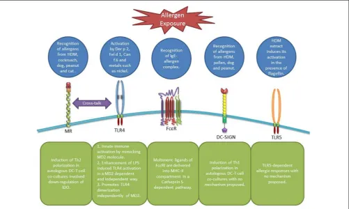
− [image:2.595.48.550.358.658.2]DCs
was shown to be partially mediated through the up-regulation of
indoleamine 2,3-dioxygenase (IDO) activity in DCs (
19
). IDO
is an enzyme that participates in tryptophan metabolism and
is involved in many immune-regulatory processes in health and
FIGURE 1 | Receptors involved in allergen recognition and uptake by DCs. MR can recognize allergens from diverse sources leading to Th2 polarization and IDO down-regulation in DCs involving a possible cross-talk with TLR4 (19). TLR4 itself can be activated by airborne allergens and diverse metals in an
disease (
21
,
22
). Further studies by the same group showed that
MR recognition of major cat allergen
Fel d
1 mediated the
produc-tion of specific IgE and IgG1 antibodies in a mouse model of allergy
(
20
). Other studies have shown that omega-1, a glycosylated T2
ribonuclease secreted by
Schistosoma mansoni
eggs, is recognized
and internalized by DCs through MR and subsequently interferes
with proteins synthesis and conditions DCs for Th2 priming (
23
).
Collectively these data highlight MR’s role in allergen recognition
and promotion of Th2-mediated immune responses.
DENDRITIC CELL-SPECIFIC INTRACELLULAR ADHESION MOLECULE 3-GRABBING NON-INTEGRIN
Dendritic cell-specific intracellular adhesion molecule 3-grabbing
non-integrin is a type-II integral transmembrane protein that
con-sists of four regions: a CRD, a hinge domain, and a transmembrane
region connected to a cytoplasmic signaling domain (
4
,
24
).
DC-SIGN is mainly expressed by antigen presenting cells (APCs) and
has been demonstrated to participate in the recognition of
aller-gens from different sources, such as peanut, HDM, pollen, and dog
(
25
–
27
).
In vitro
studies have been shown that DC-SIGN
recog-nition and uptake of Der p 1 induces Th1 cell differentiation. On
the contrary, DC-SIGN deficient DCs bias the response toward a
Th2 profile (
27
). This is opposite to previous observations in MR
−DCs which seem to support Th1 differentiation (
19
). Interestingly,
it has also been shown that Der p 1, using its enzymatic activity,
can cleave (
28
) and induce the down-regulation (
29
) of cell
sur-face DC-SIGN but not MR (
28
). In this context, we have previously
proposed that the Th1/Th2 balance in response to allergen
expo-sure can be determined by the cross-talk between these receptors
and the level of their expression on DCs (
4
,
27
). Accordingly, it is
important to note that DCs from asthmatic patients show lower
expression of DC-SIGN (
29
), which is in contrast to the high levels
of MR expression reported in atopic individuals (
17
,
18
).
TOLL-LIKE RECEPTORS
The TLRs are type-I integral membrane receptors, each with an
N-terminal ligand recognition domain constructed of tandem
copies of a leucine-rich repeat motif, a single transmembrane helix,
which participates in nucleic acid pathogen-associated molecular
patterns (PAMPs) recognition, and a C-terminal cytoplasmic
sig-naling domain known as Toll IL-1 receptor domain (
30
). There
are more than 10 different TLRs identified in humans so far (
31
),
with some of them being involved in allergen recognition or
path-ways that induces allergic responses. Within this context, mainly
three different mechanisms have been proposed. Der p 2, a major
allergen from HDM with lipid-binding activity, has been shown
to induce signaling through TLR2 (
32
) and TLR4 (
33
) depending
on the cell type involved. Due to its high homology with MD2,
which participates in the recognition of lipopolysaccharide (LPS)
by TLR4, Der p 2 forms a complex with TLR4 that signals
simi-larly to MD2/TLR4 complex inducing innate immune activation
(
33
). In addition, HDM extracts contaminated with flagellin can
induce TLR5-dependent allergic responses in mice, however, the
mechanism is still unclear (
34
). The second mechanism involves
sensitization by nickel, which may not be relevant in the context of
airway sensitization but highlights the importance of TLR4 on DCs
in allergic reactions. Nickel and other bivalent metals such as cobalt
induce a lipid-independent activation of TLR4, which is
depen-dent on the presence of two histidine residues, promoting TLR4
dimerization and subsequent receptor activation independently of
MD2 (
35
,
36
). Finally, the last mechanism involves allergens such
as Fel d 1 and Can f 6 that belong to lipocalin family and cause
enhanced LPS-induced TLR4 activation in an MD2 dependent and
independent manner respectively (
37
).
HIGH-AFFINITY IGE RECEPTOR
Fc
ε
RI is a multimeric cell surface receptor that binds IgE with
high-affinity. In humans, this receptor can be expressed by mast
cells, basophils, eosinophils, platelets, monocytes, and DCs;
how-ever, in the last four cell types it adopts a trimeric (
αγ
2) structure
instead of the classical tetrameric (
αβγ
2) structure (
38
–
43
). It
has been previously suggested that cell-bound IgE participates in
the presentation of aero-allergens by Langerhans cells (
44
). In
addition, the presence of Fc
ε
RI on Langerhans cells maximizes
antigen uptake via specific IgE and subsequent presentation to
T cells (
45
). In monocytes and peripheral blood DCs, Fc
ε
RI has
been shown to mediate IgE-dependent allergen presentation (
41
,
46
). Further studies in DCs demonstrated that multimeric ligands
of Fc
ε
RI are delivered into a major histocompatibility complex
class II compartment in a Cathepsin S-dependent pathway (
47
).
In vivo
experiments with a transgenic mouse model with
human-like Fc
ε
RI expression in DCs showed that after allergen capture
DCs instructed naïve T cells to differentiate into allergen-specific
Th2 cells at the site of allergen exposure (
48
). Taking into account
the fact that higher expression of Fc
ε
RI has been detected in atopic
individuals (
40
,
42
), this can lower the atopic individual’s
thresh-old to mount allergen-specific T cell responses. All this highlights
the fact that the high-affinity IgE receptor could play an important
role in capturing allergens by DCs and subsequent presentation to
T cell particularly in previously sensitized individuals with high
levels of specific IgE; however, the precise role of IgE receptors and
the role of other stimulatory and inhibitory Fc receptors (
49
,
50
)
in allergen presentation still need to be fully understood.
ALLERGEN RECOGNITION BY EPITHELIAL CELLS
Airway epithelial cells constitute the first line of defense against
pathogens and allergens by not only forming a physical barrier,
but also through expressing a wide range of PRR, such as TLRs,
CLRs, and protease activated receptor (PARs) (
Figure 2
). These
receptors enable AECs to recognize microbial motifs and
environ-mental allergens which lead to a cascade of events culminating
in the release of cytokines, chemokine ligands, and danger signals
which recruit and activate other immune cells.
PROTEASE ACTIVATED RECEPTORS
FIGURE 2 | Receptors involved in allergen recognition and uptake by AECs. TLR4 can be activated by pollen allergens and activate TSLP-OX40L signaling pathway (88). PAR-2 is activated by different allergens from HDM, cockroach, and fungus inducing the production of cytokines and chemokines
that modulate DC behavior (60,61,64–67). Dectin-1 is able to recognize allergens from HDM and induce the secretion of CCL20 (90). In addition, the secretion of TSLP induced by Der p 1 in AECs is thought to be
carbohydrate-dependent, however, no receptor has been identified (92).
linked to tissue damage (
51
,
52
). In addition, endothelial cells,
epithelial cells, fibroblasts, and immune cells such as lymphocytes,
monocytes, mast cells, neutrophils, eosinophils, macrophages, and
DCs have been shown to express functional PAR-2 (
53
,
54
). Due to
its ability to respond to serine proteases, proteolytic allergens from
diverse sources such as HDM, cockroach, pollen, or mold can act
as exogenous activators of PAR-2 with implications in allergy and
asthma.
Different cysteine and serine proteases from HDM, pollen, and
the fungus
Penicillium
have been shown to increase epithelial
permeability and disrupt the tight junctions by mainly
target-ing the transmembrane adhesion proteins occludin and zonula
occludens-1 (
9
–
15
). In addition, it has been demonstrated that
allergen-induced cytokine production, cell detachment and
mor-phological changes in AECs is largely dependent on allergens’
protease activity (
13
,
55
–
58
). Protease-dependant induction of
IL-25 and thymic stromal lymphopoietin (TSLP) have been shown to
be mediated by Erk and p38 MAPK pathways (
58
). More recently, it
was demonstrated, in an
in vivo
model, that IL-33 also contributes
to protease-dependent allergic airway inflammation (
59
).
Main allergens from HDM can activate PAR-2 and induce the
release of pro-inflammatory cytokines (
60
,
61
). However, there is
some contradictory results showing that Der p 1-induction of
IL-8 and IL-6 is independent of PAR-2 activation and dependent on
nuclear factor
κ
B (NF-
κ
B) and Erk signaling pathways (
62
,
63
). In
the case of allergens from the German cockroach and the fungus
Penicillium
, this effect has been demonstrated to be mediated by
the activation of Erk (
64
,
65
). In an
in vivo
model of allergy, only
when the allergens are administered through the mucosa,
cock-roach proteases regulate chemokine production and DC
recruit-ment in a PAR-2-dependent way (
66
,
67
). Furthermore, it was
demonstrated that cockroach proteases augmented tumor necrosis
factor (TNF)-
α
-induction of the matrix metalloproteinases-9, an
enzyme that has been implicated in the pathogenesis of bronchial
asthma (
68
,
69
), by a mechanism linking PAR-2, Erk, and AP-1
acti-vation (
70
). More recently, PAR-2-mediated allergic sensitization
was shown to be associated with TNF signaling pathways (
71
).
in allergen-mediated cytokine production, it has also been shown
that IL-8 production by AECs in response to allergens from the
fungus
Penicillium
is dependent on PAR-1 and PAR-2 via
activa-tion of ERK1/2 (
65
). In addition, proteases from different fungal
allergens induce the release of pro-inflammatory cytokines from
human nasal polyp epithelial cells, leading to eosinophil and
neu-trophil migration, in a mechanism that could involve PAR-2 and
PAR-3 (
75
).
TOLL-LIKE RECEPTOR-4
As previously described, TLRs are widely expressed by both APCs
and epithelial cells and recognize conserved microbial structures
and as such play a key role in controlling adaptive immune
responses. LPS is recognized by TLR4 with the participation of
the accessory proteins including CD14, LPS binding protein, and
MD2. This leads to the recruitment of the signaling adapter
pro-tein MyD88, the activation of the transcription NF-
κ
B among
others, and the expression of pro-inflammatory cytokines (
76
).
TLR4 (
77
–
80
), MyD88 (
79
,
81
,
82
), and NF-
κ
B (
83
–
86
) have been
shown to be crucial in the elicitation of allergic Th2 immune
responses. Different studies using knock-out mice have
demon-strated the importance of TLR4 expression in both hematopoietic
radiosensitive and structural radioresistant cells in the induction
of TLR4-dependent Th2 responses to intranasal allergens in the
presence of endotoxin (
77
–
80
). In addition, it has been shown that
the LPS dosage is crucial in driving either Th1 or Th2 responses,
with lower levels of LPS inducing Th2 responses to inhaled
aller-gens in a mouse model of allergic sensitization (
87
). Recently, it
was shown that short ragweed pollen acts as a TLR4 agonist,
initi-ating TLR4-dependet TSLP/OX40L signaling pathway, triggering
Th2 allergic inflammation (
88
).
C-TYPE LECTIN RECEPTORS
C-Type Lectin Receptors are receptors that recognize
oligosac-charide moieties among other molecular patterns on antigens
including allergens (
89
). CLR’s role in allergen recognition and
uptake by DCs is well established (
4
); moreover, they have also
been shown to participate in allergen recognition by AECs. It
was demonstrated that HDM induction of CCL20 by AECs was
not protease or TLR4/2 dependant; however, it was mediated by
β
-glycan moieties within HDM extract. This effect was specific
for HDM because other allergens, such as cockroach and
rag-weed, failed to induce this response (
90
). The authors suggest the
involvement of the dectin-1/Syk signaling pathway, since Syk
inhi-bition abrogated the HDM-induced CCL20 production (
91
). More
recently, our group demonstrated that TSLP secretion by AECs
upon stimulation with Der p 1 was at least partly
carbohydrate-dependent. In addition, DC uptake of deglycosylated Der p 1
was considerably decreased compared with its natural
(glycosy-lated) counterpart, indicating that glycosylation of allergens plays
a crucial role in their recognition by immune and non-immune
cells (
92
).
CROSS-TALK BETWEEN DENDRITIC AND EPITHELIAL CELLS
Due to their strategic location at the interface of external-internal
environments, AECs are able to modulate and coordinate immune
responses. Ample data have shown that the cross-talk between DCs
and AECs is crucial in driving allergen-induced Th2 responses (
6
,
7
). As previously described, the ligation of different PRR on AECs
results in the secretion of chemokines (
93
–
95
), that attract DCs,
and cytokines that induce DC maturation and activation.
How-ever, there are some contradictory results indicating that inducible
signals driven by LPS in non-hematopoietic tissues such as AECs
do not play an essential role in DCs activation (
96
). On the other
hand other studies have demonstrated that AECs can induce DC
maturation after LPS inhalation (
81
). Nevertheless a range of
cytokines including TSLP, IL-33, IL-25, IL-1
β
, and granulocyte
macrophage colony-stimulating factor (GM-CSF) are known to
be secreted by AECs after allergen challenge (
6
,
7
) which are able
to modulate DCs function (
Figure 3
). Here we focus on TSLP as a
key cytokine in initiation and maintenance of allergic responses.
ROLE OF TSLP
Thymic stromal lymphopoietin is a 140-amino acid
four-helix-bundle cytokine that belongs to the IL-2 family of cytokines. This
cytokine is mainly produced by AECs and is able to modulate
DC by binding to its receptor complex composed by the TSLP
receptor (TSLPR) and the IL-7 receptor (IL-7R) (
97
). This induces
the production of Th2-attracting chemokines such as CCL22 and
CCL17, and primes naïve T cells to produce IL-5, IL-4, TNF-
α
,
IL-13, whereas down-regulate IL-10 and IFN-
γ
(
98
,
99
) even in
the absence of DCs in an IL-4 dependant way (
100
). TSLP induces
the expression of OX40L in DCs which in turn has been shown to
trigger Th2 cell polarization in the absence of IL-12 (
101
).
Con-versely, TSLP is also able to induce IL-12 secretion after CD40
ligation in DCs however still maintaining its Th2-polarizing effect
(
102
). Furthermore, DCs activated with TSLP were able to induce
the expansion of Th2 memory cells and help to maintain their
phenotype (
103
).
Human epithelial cells can produce TSLP in response to diverse
stimuli, such as microbial products, physical injury, ambient
par-ticulate matter, protease allergens, and either pro-inflammatory or
Th2-polarizing cytokines (
58
,
104
–
108
). Protease allergens induce
TSLP in a PAR-2-dependent way (
109
) with the involving of
MAPK signaling pathway (
58
) however inflammatory cytokines
induction of TSLP is NF-
κ
B signaling dependent (
104
,
106
). In
addition to DCs, TSLP can also activate mast cells and CD34
+
blood hematopoietic progenitor cells to produce Th2 cytokines
and in that way induce the innate phase of allergic immune
responses (
105
,
110
). Finally, TSLP can interfere with
regula-tory T cell development impairing the balance between tolerance
and inflammation (
111
). Keratinocytes too can secrete functional
TSLP after stimulation with pro-inflammatory or Th2-driven
cytokines, and induces DC activation in human skin lesions of
atopic dermatitis (
112
). Recently, it was demonstrated that DCs
can also produce TSLP in response to TLR stimulation. Moreover,
interestingly DCs from mice challenged with HDM extract express
higher mRNA levels of TSLP than epithelial cells (
113
).
FIGURE 3 | Role of AECs in allergen sensitization. AECs can directly recognize allergens by PARs, TLRs or CLRs, and through NF-κB signaling, they induce the production of the cytokines (TSLP, GM-CSF, IL-33, IL-25, IL-1β), chemokine ligands (CCL2, CCL20), and danger signals (adenosine triphosphate, uric acid). TSLP has diverse effects on DCs and mast cells, which in absence of the cytokine IL-12 can lead to
recruitment of Th2 cells and (eosinophils, Th2 polarization, and the onset of allergy or allergic asthma. Moreover, IL-25 can activate Th2 cells to produce pro-allergenic cytokines. On the other hand, DCs are able to form tight junction with AECs, and allergen can digest the tight junction and both phenomena contribute to facilitate the allergen sensitization process (6,7).
allergic conjunctivitis it was demonstrated that after topical
aller-gen challenge mucosal epithelial cells produce high levels of TSLP
compared with controls leading to induction of allergic
inflam-mation through the TSLP-OX40L signaling pathway (
118
). More
recently, it was shown that the soluble TSLPS antagonist,
com-prised of the extracellular domain of the murine TSLPR and an
IgG2a Fc tail, reduced the severity of airway inflammation by
regulating DC function (
119
).
CONCLUSION
Dendritic cells are professional APCs and sentinels of the immune
system that efficiently sample allergens in the airways leading to a
cascade of events that culminates in the induction of Th2 type
immune responses. DCs are able to recognize and internalize
allergens from diverse sources through expression of a plethora
of receptors such as CLRs, TLRs, and Fc
ε
R. Recently, the role of
AECs as key players in the modulation and induction of DCs in
the airways has been highlighted. Like DCs, AECs are able to
rec-ognize allergens through several PRRs including PARs, CLRs, and
TLRs. This further leads to the production of different cytokines,
chemokines, and danger signals with the ability to initiate and
propagate immune responses to allergens. Allergens have diverse
molecular features such as specific oligosaccharide moieties,
pro-tease activity, lipid-binding properties, among others that can
facilitate their recognition by immune and non-immune cells and
contributes to their “allergenicity.” Better understanding of the
molecular basis of early events at the interface of allergens and
their receptors and the key soluble mediators/signaling pathways
involved could lead to development of more effective
therapeu-tic strategies for allergic diseases including allergic asthma. For
instance, due to the contribution of TLRs and CLRs in the
recogni-tion of allergen by both DCs and AECs, agonist and antagonists to
those receptors may provide new therapeutic targets to modulate
allergic responses. In addition, different studies have highlighted
the role of sugar moieties on allergens in their recognition and
internalization by immune cells. Accordingly, different
“glyco-forms” of allergens with immunoregulatory properties could be
developed and used in allergen-specific immunotherapy
strate-gies. Finally, diverse intracellular and extracellular molecules have
been implicated in the process of allergen recognition and
sen-sitization. Further studies to decipher these mechanisms could
pave the way for the rational design of more effective therapeutic
entities for the treatment of allergic diseases.
ACKNOWLEDGMENTS
F. Salazar is a recipient of a PhD scholarship from the National
Commission for Scientific and Technological Research
(CONI-CYT), Chile.
REFERENCES
1. Paul WE, Zhu J. How are T(H)2-type immune responses initiated and ampli-fied?Nat Rev Immunol(2010)10(4):225–35. doi:10.1038/nri2735
2. Karp CL. Guilt by intimate association: what makes an allergen an allergen? J Allergy Clin Immunol(2010)125(5):955–60. doi:10.1016/j.jaci.2010.03.002 3. Gill MA. The role of dendritic cells in asthma.J Allergy Clin Immunol(2012)
129(4):889–901. doi:10.1016/j.jaci.2012.02.028
4. Salazar F, Sewell HF, Shakib F, Ghaemmaghami AM. The role of lectins in allergic sensitization and allergic disease.J Allergy Clin Immunol(2013)
132(1):27–36. doi:10.1016/j.jaci.2013.02.001
5. Shakib F, Ghaemmaghami AM, Sewell HF. The molecular basis of allergenicity. Trends Immunol(2008)29(12):633–42. doi:10.1016/j.it.2008.08.007 6. Willart M, Hammad H. Lung dendritic cell-epithelial cell crosstalk in Th2
responses to allergens.Curr Opin Immunol(2011)23(6):772–7. doi:10.1016/j. coi.2011.09.008
7. Hammad H, Lambrecht BN. Dendritic cells and epithelial cells: linking innate and adaptive immunity in asthma.Nat Rev Immunol (2008)8(3):193–204. doi:10.1038/nri2275
8. Sung SS, Fu SM, Rose CE Jr, Gaskin F, Ju ST, Beaty SR. A major lung CD103 (alphaE)-beta7 integrin-positive epithelial dendritic cell population
expressing Langerin and tight junction proteins.J Immunol(2006)176(4): 2161–72.
9. Herbert CA, King CM, Ring PC, Holgate ST, Stewart GA, Thompson PJ, et al. Augmentation of permeability in the bronchial epithelium by the house dust mite allergen Der p1.Am J Respir Cell Mol Biol(1995)12(4):369–78. doi:10.1165/ajrcmb.12.4.7695916
10. Roche N, Chinet TC, Belouchi NE, Julie C, Huchon GJ. Dermatophagoides pteronyssinus and bioelectric properties of airway epithelium: role of cysteine proteases.Eur Respir J (2000)16(2):309–15. doi:10.1034/j.1399-3003.2000. 16b20.x
11. Wan H, Winton HL, Soeller C, Gruenert DC, Thompson PJ, Cannell MB, et al. Quantitative structural and biochemical analyses of tight junction dynamics following exposure of epithelial cells to house dust mite allergen Der p 1.Clin Exp Allergy(2000)30(5):685–98. doi:10.1046/j.1365-2222.2000.00820.x 12. Wan H, Winton HL, Soeller C, Taylor GW, Gruenert DC, Thompson PJ, et al.
The transmembrane protein occludin of epithelial tight junctions is a func-tional target for serine peptidases from faecal pellets ofDermatophagoides pteronyssinus.Clin Exp Allergy(2001)31(2):279–94. doi:10.1046/j.1365-2222. 2001.00970.x
13. Tai HY, Tam MF, Chou H, Peng HJ, Su SN, Perng DW, et al. Pen ch 13 aller-gen induces secretion of mediators and degradation of occludin protein of human lung epithelial cells.Allergy(2006)61(3):382–8. doi:10.1111/j.1398-9995.2005.00958.x
14. Runswick S, Mitchell T, Davies P, Robinson C, Garrod DR. Pollen pro-teolytic enzymes degrade tight junctions.Respirology (2007)12(6):834–42. doi:10.1111/j.1440-1843.2007.01175.x
15. Vinhas R, Cortes L, Cardoso I, Mendes VM, Manadas B, Todo-Bom A, et al. Pollen proteases compromise the airway epithelial barrier through degrada-tion of transmembrane adhesion proteins and lung bioactive peptides.Allergy (2011)66(8):1088–98. doi:10.1111/j.1398-9995.2011.02598.x
16. Taylor PR, Gordon S, Martinez-Pomares L. The mannose receptor: linking homeostasis and immunity through sugar recognition.Trends Immunol(2005)
26(2):104–10. doi:10.1016/j.it.2004.12.001
17. Deslee G, Charbonnier AS, Hammad H, Angyalosi G, Tillie-Leblond I, Manto-vani A, et al. Involvement of the mannose receptor in the uptake of Der p 1, a major mite allergen, by human dendritic cells.J Allergy Clin Immunol(2002)
110(5):763–70. doi:10.1067/mai.2002.129121
18. Kayserova J, Zentsova-Jaresova I, Budinsky V, Rozkova D, Kopecka J, Vernerova E, et al. Selective increase in blood dendritic cell antigen-3-positive dendritic cells in bronchoalveolar lavage fluid in allergic patients.Scand J Immunol(2012)
75(3):305–13. doi:10.1111/j.1365-3083.2011.02649.x
19. Royer PJ, Emara M, Yang C, Al-Ghouleh A, Tighe P, Jones N, et al. The mannose receptor mediates the uptake of diverse native allergens by dendritic cells and determines allergen-induced T cell polarization through modulation of IDO activity.J Immunol(2010)185(3):1522–31. doi:10.4049/jimmunol.1000774 20. Emara M, Royer PJ, Abbas Z, Sewell HF, Mohamed GG, Singh S, et al.
Recog-nition of the major cat allergen Fel d 1 through the cysteine-rich domain of the mannose receptor determines its allergenicity.J Biol Chem (2011)
286(15):13033–40. doi:10.1074/jbc.M111.220657
21. Munn DH, Mellor AL. Indoleamine 2,3 dioxygenase and metabolic control of immune responses.Trends Immunol(2012)34(3):137–43. doi:10.1016/j.it. 2012.10.001
22. Orabona C, Pallotta MT, Grohmann U. Different partners, opposite outcomes: a new perspective of IDO’s immunobiology.Mol Med(2012)18(1):834–42. doi:10.2119/molmed.2012.00029
23. Everts B, Hussaarts L, Driessen NN, Meevissen MH, Schramm G, van der Ham AJ, et al. Schistosome-derived omega-1 drives Th2 polarization by suppressing protein synthesis following internalization by the mannose receptor.J Exp Med (2012)209(10):1753–67. doi:10.1084/jem.20111381
24. Zhou T, Chen Y, Hao L, Zhang Y. DC-SIGN and immunoregulation.Cell Mol Immunol(2006)3(4):279–83.
25. Shreffler WG, Castro RR, Kucuk ZY, Charlop-Powers Z, Grishina G, Yoo S, et al. The major glycoprotein allergen fromArachis hypogaea, Ara h 1, is a ligand of dendritic cell-specific ICAM-grabbing nonintegrin and acts as a Th2 adjuvant in vitro.J Immunol(2006)177(6):3677–85.
dendritic cells.J Biol Chem(2010)285(11):7903–10. doi:10.1074/jbc.M109. 058370
27. Emara M, Royer PJ, Mahdavi J, Shakib F, Ghaemmaghami AM. Retagging identifies dendritic cell-specific intercellular adhesion molecule-3 (ICAM3)-grabbing non-integrin (DC-SIGN) protein as a novel receptor for a major allergen from house dust mite.J Biol Chem(2012)287(8):5756–63. doi:10. 1074/jbc.M111.312520
28. Furmonaviciene R, Ghaemmaghami AM, Boyd SE, Jones NS, Bailey K, Willis AC, et al. The protease allergen Der p 1 cleaves cell surface DC-SIGN and DC-SIGNR: experimental analysis of in silico substrate identification and implications in allergic responses.Clin Exp Allergy (2007)37(2):231–42. doi:10.1111/j.1365-2222.2007.02651.x
29. Huang HJ, Lin YL, Liu CF, Kao HF, Wang JY. Mite allergen decreases DC-SIGN expression and modulates human dendritic cell differentiation and function in allergic asthma.Mucosal Immunol(2011)4(5):519–27. doi:10.1038/mi.2011.17 30. Botos I, Segal DM, Davies DR. The structural biology of toll-like receptors.
Structure(2011)19(4):447–59. doi:10.1016/j.str.2011.02.004
31. O’Neill LA, Golenbock D, Bowie AG. The history of toll-like receptors – redefin-ing innate immunity.Nat Rev Immunol(2013)13(6):453–60. doi:10.1038/ nri3446
32. Chiou YL, Lin CY. Der p2 activates airway smooth muscle cells in a TLR2/MyD88-dependent manner to induce an inflammatory response.J Cell Physiol(2009)220(2):311–8. doi:10.1002/jcp.21764
33. Trompette A, Divanovic S, Visintin A, Blanchard C, Hegde RS, Madan R, et al. Allergenicity resulting from functional mimicry of a Toll-like receptor complex protein.Nature(2009)457(7229):585–8. doi:10.1038/nature07548 34. Wilson RH, Maruoka S, Whitehead GS, Foley JF, Flake GP, Sever ML,
et al. The toll-like receptor 5 ligand flagellin promotes asthma by prim-ing allergic responses to indoor allergens.Nat Med(2012)18(11):1705–10. doi:10.1038/nm.2920
35. Schmidt M, Raghavan B, Muller V, Vogl T, Fejer G, Tchaptchet S, et al. Crucial role for human Toll-like receptor 4 in the development of contact allergy to nickel.Nat Immunol(2010)11(9):814–9. doi:10.1038/ni.1919
36. Raghavan B, Martin SF, Esser PR, Goebeler M, Schmidt M. Metal allergens nickel and cobalt facilitate TLR4 homodimerization independently of MD2. EMBO Rep(2012)13(12):1109–15. doi:10.1038/embor.2012.155
37. Herre J, Gronlund H, Brooks H, Hopkins L, Waggoner L, Murton B, et al. Allergens as immunomodulatory proteins: the cat dander protein Fel d 1 enhances TLR activation by lipid ligands.J Immunol(2013)191(4):1529–35. doi:10.4049/jimmunol.1300284
38. Kinet JP. The high-affinity IgE receptor (Fc epsilon RI): from physiology to pathology.Annu Rev Immunol (1999)17:931–72. doi:10.1146/annurev. immunol.17.1.931
39. Bieber T, de la Salle H, Wollenberg A, Hakimi J, Chizzonite R, Ring J, et al. Human epidermal Langerhans cells express the high affinity receptor for immunoglobulin E (Fc epsilon RI).J Exp Med (1992)175(5):1285–90. doi:10.1084/jem.175.5.1285
40. Maurer D, Fiebiger E, Reininger B, Wolff-Winiski B, Jouvin MH, Kilgus O, et al. Expression of functional high affinity immunoglobulin E receptors (Fc epsilon RI) on monocytes of atopic individuals.J Exp Med(1994)179(2):745–50. doi:10.1084/jem.179.2.745
41. Maurer D, Fiebiger S, Ebner C, Reininger B, Fischer GF, Wichlas S, et al. Periph-eral blood dendritic cells express Fc epsilon RI as a complex composed of Fc epsilon RI alpha- and Fc epsilon RI gamma-chains and can use this receptor for IgE-mediated allergen presentation.J Immunol(1996)157(2):607–16. 42. Tunon-De-Lara JM, Redington AE, Bradding P, Church MK, Hartley JA,
Sem-per AE, et al. Dendritic cells in normal and asthmatic airways: expression of the alpha subunit of the high affinity immunoglobulin E receptor (Fc epsilon RI-alpha).Clin Exp Allergy(1996)26(6):648–55. doi:10.1046/j.1365-2222.1996. 1095481.x
43. Novak N, Allam JP, Hagemann T, Jenneck C, Laffer S, Valenta R, et al. Charac-terization of FcepsilonRI-bearing CD123 blood dendritic cell antigen-2 plas-macytoid dendritic cells in atopic dermatitis.J Allergy Clin Immunol(2004)
114(2):364–70. doi:10.1016/j.jaci.2004.05.038
44. Mudde GC, Van Reijsen FC, Boland GJ, de Gast GC, Bruijnzeel PL, Bruijnzeel-Koomen CA. Allergen presentation by epidermal Langerhans’ cells from patients with atopic dermatitis is mediated by IgE. Immunology (1990)
69(3):335–41.
45. Bieber T. Fc epsilon RI on human epidermal Langerhans cells: an old receptor with new structure and functions.Int Arch Allergy Immunol(1997)113 (1-3):30–4. doi:10.1159/000237500
46. Maurer D, Ebner C, Reininger B, Fiebiger E, Kraft D, Kinet JP, et al. The high affinity IgE receptor (Fc epsilon RI) mediates IgE-dependent allergen presen-tation.J Immunol(1995)154(12):6285–90.
47. Maurer D, Fiebiger E, Reininger B, Ebner C, Petzelbauer P, Shi GP, et al. Fc epsilon receptor I on dendritic cells delivers IgE-bound multivalent antigens into a cathepsin S-dependent pathway of MHC class II presentation.J Immunol (1998)161(6):2731–9.
48. Sallmann E, Reininger B, Brandt S, Duschek N, Hoflehner E, Garner-Spitzer E, et al. High-affinity IgE receptors on dendritic cells exacerbate Th2-dependent inflammation.J Immunol(2011)187(1):164–71. doi:10.4049/ jimmunol.1003392
49. Wigginton SJ, Furtado PB, Armour KL, Clark MR, Robins A, Emara M, et al. An immunoglobulin E-reactive chimeric human immunoglobulin G1 anti-idiotype inhibits basophil degranulation through cross-linking of FcepsilonRI with FcgammaRIIb.Clin Exp Allergy(2008)38(2):313–9. doi:10.1111/j.1365-2222.2007.02896.x
50. Nakamura A, Kubo T, Takai T. Fc receptor targeting in the treatment of allergy, autoimmune diseases and cancer.Adv Exp Med Biol(2008)640:220–33. doi:10.1007/978-0-387-09789-3_17
51. Lee SE, Jeong SK, Lee SH. Protease and protease-activated receptor-2 signaling in the pathogenesis of atopic dermatitis.Yonsei Med J(2010)51(6):808–22. doi:10.3349/ymj.2010.51.6.808
52. Arora P, Ricks TK, Trejo J. Protease-activated receptor signalling, endo-cytic sorting and dysregulation in cancer.J Cell Sci(2007)120(Pt 6):921–8. doi:10.1242/jcs.03409
53. Steinhoff M, Corvera CU, Thoma MS, Kong W, McAlpine BE, Caughey GH, et al. Proteinase-activated receptor-2 in human skin: tissue distribution and activation of keratinocytes by mast cell tryptase.Exp Dermatol(1999)
8(4):282–94. doi:10.1111/j.1600-0625.1999.tb00383.x
54. Rattenholl A, Steinhoff M. Proteinase-activated receptor-2 in the skin: recep-tor expression, activation and function during health and disease.Drug News Perspect(2008)21(7):369–81. doi:10.1358/dnp.2008.21.7.1255294
55. Tomee JF, van Weissenbruch R, de Monchy JG, Kauffman HF. Interactions between inhalant allergen extracts and airway epithelial cells: effect on cytokine production and cell detachment.J Allergy Clin Immunol(1998)102(1):75–85. doi:10.1016/S0091-6749(98)70057-0
56. King C, Brennan S, Thompson PJ, Stewart GA. Dust mite proteolytic allergens induce cytokine release from cultured airway epithelium.J Immunol(1998)
161(7):3645–51.
57. Kauffman HF, Tomee JF, van de Riet MA, Timmerman AJ, Borger P. Protease-dependent activation of epithelial cells by fungal allergens leads to morpho-logic changes and cytokine production.J Allergy Clin Immunol(2000)105(6 Pt 1):1185–93. doi:10.1067/mai.2000.106210
58. Yu HS, Angkasekwinai P, Chang SH, Chung Y, Dong C. Protease allergens induce the expression of IL-25 via Erk and p38 MAPK pathway.J Korean Med Sci(2010)25(6):829–34. doi:10.3346/jkms.2010.25.6.829
59. Kamijo S, Takeda H, Tokura T, Suzuki M, Inui K, Hara M, et al. IL-33-mediated innate response and adaptive immune cells contribute to maximum responses of protease allergen-induced allergic airway inflammation.J Immunol(2013)
190(9):4489–99. doi:10.4049/jimmunol.1201212
60. Sun G, Stacey MA, Schmidt M, Mori L, Mattoli S. Interaction of mite aller-gens Der p3 and Der p9 with protease-activated receptor-2 expressed by lung epithelial cells.J Immunol(2001)167(2):1014–21.
61. Asokananthan N, Graham PT, Stewart DJ, Bakker AJ, Eidne KA, Thompson PJ, et al. House dust mite allergens induce proinflammatory cytokines from respiratory epithelial cells: the cysteine protease allergen, Der p 1, activates protease-activated receptor (PAR)-2 and inactivates PAR-1.J Immunol(2002)
169(8):4572–8.
62. Adam E, Hansen KK, Astudillo Fernandez O, Coulon L, Bex F, Duhant X, et al. The house dust mite allergen Der p 1, unlike Der p 3, stimulates the expression of interleukin-8 in human airway epithelial cells via a proteinase-activated receptor-2-independent mechanism.J Biol Chem(2006)281(11):6910–23. doi:10.1074/jbc.M507140200
protease-dependent and protease-independent mechanisms.Clin Mol Allergy (2006)4:5. doi:10.1186/1476-7961-4-5
64. Page K, Strunk VS, Hershenson MB. Cockroach proteases increase IL-8 expres-sion in human bronchial epithelial cells via activation of protease-activated receptor (PAR)-2 and extracellular-signal-regulated kinase.J Allergy Clin Immunol(2003)112(6):1112–8. doi:10.1016/j.jaci.2003.08.050
65. Chiu LL, Perng DW, Yu CH, Su SN, Chow LP. Mold allergen, pen C 13, induces IL-8 expression in human airway epithelial cells by activating protease-activated receptor 1 and 2.J Immunol(2007)178(8):5237–44.
66. Page K, Ledford JR, Zhou P, Dienger K, Wills-Karp M. Mucosal sensitization to German cockroach involves protease-activated receptor-2.Respir Res(2010)
11:62. doi:10.1186/1465-9921-11-62
67. Day SB, Ledford JR, Zhou P, Lewkowich IP, Page K. German cockroach pro-teases and protease-activated receptor-2 regulate chemokine production and dendritic cell recruitment.J Innate Immun(2012)4(1):100–10. doi:10.1159/ 000329132
68. Lee YC, Lee HB, Rhee YK, Song CH. The involvement of matrix metalloproteinase-9 in airway inflammation of patients with acute asthma.Clin Exp Allergy(2001)31(10):1623–30. doi:10.1046/j.1365-2222.2001.01211.x 69. Kelly EA, Busse WW, Jarjour NN. Increased matrix metalloproteinase-9 in the
airway after allergen challenge.Am J Respir Crit Care Med(2000)162(3 Pt 1):1157–61. doi:10.1164/ajrccm.162.3.9908016
70. Page K, Hughes VS, Bennett GW, Wong HR. German cockroach proteases reg-ulate matrix metalloproteinase-9 in human bronchial epithelial cells.Allergy (2006)61(8):988–95. doi:10.1111/j.1398-9995.2006.01103.x
71. Ebeling C, Lam T, Gordon JR, Hollenberg MD, Vliagoftis H. Proteinase-activated receptor-2 promotes allergic sensitization to an inhaled antigen through a TNF-mediated pathway.J Immunol(2007)179(5):2910–7. 72. Antony AB, Tepper RS, Mohammed KA. Cockroach extract antigen increases
bronchial airway epithelial permeability. J Allergy Clin Immunol (2002)
110(4):589–95. doi:10.1067/mai.2002.127798
73. Capetandes A, Zhuang M, Haque FN, Xie L, Frieri M. Vascular endothe-lial growth factor is increased by human pulmonary cells stimulated with Dermatophagoides sp. extract. Allergy Asthma Proc (2007) 28(3):324–30. doi:10.2500/aap.2007.28.2999
74. Heijink IH, van Oosterhout A, Kapus A. Epidermal growth factor receptor sig-nalling contributes to house dust mite-induced epithelial barrier dysfunction. Eur Respir J(2010)36(5):1016–26. doi:10.1183/09031936.00125809 75. Shin SH, Lee YH, Jeon CH. Protease-dependent activation of nasal
polyp epithelial cells by airborne fungi leads to migration of eosinophils and neutrophils. Acta Otolaryngol (2006) 126(12):1286–94. doi:10.1080/ 00016480500395179
76. Takeda K, Kaisho T, Akira S. Toll-like receptors.Annu Rev Immunol(2003)
21:335–76. doi:10.1146/annurev.immunol.21.120601.141126
77. Dabbagh K, Dahl ME, Stepick-Biek P, Lewis DB. Toll-like receptor 4 is required for optimal development of Th2 immune responses: role of dendritic cells. J Immunol(2002)168(9):4524–30.
78. Hammad H, Chieppa M, Perros F, Willart MA, Germain RN, Lambrecht BN. House dust mite allergen induces asthma via Toll-like receptor 4 triggering of airway structural cells.Nat Med(2009)15(4):410–6. doi:10.1038/nm.1946 79. Phipps S, Lam CE, Kaiko GE, Foo SY, Collison A, Mattes J, et al. Toll/IL-1
signaling is critical for house dust mite-specific helper T cell type 2 and type 17 [corrected] responses.Am J Respir Crit Care Med(2009)179(10):883–93. doi:10.1164/rccm.200806-974OC
80. Tan AM, Chen HC, Pochard P, Eisenbarth SC, Herrick CA, Bottomly HK. TLR4 signaling in stromal cells is critical for the initiation of allergic Th2 responses to inhaled antigen.J Immunol(2010)184(7):3535–44. doi:10.4049/jimmunol. 0900340
81. Noulin N, Quesniaux VF, Schnyder-Candrian S, Schnyder B, Maillet I, Robert T, et al. Both hemopoietic and resident cells are required for MyD88-dependent pulmonary inflammatory response to inhaled endotoxin.J Immunol(2005)
175(10):6861–9.
82. Piggott DA, Eisenbarth SC, Xu L, Constant SL, Huleatt JW, Herrick CA, et al. MyD88-dependent induction of allergic Th2 responses to intranasal antigen. J Clin Invest(2005)115(2):459–67. doi:10.1172/JCI200522462
83. Poynter ME, Cloots R, van Woerkom T, Butnor KJ, Vacek P, Taatjes DJ, et al. NF-kappa B activation in airways modulates allergic inflammation but not hyperresponsiveness.J Immunol(2004)173(11):7003–9.
84. Skerrett SJ, Liggitt HD, Hajjar AM, Ernst RK, Miller SI, Wilson CB. Res-piratory epithelial cells regulate lung inflammation in response to inhaled endotoxin. Am J Physiol Lung Cell Mol Physiol (2004) 287(1):L143–52. doi:10.1152/ajplung.00030.2004
85. Sheller JR, Polosukhin VV, Mitchell D, Cheng DS, Peebles RS, Blackwell TS. Nuclear factor kappa B induction in airway epithelium increases lung inflammation in allergen-challenged mice.Exp Lung Res(2009)35(10):883–95. doi:10.3109/01902140903019710
86. Ather JL, Hodgkins SR, Janssen-Heininger YM, Poynter ME. Airway epithelial NF-kappaB activation promotes allergic sensitization to an innocuous inhaled antigen.Am J Respir Cell Mol Biol(2011)44(5):631–8. doi:10.1165/rcmb.2010-0106OC
87. Eisenbarth SC, Piggott DA, Huleatt JW, Visintin I, Herrick CA, Bottomly K. Lipopolysaccharide-enhanced, toll-like receptor 4-dependent T helper cell type 2 responses to inhaled antigen.J Exp Med (2002)196(12):1645–51. doi:10.1084/jem.20021340
88. Li DQ, Zhang L, Pflugfelder SC, De Paiva CS, Zhang X, Zhao G, et al. Short ragweed pollen triggers allergic inflammation through Toll-like receptor 4-dependent thymic stromal lymphopoietin/OX40 ligand/OX40 signaling path-ways.J Allergy Clin Immunol(2011)128:1318.e–25.e. doi:10.1016/j.jaci.2011. 06.041
89. Sancho D, Reis e Sousa C. Signaling by myeloid C-type lectin receptors in immunity and homeostasis.Annu Rev Immunol(2012)30:491–529. doi:10. 1146/annurev-immunol-031210-101352
90. Nathan AT, Peterson EA, Chakir J, Wills-Karp M. Innate immune responses of airway epithelium to house dust mite are mediated through beta-glucan-dependent pathways.J Allergy Clin Immunol(2009)123(3):612–8. doi:10.1016/ j.jaci.2008.12.006
91. Brown GD, Herre J, Williams DL, Willment JA, Marshall AS, Gordon S. Dectin-1 mediates the biological effects of beta-glucans.J Exp Med(2003)
197(9):1119–24. doi:10.1084/jem.20021890
92. Al-Ghouleh A, Johal R, Sharquie IK, Emara M, Harrington H, Shakib F, et al. The glycosylation pattern of common allergens: the recognition and uptake of Der p 1 by epithelial and dendritic cells is carbohydrate dependent.PLoS ONE (2012)7(3):e33929. doi:10.1371/journal.pone.0033929
93. Reibman J, Hsu Y, Chen LC, Bleck B, Gordon T. Airway epithelial cells release MIP-3alpha/CCL20 in response to cytokines and ambient particulate mat-ter.Am J Respir Cell Mol Biol(2003)28(6):648–54. doi:10.1165/rcmb.2002-0095OC
94. Pichavant M, Charbonnier AS, Taront S, Brichet A, Wallaert B, Pestel J, et al. Asthmatic bronchial epithelium activated by the proteolytic allergen Der p 1 increases selective dendritic cell recruitment.J Allergy Clin Immunol(2005)
115(4):771–8. doi:10.1016/j.jaci.2004.11.043
95. Stumbles PA, Strickland DH, Pimm CL, Proksch SF, Marsh AM, McWilliam AS, et al. Regulation of dendritic cell recruitment into resting and inflamed airway epithelium: use of alternative chemokine receptors as a function of inducing stimulus.J Immunol(2001)167(1):228–34.
96. Nolte MA, Leibundgut-Landmann S, Joffre O, Reis e Sousa C. Dendritic cell quiescence during systemic inflammation driven by LPS stimulation of radiore-sistant cells in vivo.J Exp Med (2007)204(6):1487–501. doi:10.1084/jem. 20070325
97. Takai T. TSLP expression: cellular sources, triggers, and regulatory mechanisms. Allergol Int(2012)61(1):3–17. doi:10.2332/allergolint.11-RAI-0395 98. Soumelis V, Reche PA, Kanzler H, Yuan W, Edward G, Homey B, et al. Human
epithelial cells trigger dendritic cell mediated allergic inflammation by produc-ing TSLP.Nat Immunol(2002)3(7):673–80. doi:10.1038/ni805
99. Larson RP, Comeau MR, Ziegler SF. Cutting edge: allergen-specific CD4 T cells respond indirectly to thymic stromal lymphopoietin to promote allergic responses in the skin.J Immunol(2013)190(9):4474–7. doi:10.4049/jimmunol. 1201677
100. Omori M, Ziegler S. Induction of IL-4 expression in CD4(+) T cells by thymic stromal lymphopoietin.J Immunol(2007)178(3):1396–404.
101. Ito T, Wang YH, Duramad O, Hori T, Delespesse GJ, Watanabe N, et al. TSLP-activated dendritic cells induce an inflammatory T helper type 2 cell response through OX40 ligand.J Exp Med(2005)202(9):1213–23. doi:10.1084/jem. 20051135
dendritic cells but maintains their Th2 priming potential.Blood (2005)
105(12):4749–51. doi:10.1182/blood-2004-09-3622
103. Wang YH, Ito T, Homey B, Watanabe N, Martin R, Barnes CJ, et al. Mainte-nance and polarization of human TH2 central memory T cells by thymic stro-mal lymphopoietin-activated dendritic cells.Immunity(2006)24(6):827–38. doi:10.1016/j.immuni.2006.03.019
104. Kato A, Favoreto S Jr, Avila PC, Schleimer RP. TLR3- and Th2 cytokine-dependent production of thymic stromal lymphopoietin in human airway epithelial cells.J Immunol(2007)179(2):1080–7.
105. Allakhverdi Z, Comeau MR, Jessup HK, Yoon BR, Brewer A, Chartier S, et al. Thymic stromal lymphopoietin is released by human epithelial cells in response to microbes, trauma, or inflammation and potently activates mast cells.J Exp Med(2007)204(2):253–8. doi:10.1084/jem.20062211
106. Lee HC, Ziegler SF. Inducible expression of the proallergic cytokine thymic stromal lymphopoietin in airway epithelial cells is controlled by NFkappaB. Proc Natl Acad Sci U S A(2007)104(3):914–9. doi:10.1073/pnas.0607305104 107. Bleck B, Tse DB, Curotto de Lafaille MA, Zhang F, Reibman J. Diesel exhaust
particle-exposed human bronchial epithelial cells induce dendritic cell mat-uration and polarization via thymic stromal lymphopoietin.J Clin Immunol (2008)28(2):147–56. doi:10.1007/s10875-007-9149-0
108. Bleck B, Grunig G, Chiu A, Liu M, Gordon T, Kazeros A, et al. MicroRNA-375 regulation of thymic stromal lymphopoietin by diesel exhaust particles and ambient particulate matter in human bronchial epithelial cells.J Immunol (2013)190(7):3757–63. doi:10.4049/jimmunol.1201165
109. Kouzaki H, O’Grady SM, Lawrence CB, Kita H. Proteases induce produc-tion of thymic stromal lymphopoietin by airway epithelial cells through protease-activated receptor-2.J Immunol(2009)183(2):1427–34. doi:10.4049/ jimmunol.0900904
110. Allakhverdi Z, Comeau MR, Smith DE, Toy D, Endam LM, Desrosiers M, et al. CD34+ hemopoietic progenitor cells are potent effectors of allergic inflamma-tion.J Allergy Clin Immunol(2009)123(2):472–8. doi:10.1016/j.jaci.2008.10. 022
111. Lei L, Zhang Y, Yao W, Kaplan MH, Zhou B. Thymic stromal lymphopoi-etin interferes with airway tolerance by suppressing the generation of antigen-specific regulatory T cells.J Immunol (2011)186(4):2254–61. doi:10.4049/ jimmunol.1002503
112. Bogiatzi SI, Fernandez I, Bichet JC, Marloie-Provost MA, Volpe E, Sastre X, et al. Cutting Edge: Proinflammatory and Th2 cytokines synergize to induce thymic stromal lymphopoietin production by human skin keratinocytes.J Immunol (2007)178(6):3373–7.
113. Kashyap M, Rochman Y, Spolski R, Samsel L, Leonard WJ. Thymic stromal lym-phopoietin is produced by dendritic cells.J Immunol(2011)187(3):1207–11. doi:10.4049/jimmunol.1100355
114. Al-Shami A, Spolski R, Kelly J, Keane-Myers A, Leonard WJ. A role for TSLP in the development of inflammation in an asthma model.J Exp Med(2005)
202(6):829–39. doi:10.1084/jem.20050199
115. Larson RP, Zimmerli SC, Comeau MR, Itano A, Omori M, Iseki M, et al. Dibutyl phthalate-induced thymic stromal lymphopoietin is required for Th2 contact hypersensitivity responses.J Immunol(2010)184(6):2974–84. doi:10.4049/jimmunol.0803478
116. Yoo J, Omori M, Gyarmati D, Zhou B, Aye T, Brewer A, et al. Spontaneous atopic dermatitis in mice expressing an inducible thymic stromal lymphopoietin transgene specifically in the skin.J Exp Med(2005)202(4):541–9. doi:10.1084/ jem.20041503
117. Zhou B, Comeau MR, De Smedt T, Liggitt HD, Dahl ME, Lewis DB, et al. Thymic stromal lymphopoietin as a key initiator of allergic airway inflamma-tion in mice.Nat Immunol(2005)6(10):1047–53. doi:10.1038/ni1247 118. Zheng X, Ma P, de Paiva CS, Cunningham MA, Hwang CS, Pflugfelder
SC, et al. TSLP and downstream molecules in experimental mouse allergic conjunctivitis.Invest Ophthalmol Vis Sci(2010)51(6):3076–82. doi:10.1167/ iovs.09-4122
119. Zhang F, Huang G, Hu B, Song Y, Shi Y. A soluble thymic stromal lymphopoi-etin (TSLP) antagonist, TSLPR-immunoglobulin, reduces the severity of aller-gic disease by regulating pulmonary dendritic cells.Clin Exp Immunol(2011)
164(2):256–64. doi:10.1111/j.1365-2249.2011.04328.x
120. Ying S, O’Connor B, Ratoff J, Meng Q, Mallett K, Cousins D, et al. Thymic stro-mal lymphopoietin expression is increased in asthmatic airways and correlates with expression of Th2-attracting chemokines and disease severity.J Immunol (2005)174(12):8183–90.
121. Shamim Z, Muller K, Svejgaard A, Poulsen LK, Bodtger U, Ryder LP. Asso-ciation between genetic polymorphisms in the human interleukin-7 receptor alpha-chain and inhalation allergy.Int J Immunogenet(2007)34(3):149–51. doi:10.1111/j.1744-313X.2007.00657.x
122. Bunyavanich S, Melen E, Wilk JB, Granada M, Soto-Quiros ME, Avila L, et al. Thymic stromal lymphopoietin (TSLP) is associated with allergic rhinitis in children with asthma.Clin Mol Allergy(2011)9:1. doi:10.1186/1476-7961-9-1
123. Harada M, Hirota T, Jodo AI, Hitomi Y, Sakashita M, Tsunoda T, et al. Thymic stromal lymphopoietin gene promoter polymorphisms are associated with sus-ceptibility to bronchial asthma.Am J Respir Cell Mol Biol(2011)44(6):787–93. doi:10.1165/rcmb.2009-0418OC
Conflict of Interest Statement:The authors declare that the research was conducted in the absence of any commercial or financial relationships that could be construed as a potential conflict of interest.
Received: 29 August 2013; accepted: 21 October 2013; published online: 04 November 2013.
Citation: Salazar F and Ghaemmaghami AM (2013) Allergen recognition by innate immune cells: critical role of dendritic and epithelial cells. Front. Immunol.4:356. doi: 10.3389/fimmu.2013.00356
This article was submitted to Immunotherapies and Vaccines, a section of the journal Frontiers in Immunology.