RESEARCH ARTICLE
Ambient DESI and LESA-MS Analysis of Proteins Adsorbed
to a Biomaterial Surface Using In-Situ Surface Tryptic
Digestion
Wei Rao,
1Adam D. Celiz,
2David J. Scurr,
2Morgan R. Alexander,
2David A. Barrett
1 1Centre for Analytical Bioscience, School of Pharmacy, University of Nottingham, Nottingham, NG7 2RD, UK
2
Laboratory of Biophysics and Surface Analysis, School of Pharmacy, University of Nottingham, Nottingham, NG7 2RD, UK
Abstract.The detection and identification of proteins adsorbed onto biomaterial surfaces under ambient conditions has significant experimental advantages but has proven to be difficult to achieve with conventional measuring technologies. In this study, we present an adaptation of desorption electrospray ionization (DESI) and liquid extraction surface analysis (LESA) mass spectrometry (MS) coupled with in-situ surface tryptic digestion to identify protein species from a biomaterial surface. Cytochrome c, myoglobin, and BSA in a combination of single and mixture spots were printed in an array format onto Permanox slides, followed by in-situ surface digestion and detection via MS. Automated tandem MS performed on surface peptides was able to identify the proteins via MASCOT. Limits of detection were determined for DESI-MS and a comparison of DESI and LESA-MS peptide spectra characteristics and sensitivity was made. DESI-MS images of the arrays were produced and analyzed with imaging multivariate analysis to automatically separate peptide peaks for each of the proteins within a mixture into distinct components. This is the first time that DESI and LESA-MS have been used for the in-situ detection of surface digested proteins on biomaterial surfaces and presents a promising proof of concept for the use of ambient MS in the rapid and automated analysis of surface proteins.
Keywords:LESA-MS, DESI-MS, Ambient MS, Protein arrays, In-situ digestion, MS imaging, MCR analysis
Received: 16 July 2013/Revised: 20 August 2013/Accepted: 20 August 2013/Published nline: 19 September 2013
Introduction
T
he identity and quantity of proteins that adsorb at the surface of biomaterials in vitro from cell culture material and in vivo from the biological milieu define the way in which cells and the body respond, respectively, to man-made materials [1]. This can vary from stem cells being encouraged to maintain their pluripotent nature [2] at the surface of protein coated polymers [3] to foreign body encapsulation of biomedical devices [4]. Identification and quantification of unknown proteins at low concentration (frequently sub monolayer) in surface mixtures of many proteins requires a technique that does not prejudge the important species (as in the case with the commonly used immuno-histochemical labeling [5]), and mass spectrometry is well suited to this approach. Much work has beenundertaken on this challenge with ultra-high vacuum analysis of proteins adsorbed to surfaces using time of flight secondary ion mass spectrometry (ToF-SIMS) [6, 7] and matrix assisted laser desorption/ionization (MALDI) MS [8, 9] with some success. The vacuum environment is, however, a significant obstacle to the analysis of the native surface protein state, and emerging ambient MS techniques offer great opportunities in this respect [10].
Desorption electrospray ionization (DESI) is an ambient MS technique first described in 2004 [11]. Since its introduc-tion, DESI has become one of the most popular MS techniques for the study of ambient surfaces, mainly because of its ease of operation, flexibility, and sensitivity. DESI-MS has been explored in the study of small molecules in a number of systems, including pharmaceuticals, lipids, plants, lung tissue, and others [12,13]. Part of the attraction of DESI-MS comes from the flexibility of the system that allows it to be used for a wide variety of experiments, and it has already been modified in a number of ways to detect analytes over large areas [14], use additives for enhanced detection [15], and for MS imaging [16]. The capacity of DESI-MS imaging has seen an increase in
Electronic supplementary material The online version of this article (doi:10.1007/s13361-013-0737-3) contains supplementary material, which is available to authorized users.
Correspondence to:David Barrett;e-mail:david.barrett@nottingham.ac.uk
popularity since the technique was first described, and numerous advances have been made in the imaging of small molecules from biological surfaces [16,17].
The mechanism of DESI-MS involves a charged droplet-pickup process in which a spray of ionized solvent is focused onto a surface using a nebulizer gas and the desorbed molecules are collected and detected within the mass spectrometer [12,18]. While this mechanism has proven to be successful in analyzing small molecules, it has proven to be more challenging to adapt the technique for the analysis of proteins. Surprisingly few studies using DESI-MS have focused in the direction of detection of proteins on surfaces, although there is growing interest within the field for further advances [19–21]. Previously, studies have been able to identify whole proteins from a surface up to the size of BSA (66.8 kDa) [22], typically as a multiply charged series of peaks within the mass spectrum in a way that is similar to classic protein detection by electrospray ionization (ESI). Protein identification within mixtures have also been attempted, with the result that the multiply charged series from three different proteins (insulin, myoglobin, and BSA) can be spatially resolved in an automated manner using imaging multivariate data analysis (MVA) [23]. Although this approach worked well within a model system, significant challenges remain before DESI-MS is able to analyze and identify proteins on a complex biomaterial or biological surface.
Another ambient analysis technique, liquid extraction surface analysis (LESA), has gathered attention of late as a powerful tool for surface MS analysis. LESA-MS is a recent technique first described in 2010 [24] and has already been used in the study of glucocorticoid receptor agonists [25], metabolites from tissue sections [26], and lipids from contact lenses [27]. LESA extracts analytes directly from a surface via the maintenance of a liquid/surface microjunction, which is then taken up and transferred into the mass spectrometer for analysis by nanospray. Compared with DESI-MS, LESA-MS has the advantage of a potentially higher sensitivity of detection as the surface extraction method is a more efficient mechanism than the droplet-pickup process; however LESA is somewhat unsuited to MS imaging as the extraction area is too large for even moderate levels of spatial resolution. LESA-MS analysis of whole proteins is in its infancy and has been applied to the analysis of hemoglobin variants on dried bloodspots [28]. A related technique, liquid-microjunction surface sampling probe (LMJ-SSP), is also an ambient surface extraction MS method that has been used in the analysis of whole proteins [29,30].
An innovative approach to MS surface protein analysis has emerged in recent years that involves in-situ tryptic digestion for the detection of proteins on surfaces [31]. This method consists of depositing trypsin solution onto a sample surface, which digests the proteins into tryptic peptides that can then be analyzed using MS techniques. This approach can be very versatile as it has the potential to resolve multiple proteins through the identification of unique peptide fragments. Tandem MS can also be performed on these peptides for sequencing and further improve the validity of identifications. In-situ surface
digestion has already been attempted with MALDI for the analysis of adenocarcinoma tissues [32], rat brain sections [33, 34], and protein chips [35], and atmospheric pressure MALDI imaging of peptides have also been applied to mouse brain tissue [36]. One of the major advantages of using DESI and LESA-MS as detection methods for surface protein digests is the ambient nature of these systems requiring little to no sample pretreat-ment, which can eliminate potentially confounding factors such as matrix and vacuum effects that may introduce artifacts in the resulting spectra. The singly-charged peptides that are produced by this process will also improve data interpretation compared with the presence of multiply charged peaks which complicate whole protein analysis. There have been previous DESI-MS studies that have looked into the detection of pre-digested tryptic peptides deposited onto a surface [37,38] or separated on a TLC plate [39], but only an exploratory examination of in-situ surface digestion of single proteins have been made to date [40]. For ambient surface extraction, LMJ-SSP has also been used to detect pre-digested tryptic peptides [41], whereas LESA-MS has been used preliminarily in studies involving in-situ surface digestion of proteins on tissue sections [42].
Both DESI and LESA-MS are readily compatible with the analysis of surface protein arrays prepared by advanced automated liquid deposition methods. Inkjet printing of protein samples and trypsin for surface digestion could offer several advantages over simple surface deposition of the enzyme, as it allows a smaller volume to be used in the analysis, uniformity of spot size, faster turnover via automation, and use within an array that allows high throughput experiments to be performed. MS protein detection from arrays is an emerging field [43,44], and both DESI and LESA-MS have the potential to become useful ambient MS techniques for array based high throughput protein analysis.
In this paper, we explore the use of in-situ tryptic digestion for the ambient detection of proteins deposited onto Permanox (Nunc, Thermo Fisher Scientific, Waltham, MA, USA) surfaces with DESI and LESA-MS. Permanox is a commer-cially available biomaterial surface that is used routinely for the culturing of human cell lines, including stem cells [45, 46]. Inkjet printing was used for the accurate and rapid deposition of protein samples and trypsin onto the surface. We compare for the first time the use of DESI and LESA-MS to detect proteins in ink-jet printed arrays and the application of tandem MS for the automated identification of proteins within a mixture. Finally, we investigate the use of DESI-MS imaging on the printed protein arrays and the ability of imaging MVA to automatically and reliably separate proteins from a mixture.
Experimental
Chemicals
Sigma-Aldrich (Gillingham, UK). Rhodamine 6G was purchased from Acros Organics (Geel, Belgium), and ammonium carbonate was acquired from BHD Chemicals (Poole, UK). Sequence grade modified trypsin was bought from Promega (Southampton, UK).
Preparation of Samples
Cytoc, Myo, and BSA were dissolved in water:acetonitrile:formic acid (50:50:0.1). Binary mixture of proteins (Cytoc+ Myo, Cyto
c+ BSA, Myo + BSA) in equimolar solutions were also dissolved in the same solvent mixture. Proteins were made up in molar concentrations for limit of detection (LOD), tandem MS, and MS imaging experiments. Rhodamine 6G guides were made up to 10 mM concentration in water:acetonitrile (80:20).
Trypsin was dissolved in 50 mM glacial acetic acid to a stock concentration of 0.5μg/μL and stored at–20 °C. Prior to usage trypsin was diluted to 0.05 μg/μL with 100 mM ammonium carbonate at room temperature.
Printed In-Situ Tryptic Digestion for DESI-MS
Limit of Detection (LOD) Studies
Protein printing was performed with a SciFlexArrayer S11 inkjet printer (Scienion AG, Berlin, Germany) using piezoelectric ink-jet capillaries. For limit of detection tests protein spots were printed onto Permanox laboratory slides, 75 × 25 mm in five columns, with decreasing concentration from the right to the left separated by 2.5 mm. Two extra columns of rhodamine 6G spots were printed as guides at both ends of the row. Each spot was formedvia piezoelectric dosing of 200 drops from the ink-jet capillary, each drop was tuned to approximately 300 pL. The diameter of each spot was approximately 300μm, which made the area they cover calculated at around 0.2826 mm2. A schematic of the setup can be seen in Figure 1a. After the proteins had dried on the surface, trypsin solution at 0.05μg/μL concentration was printed on top of the protein spots with 300 drops for each spot. Drop dispensing parameters for the proteins and trypsin were calibrated before each run to ensure that the analytes were uniformly deposited. The printed slide was immediately placed within a humidity chamber made from a petri dish with dampened paper placed along the rim (Figure1b), which was then sealed to make sure the trypsin solution remained at the surface. These slides were incubated at room temperature for 18 h and then left to dry.
Detection of peptides was performed with the Prosolia DESI 2D Omnispray system (Prosolia, USA) with a slight modification to the MS inlet described previously [23] on a Thermo Velos LTQ mass spectrometer (Thermo, USA). For limit of detection work the DESI parameters were setup as follows: DESI sprayer to surface angle 50°, DESI tip-to-surface distance 0.5 mm, DESI tip-to-MS inlet 2.5 mm, Velos MS inlet to surface ~0 mm, and Velos MS inlet to surface angle at 10°. N2nebulising gas was
used at 120 psi. Sprayer solution was made up of 50 % H2O,
50 % acetonitrile and introduced to the surface at a rate of 1μL/ min. DESI raster speed was set to 500μm/s. Settings for the mass
spectrometer were as follows: positive ion mode, mass range 400–2000 m/z, 1 microscan, 100 ms max injection time, AGC mode on, capillary temperature 300 °C, 5 kV voltage.
In-Situ Tryptic Digestion for Data-Dependent
Analysis (DDA) Tandem MS for DESI
and LESA-MS
For tandem MS experiments proteins spots were printed in a similar manner to the LOD analysis with the exception that all of the protein spots were printed at 0.1 mM (Figure1a). Conditions for trypsin deposition and incubation were also the same.
For DESI-MS the parameters were similar to the LOD experiments with the exception that the DESI raster speed was set at 250μm/s. Each data set was collected over a run of five protein spots.
LESA-MS was performed on a TriVersa NanoMate with the LESA module (Advion, Ithaca, NY, USA) on a Thermo LTQ Velos mass spectrometer. The height of the LESA tip was calibrated before each run. The LESA instrument was setup as follows: 50 % H2O, 50 % acetonitrile extraction solution, total
solvent volume 2.5μL, deposit volume 0.7μL, aspirate volume 0.8 μL, repeat 7 times, positive mode, voltage 1.8 kV, gas pressure 0.5 psi. Each data set was collected from a single protein spot.
The mass spectrometer was set to data-dependent analysis (DDA) acquisition mode with rapid switching between scan and tandem MS modes. The parameters were as follows: first segment, positive ion mode, mass range m/z 400–2000, 1 microscan, 100 ms max injection time, AGC mode on, capillary temperature and voltage was 300 °C and 5 kV for DESI and 190 °C (from manufacturer’s recommendations) and 0 kV for LESA. Second segment with DDA- dynamic exclusion on, repeat count 1, repeat duration 30 s, exclusion list 500 (maximum), exclusion duration for the entire run, tandem MS isolation width 2 mass units, normalized collision energy 35.0 (arbitrary units), which ensures that each peak is analysed once with tandem MS with no repeats.
RAW data files for DDA were converted to .mzML format with peak picking selected using MSconvert (free-ware, http://proteowizard.sourceforge.net/). The resulting files were searched with MASCOT MS/MS ions search online (http://www.matrixscience.com/) against the follow-ing parameters: enzyme trypsin, database SwissProt, taxon-omy all, modifications none, peptide tolerance 2.0m/z, MS/ MS tolerance 0.6m/z, peptide charge + 1, + 2, and + 3.
DESI-MS Imaging of Tryptically Digested Protein
Mixtures
solutions were printed at 200 drops per spot and were left to dry before digestion. Trypsin solution (0.05μg/μL) was printed on top of the protein spots at 300 drops per spot, then immediately incubated in a humidified chamber as described for LOD experiments. After incubation, the spots were allowed to dry at room temperature before imaging.
DESI-MS imaging of the protein arrays was performed at 100μm resolution with a final dimension of 14.5 × 10 mm. The following parameters were used for the DESI and mass spectrometer during the imaging process: DESI sprayer to
surface angle 50°, DESI tip-to-surface distance 0.5 mm, DESI tip-to-MS inlet 2.5 mm, LTQ Velos MS inlet to surface ~0 mm, Velos MS inlet to surface angle at 10°, N2nebulising gas at
120 psi, sprayer solution 50 % H2O, 50 % AcCN, spray rate
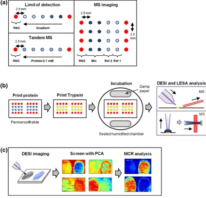
[image:4.595.75.501.72.480.2]0.5 μL/min, DESI raster speed was set to 500 μm/s. MS settings were as follows: positive ion mode, 5 kV voltage, mass range 400–2000m/z, 1 microscan, 110 ms max injection time, AGC mode off. Prosolia Firefly ver.1.2.0.1 software (Prosolia, Indianapolis, IN, USA) was used to convert scanned data into Analyse 7.5 format images, and Figure 1. Schematics for DESI and LESA-MS experiments. (a) Diagram of general DESI setup. The spray is focused onto the analysis
area by a nebulizing gas, which takes analytes into the MS inlet form/zdetermination. (b) Setup of protein printing for the various surface
digestion experiments. In limit of detection experiments the protein spots were printed in a concentration gradient of five spots from the right to left. For tandem MS five protein spots of 0.1 mM were printed in a row. For MS imaging four protein spots were printed with two single protein reference spots (Ref [1] and Ref [2]) on the right and two double protein mixture spots (Mix) on the left, which was repeated for four rows. All rows of proteins contained two rhodamine 6G (R6G) spots at each end as guides. All spots were printed
2.5 mm apart from each other. (c) Procedure for surface digestion. Proteins printed on slides were left to dry and another layer of trypsin
was printed on top of the previous spots. The slides were then placed within a sealed incubation chamber for 18 h. After incubation the
spots were allowed to dry and analyzed with DESI and LESA-MS. (d) Imaging MVA of DESI-MS images. After acquisition DESI-MS
Biomap (freeware, http://www.maldi-msi.org/) was used to produce images of individual m/z values.
MCR Analysis of DESI-MS Images
Multivariate curve resolution (MCR) analysis is an imaging MVA technique and was applied to the DESI-MS images of tryptically digested proteins previously described [23]. MS images were mass binned to 1m/zunit, cropped to isolate area of interest and reduce file size, and converted into a file format compatible with MCR analysis using in-house software.
PLS_Toolbox (ver. 5.2; Eigenvector Research, Manson, WA, USA) for Matlab (Mathsworks, Inc., Natick, MA, USA) was used to analyse the converted files with MCR. Imaging PCA analysis was performed as a precursor to evaluate the number of components for MCR analysis (Figure1c). For these data, mean centering was not carried out as MCR contains a non-negativity constraint that requires the data to have positive values.
Results and Discussion
Optimization of Surface Tryptic Digestion
for DESI and LESA-MS on Biomaterial Surfaces
Previously we have achieved some success with the surface detection of whole proteins and protein mixtures with DESI-MS [23]. However, detection was difficult for proteins of
large MW (966.8 kDa) as they could only be detected as a multiply charged series of spectra, which were difficult to interpret. Separation of proteins within a mixture was possible but required the use of MVA. Lastly, the sensitivity of detection was limited and only a relatively large concentration (960 nmol/mm2) of proteins of high molecular mass was detectable on the surface by DESI-MS [22].
[image:5.595.60.558.413.708.2]Surface digestion presents a new direction for the analysis of proteins with DESI and LESA-MS, which involves the deposition of trypsin onto surface proteins to achieve in-situ digestion. During optimization, it was found that an incubation time of 18 h at room temperature was sufficient to ensure that most of the proteins had undergone digestion on the surface (data not shown). The DESI and LESA-MS solvent mixtures were optimized by testing combinations of methanol, acetoni-trile, isopropanol and water with various amounts of formic acid as additives (data not shown). A combination of acetonitrile:water (1:1) was found to be the optimal solvent composition for the detection of surface peptides for both techniques. Formic acid addition gave minimal changes to the ionization efficiency and perhaps affected peptides less than other types of analytes under ambient conditions. Note that this solvent mixture is much more typical of solvents used for small molecule ambient DESI-MS (within them/z400–2000 range) compared with our previous findings for DESI-MS of whole proteins, which involved solvents such as isopropanol and high amounts of formic acid [23].
Figure 2. Mass spectra for the DESI-MS analysis of in-situ surface tryptic digestion of (a) Cytoc(green), (b) Myo (red), and (c)
Single protein and protein mixture solutions at different concentrations were printed in arrays onto Permanox cell culture slides. During optimization it was determined that 200 drops per protein (at approximately 300 pL per drop) was optimal for deposition onto the surface. For the printing of trypsin, 300 drops per spot was optimal for deposition onto the protein spots in order to ensure that they did not dry out during incubation. Rhodamine 6G solution was printed at the ends of each column as a visual guide for alignment to improve sample handling.
DESI-MS Detection of Tryptically Digested
Surface Proteins with LOD Analysis
Three proteins, Cytoc(12.5 kDa), Myo (17.7 kDa, and BSA (66.8 kDa) were tested for surface digestion by deposition onto Permanox slides. The mass spectra produced for these proteins showed efficient digestion by the trypsin. For Cyto
c, DESI-MS was able to detect peaks corresponding to six tryptic peptides (Figure2a), whereas for Myo, eight peptide peaks were detected (Figure2b). BSA is a large protein that was previously difficult to detect in its intact form using DESI-MS but was readily detected using in-situ tryptic digestion (Figure 2c). Peptide sequences are presented in Supplementary Figure1. Most of the peptides detected were singly charged species with a small number of doubly charged species, which is in line with previous studies [37]. Next we tested the ability of surface tryptic digestion for the analysis of protein mixtures printed onto Permanox surface, which was applied to Cytoc+ Myo, Cytoc+ BSA, and Myo + BSA protein mixtures. Individual peptide peaks belonging to each protein within the mixtures can be clearly distinguished from their spectra as shown for Cytoc+ Myo, Cytoc+ BSA, and Myo + BSA in Figure2d, e, and f, respectively. A reduction of peptide species detected for each protein was observed compared with single protein analysis, which may have been due to the effects of ion suppression of the MS signal caused by the presence of extra tryptic peptides. Several peptide species were still clearly observed in all of the mixture samples (peptide sequences in Supplementary Figure1). The presence of multiple peptides per protein increases the confidence of identification for the proteins within the mixtures. The spectra from these tryptically digested protein mixtures mark an improvement in interpretation over previous DESI-MS analysis of whole proteins where multiply charged series from several proteins were presented within the same spectra [23].
LOD analysis was carried out with each of the single proteins as well as the binary protein mixtures. The criteria of at least two peptide peaks per protein at intensities three times higher than the background was used for confirmation of detection for all of the protein samples examined. All of the single proteins and mixtures were detected on the Permanox slides at the pmol/mm2 level surface concentration, with Myo, Cytoc+ Myo, and Cyto
c+ BSA detected at 4.2 pmol/mm2and Cytoc, BSA, and Myo +
BSA detected at 2.1 pmol/mm2. Table
[image:6.595.315.431.80.741.2]Tandem MS of Peptides from Surface Digested
Proteins with DESI and LESA-MS
While it is possible to identify proteins purely from peptide mass via peptide mass fingerprinting, the specificity of identifications would be greatly improved if the tryptic peptide sequences were known via tandem MS. To this end we had set up the DESI and LESA instruments to perform DDA tandem MS analysis of digested protein spots for single proteins (Cyto
c, Myo and BSA) and binary protein mixtures (Cytoc+ Myo, Cytoc+ BSA, Myo + BSA) so that sequence information of the individual peptides can be elucidated in an automated manner. The tandem MS experiments for each of the samples produced a large number of spectra that included many of the surface digested peptides. After MASCOT analysis all of the proteins from each of the single protein and protein mixtures were correctly identified (Table1). A previous report for BSA tandem MS by ESI was able to achieve a sequence coverage of 33 % [47], which is a much higher result than was observed by DESI-MS (16 %), with LESA-MS (27 %) approaching this level of identification. In depth information of the protein identifications can be found in Supplementary Figures2–5.
LESA-MS gave better MASCOT scores and sequence coverage than DESI-MS for each of the proteins tested within their respective samples (Table1) and was able to perform much better in the detection of tryptic peptides, especially considering that the DESI-MS analysis was carried out over a row of five protein spots, whereas only one spot was used for each
LESA-MS run. The peak intensities for LESA-LESA-MS analyzed peptides were in general two orders of magnitude higher than their equivalent with DESI-MS (data not shown), which may have been due to LESA-MS having a more efficient desorption/ surface extraction process that allows a higher amount of peptides to be analyzed within the MS. There were also a higher number of multiply charged peptides observed for LESA-MS
compared with DESI-MS (Table 2). LESA-MS has an
ionization mechanism that is similar to nano ESI-MS since it uses the Advion Nanomate chip system [48], while DESI-MS of peptides was postulated to occur via a gas phase proton transfer system proposed by Kaur-Atwal et al. [37], which may explain the reason for the observed lower efficiency of multiple charging and ionization from DESI-MS.
DESI-MS Imaging of In-Situ Surface Digested
Protein Arrays
[image:7.595.54.555.90.207.2]MS imaging of tryptically digested protein arrays was performed for the first time using DESI-MS on Permanox surfaces. Proteins were printed in an array format with two single protein reference spots followed by two mixture spots containing those proteins with rhodamine 6G spot guides at each end, which was then repeated for four rows (Figure1a). Protein arrays of Cytoc+ Myo, Cytoc+ BSA, and Myo + BSA were produced with this layout, which were then imaged using DESI-MS at a spatial resolution of 100 μm.
Table 2.The Level of Charge for Peptides Observed via DESI and LESA DDA Tandem MS Analysis for Each Surface Digested Protein
Sample for DDA First protein Second protein
DESI charges LESA charges DESI charges LESA charges
+1 +2 +3 +1 +2 +3 +1 +2 +3 +1 +2 +3
Cytoc 5 0 0 6 9 0
Myo 3 1 0 6 8 7
BSA 6 4 0 14 14 4
Cytoc+ Myo 2 1 0 3 3 0 3 3 0 2 5 3
Cytoc+ BSA 5 4 0 4 5 0 9 3 0 15 14 2
Myo + BSA 7 6 1 8 12 5 10 5 0 6 11 2
Figure 3. DESI-MS imaging experiments for binary combinations of the proteins Cytoc+ BSA (Cytocis indicated green and
BSA is blue). The protein spots layout is presented with the images of three selected peptide peaks showingm/zvalues and
[image:7.595.148.459.574.702.2]Data are presented for the Cyto c+ BSA MS images, with the MS images for other two protein combinations are presented in Supplementary Figures. From the resulting DESI-MS images, multiple peptide peaks for each individual protein can be observed within the mixture spots as well as their respective reference spots, which were used to spatially separate the surface proteins within the array (Figure 3, Supplementary Figure 6). For Cyto c the peptides IFVQK (m/z 635.3), MIFAGIK (m/z 781.4) and TGPNLHGLFGR (m/z 1170.1) clearly separated this protein within the reference and mixture spots, whereas for BSA the peptides MPCTEDYLSLILNR (m/z834.5 + 2 charge), CCTESLVNR (m/z1025.7), and LGEYGFQNALIVR (m/z1480.7) gave the best results. Other peptide peaks for each of the proteins were also able to discriminate their spatial distribution with the arrays, which allows the option to use multiple peptides to further improve the spatial separation of proteins.
Imaging MVA of Surface Digested Protein Arrays
MCR analysis is a powerful imaging MVA technique that has the ability to separate common features within a complex MS image. MCR has been applied to DESI-MS images of whole proteins and was demonstrated to be able to separate the complex spectra of several overlaid multiply charged series of peaks in a spatially defined manner [23]. MCR was performed here on each of the three DESI-MS images of the protein mixture arrays (Cytoc+ Myo, Cyto c+ BSA, and Myo + BSA) to deconvolute their spectra and automatically assign spatial information for each of the proteins. Data for the images needed to be cropped to focus on the area of interest and mass binned to 1m/zto reduce file size for compatibility with the analysis software. For each of the MS data-sets the initial PCA analysis determined four distinct components.
A selection of the MCR component images and associated loadings for Cytoc+ BSA are shown in Figure4. The loadings of components from the MCR analyses represent the particularm/z
values associated with the image of the component. The plotted loadings for the components visually resemble the shape of a typical MS spectrum, which allows for an easy and straightfor-ward method for the interpretation of the results. For the Cytoc+ BSA protein array MCR was able to spatially separate Cytocand BSA peaks into distinct components, with the loadings identifying the major peptide species present for each of the proteins. MCR analysis also separated two other components that contained peaks for the background (Supplementary Figure 7). MCR analyses for the Cytoc+ Myo and Myo + BSA protein arrays were also able to spatially separate each of the proteins into distinct components (Supplementary Figures 8 and 9). The application of MCR allowed easier separation of protein peaks over manual analysis and has the potential to greatly improve the interpretation of protein array DESI-MS images. MCR analysis of surface digested proteins was able to give a more clearly defined separation of its components than was previously achieved with MS images of whole proteins [23]. The combination of MS imaging and MVA offers a potentially automated method for the identification of proteins from culture media deposited onto biomaterial surfaces; however, the detection of unknown proteins with this method may prove to be challenging as protein concentrations may vary greatly in the sample.
Conclusion
[image:8.595.108.474.70.270.2]surface proteins with MS. We have shown the ability for DESI and LESA-MS to conduct ambient detection, identification, and separation of model protein species from mixtures. The results obtained here gave marked improvements over DESI-MS of whole (undigested) proteins [23], and proteins of any size that are amenable to tryptic digestion can theoretically be analyzed in this manner. LESA-MS was also shown to be a more sensitive technique compared with DESI-MS and holds potential as a tool for the rapid screening of surface proteins. Advances in in-situ digestion, such as microwave-assisted surface digestion [35], could drastically reduce the time needed for this step and increase the speed of automation for large scale studies. The analysis time per sample for both of these techniques would be measured in minutes, which can give a rapid turnover for screening potentially more than 20 samples per hour. Problems of sample carryover between runs should be minimal; DESI-MS may accumulate some carryover within the MS inlet over very long periods of operation that can be removed by regular washing, whereas LESA-MS theoretically does not have any carryover problems because of the single-use nature of its tips and nanospray nozzles. DESI-MS imaging of surface digested proteins was shown to be possible in this model system, but applications to a biological surface may be difficult owing to variable surface protein concentration and possible ion suppression effects from other analytes. Ambient MS detection of proteins has gained interest in recent years but is at present in an embryonic stage of development [19], and the work described in this study presents a promising new direction for this type of analysis.
The complex milieu of proteins present within culture media currently being employed for applications such as human pluripotent stem cell (hPSC) culture makes detection of individual surface proteins adsorbed to the biomaterial surface very challenging. Understanding the specific roles of these adsorbed proteins in the process of cell attachment and growth within a culture medium will enable the design of better biomaterials. We have presented an initial examination of ambient surface protein detection with DESI and LESA-MS and have shown the ability of these methods to identify model proteins from a commercially available cell culture surface (Permanox). The next stage of work will be to apply our methods to identify proteins deposited by cell culture media onto these surfaces. Through these advances, we hope to one day be able to elucidate key media adsorbents that would allow for the design of better materials and optimized media for biological cell culture.
Acknowledgments
The authors acknowledge support from the UK Engineering and Physical Sciences Research Council (EPSRC) grant number EP/H045384/1.
Open Access
This article is distributed under the terms of the Creative Commons Attribution License which permits any use,
distribution, and reproduction in any medium, provided the original author(s) and the source are credited.
References
1. Castner, D.G., Ratner, B.D.: Biomedical surface science: Foundations to frontiers. Surf. Sci.500, 28–60 (2002)
2. Mei, Y., Saha, K., Bogatyrev, S.R., Yang, J., Hook, A.L., Kalcioglu, Z.I., Cho, S.W., Mitalipova, M., Pyzocha, N., Rojas, F., Van Vliet, K.J., Davies, M.C., Alexander, M.R., Langer, R., Jaenisch, R., Anderson, D.G.: Combinatorial development of biomaterials for clonal growth of human pluripotent stem cells. Nat. Mater.9, 768–778 (2010) 3. Saha, K., Mei, Y., Reisterer, C.M., Pyzocha, N.K., Yang, J., Muffat, J.,
Davies, M.C., Alexander, M.R., Langer, R., Anderson, D.G., Jaenisch, R.: Surface-engineered substrates for improved human pluripotent stem cell culture under fully defined conditions. Proc. Natl. Acad. Sci. U. S. A.108, 18714–18719 (2011)
4. Liu, L.Y., Chen, G., Chao, T., Ratner, B.D., Sage, E.H., Jiang, S.Y.: Reduced foreign body reaction to implanted biomaterials by surface treatment with oriented osteopontin. J. Biomater. Sci. Polym. Ed.19, 821–835 (2008) 5. Wagner, M.S., Horbett, T.A., Castner, D.G.: Characterizing multicomponent
adsorbed protein films using electron spectroscopy for chemical analysis, time-of-flight secondary ion mass spectrometry, and radiolabeling: capabil-ities and limitations. Biomaterials24, 1897–1908 (2003)
6. Lhoest, J.B., Wagner, M.S., Tidwell, C.D., Castner, D.G.: Character-ization of adsorbed protein films by time of flight secondary ion mass spectrometry. J. Biomed. Mater. Res.57, 432–440 (2001)
7. Muramoto, S., Graham, D.J., Wagner, M.S., Lee, T.G., Moon, D.W., Castner, D.G.: ToF-SIMS analysis of adsorbed proteins: principal component analysis of the primary ion species effect on the protein fragmentation patterns. J. Phys. Chem. C115, 24247–24255 (2011) 8. McCombie, G., Staab, D., Stoeckli, M., Knochenmuss, R.: Spatial and
spectral correlations in MALDI mass spectrometry images by clustering and multivariate analysis. Anal. Chem.77, 6118–6124 (2005) 9. Deininger, S.O., Ebert, M.P., Futterer, A., Gerhard, M., Rocken, C.:
MALDI imaging combined with hierarchical clustering as a new tool for the interpretation of complex human cancers. J. Proteome Res.7, 5230–5236 (2008)
10. Stein, M.J., Lo, E., Castner, D.G., Ratner, B.D.: Plasma pencil atmospheric mass spectrometry detection of positive ions from micronutrients emitted from surfaces. Anal. Chem.84, 1572–1578 (2012)
11. Takats, Z., Wiseman, J.M., Gologan, B., Cooks, R.G.: Mass spectrom-etry sampling under ambient conditions with desorption electrospray ionization. Science306, 471–473 (2004)
12. Ifa, D.R., Wu, C., Ouyang, Z., Cooks, R.G.: Desorption electrospray ionization and other ambient ionization methods: current progress and preview. Analyst135, 669–681 (2010)
13. Jackson, A.U., Tata, A., Wu, C., Perry, R.H., Haas, G., West, L., Cooks, R.G.: Direct analysis of Stevia leaves for diterpene glycosides by desorption electrospray ionization mass spectrometry. Analyst134, 867–874 (2009) 14. Soparawalla, S., Salazar, G.A., Sokol, E., Perry, R.H., Cooks, R.G.:
Trace detection of non-uniformly distributed analytes on surfaces using mass transfer and large-area desorption electrospray ionization (DESI) mass spectrometry. Analyst135, 1953–1960 (2010)
15. Wu, C.P., Ifa, D.R., Manicke, N.E., Cooks, R.G.: Rapid, direct analysis of cholesterol by charge labeling in reactive desorption electrospray ionization. Anal. Chem.81, 7618–7624 (2009)
16. Vickerman, J.C.: Molecular imaging and depth profiling by mass spectrometry—SIMS, MALDI, or DESI? Analyst136, 2199–2217 (2011) 17. Gerbig, S., Golf, O., Balog, J., Denes, J., Baranyai, Z., Zarand, A., Raso, E., Timar, J., Takats, Z.: Analysis of colorectal adenocarcinoma tissue by desorption electrospray ionization mass spectrometric imag-ing. Anal. Bioanal. Chem.403, 2315–2325 (2012)
18. Gao, L., Li, G., Cyriac, J., Nie, Z., Cooks, R.G.: Imaging of surface charge and the mechanism of desorption electrospray ionization mass spectrometry. J. Phys. Chem.114, 5331–5337 (2011)
19. Yao, Z.P.: Characterization of proteins by ambient mass spectrometry. Mass Spectrom. Rev.31, 437–447 (2012)
21. Douglass, K.A., Venter, A.R.: Protein analysis by desorption electrospray ionization mass spectrometry and related methods. J. Mass Spectrom.48, 553–560 (2013)
22. Shin, Y.S., Drolet, B., Mayer, R., Dolence, K., Basile, F.: Desorption electrospray ionization-mass spectrometry of proteins. Anal. Chem.79, 3514–3518 (2007)
23. Rao, W., Scurr, D.J., Burston, J., Alexander, M.R., Barrett, D.A.: Use of imaging multivariate analysis to improve biochemical and anatomical discrimination in desorption electrospray ionisation mass spectrometry imaging. Analyst137, 3946–3953 (2012)
24. Kertesz, V., Van Berkel, G.J.: Fully automated liquid extraction-based surface sampling and ionization using a chip-based robotic nanoelectrospray platform. J. Mass Spectrom.45, 252–260 (2010) 25. Marshall, P., Toteu-Djomte, V., Bareille, P., Perry, H., Brown, G.,
Baumert, M., Biggadike, K.: Correlation of skin blanching and percutaneous absorption for glucocorticoid receptor agonists by ma-trix-assisted laser desorption ionization mass spectrometry imaging and liquid extraction surface analysis with nanoelectrospray ionization mass spectrometry. Anal. Chem.82, 7787–7794 (2010)
26. Parson, W.B., Koeniger, S.L., Johnson, R.W., Erickson, J., Tian, Y., Stedman, C., Schwartz, A., Tarcsa, E., Cole, R., Van Berkel, G.J.: Analysis of chloroquine and metabolites directly from whole-body animal tissue sections by liquid extraction surface analysis (LESA) and tandem mass spectrometry. J. Mass Spectrom.47, 1420–1428 (2012) 27. Brown, S.H.J., Huxtable, L.H., Willcox, M.D.P., Blanksby, S.J., Mitchell,
T.W.: Automated surface sampling of lipids from worn contact lenses coupled with tandem mass spectrometry. Analyst138, 1316–1320 (2013) 28. Edwards, R.L., Creese, A.J., Baumert, M., Griffiths, P., Bunch, J., Cooper, H.J.: Hemoglobin variant analysis via direct surface sampling of dried blood spots coupled with high-resolution mass spectrometry. Anal. Chem.83, 2265–2270 (2011)
29. Van Berkel, G.J., Ford, M.J., Doktycz, M.J., Kennel, S.J.: Evaluation of a surface-sampling probe electrospray mass spectrometry system for the analysis of surface-deposited and affinity-captured proteins. Rapid Commun. Mass Spectrom.20, 1144–1152 (2006)
30. Van Berkel, G.J., Kertesz, V.: Continuous-flow liquid microjunction surface sampling probe connected on-line with high-performance liquid chromatog-raphy/mass spectrometry for spatially resolved analysis of small molecules and proteins. Rapid Commun. Mass Spectrom.27, 1329–1334 (2013) 31. Casadonte, R., Caprioli, R.M.: Proteomic analysis of formalin-fixed
paraffin-embedded tissue by MALDI imaging mass spectrometry. Nat. Protoc.6, 1695–1709 (2011)
32. Stauber, J., MacAleese, L., Franck, J., Claude, E., Snel, M., Kaletas, B.K., Wiel, I.M.V.D., Wisztorski, M., Fournier, I., Heeren, R.M.A.: On-tissue protein identification and imaging by MALDI-ion mobility mass spectrometry. J. Am. Soc. Mass Spectrom.21, 338–347 (2010) 33. Djidja, M.C., Francese, S., Loadman, P.M., Sutton, C.W., Scriven, P.,
Claude, E., Snel, M.F., Franck, J., Salzet, M., Clench, M.R.: Detergent addition to tryptic digests and ion mobility separation prior to MS/MS improves peptide yield and protein identification for in situ proteomic investigation of frozen and formalin-fixed paraffin-embedded adeno-carcinoma tissue sections. Proteomics9, 2750–2763 (2009)
34. Groseclose, M.R., Andersson, M., Hardesty, W.M., Caprioli, R.M.: Identifi-cation of proteins directly from tissue: in situ tryptic digestions coupled with imaging mass spectrometry. J. Mass Spectrom.42, 254–262 (2007) 35. Ha, N.Y., Kim, S.H., Lee, T.G., Han, S.: Rapid characterization of
protein chips using microwave-assisted protein tryptic digestion and MALDI mass spectrometry. Langmuir27, 10098–10105 (2011) 36. Schober, Y., Guenther, S., Spengler, B., Rompp, A.: High-resolution
matrix-assisted laser desorption/ionization imaging of tryptic peptides from tissue. Rapid Commun. Mass Spectrom.26, 1141–1146 (2012) 37. Kaur-Atwal, G., Weston, D.J., Green, P.S., Crosland, S., Bonner, P.L.,
Creaser, C.S.: Analysis of tryptic peptides using desorption electrospray ionisation combined with ion mobility spectrometry/mass spectrometry. Rapid Commun. Mass Spectrom.21, 1131–1138 (2007)
38. Takats, Z., Wiseman, J. M., Ifa, D. R., Cooks, R. G.: Desorption electrospray ionization (DESI) analysis of tryptic digests/peptides. CSH Protoc.2008, pdb prot4993 (2008)
39. Pasilis, S.P., Kertesz, V., Van Berkel, G.J., Schulz, M., Schorcht, S.: Using HPTLC/DESI-MS for peptide identification in 1D separations of tryptic protein digests. Anal. Bioanal. Chem.391, 317–324 (2008) 40. Heaton, K., Solazzo, C., Collins, M.J., Thomas-Oates, J., Bergstrom,
E.T.: Towards the application of desorption electrospray ionisation mass spectrometry (DESI-MS) to the analysis of ancient proteins from artefacts. J. Archaeol. Sci.36, 2145–2154 (2009)
41. Emory, J.F., Walworth, M.J., Van Berkel, G.J., Schulz, M., Minarik, S.: Direct analysis of reversed-phase high-performance thin layer chroma-tography separated tryptic protein digests using a liquid microjunction surface sampling probe/electrospray ionization mass spectrometry system. Eur. J. Mass Spectrom.16, 21–33 (2010)
42. Quanico, J., Franck, J., Dauly, C., Strupat, K., Dupuy, J., Day, R., Salzet, M., Fournier, I., Wisztorski, M.: Development of liquid microjunction extraction strategy for improving protein identification from tissue sections. J. Proteome79, 200–218 (2013)
43. Su, Y., Zhu, Y., Fang, Q.: A multifunctional microfluidic droplet-array chip for analysis by electrospray ionization mass spectrometry. Lab. Chip.13, 1876–1882 (2013)
44. Adler, B., Bostrom, T., Ekstrom, S., Hober, S., Laurell, T.: Miniaturized and automated high-throughput verification of proteins in the ISET platform with MALDI MS. Anal. Chem.84, 8663–8669 (2012) 45. Martinez-Osorio, H., Calonge, M., Corell, A., Reinoso, R., Lopez, A.,
Fernandez, I., San Jose, E.G., Diebold, Y.: Characterization and short-term culture of cells recovered from human conjunctival epithelium by minimally invasive means. Mol. Vis.15, 2185–2195 (2009)
46. Ho, M., Yu, D., Davidsion, M.C., Silva, G.A.: Comparison of standard surface chemistries for culturing mesenchymal stem cells prior to neural differentiation. Biomaterials27, 4333–4339 (2006)
47. Walcher, W., Franze, T., Weller, M.G., Poschl, U., Huber, C.G.: Liquid- and gas-phase nitration of bovine serum albumin studied by LC-MS and LC-MS/MS using monolithic columns. J. Proteome Res.2, 534–542 (2003)