The Scottish Structural Proteomics Facility: targets, methods
and outputs
Muse Oke•Lester G. Carter•Kenneth A. Johnson •Huanting Liu•Stephen A. McMahon • Xuan Yan•Melina Kerou•Nadine D. Weikart •Nadia Kadi•Md. Arif Sheikh•Stefan Schmelz• Mark Dorward• Michal Zawadzki•Christopher Cozens•Helen Falconer• Helen Powers• Ian M. Overton• C. A. Johannes van Niekerk•Xu Peng•Prakash Patel•Roger A. Garrett•
David Prangishvili•Catherine H. Botting• Peter J. Coote•David T. F. Dryden•Geoffrey J. Barton• Ulrich Schwarz-Linek•Gregory L. Challis•Garry L. Taylor•Malcolm F. White•
James H. Naismith
Received: 9 March 2010 / Accepted: 6 April 2010 / Published online: 24 April 2010 ÓThe Author(s) 2010. This article is published with open access at Springerlink.com
Abstract The Scottish Structural Proteomics Facility was funded to develop a laboratory scale approach to high throughput structure determination. The effort was suc-cessful in that over 40 structures were determined. These structures and the methods harnessed to obtain them are reported here. This report reflects on the value of auto-mation but also on the continued requirement for a high
degree of scientific and technical expertise. The efficiency of the process poses challenges to the current paradigm of structural analysis and publication. In the 5 year period we published ten peer-reviewed papers reporting structural data arising from the pipeline. Nevertheless, the number of structures solved exceeded our ability to analyse and publish each new finding. By reporting the experimental details and depositing the structures we hope to maximize the impact of the project by allowing others to follow up the relevant biology.
Keywords High-throughputProtein crystallography Structural proteomicsSSPF
Muse Oke, Lester G. Carter, Kenneth A. Johnson, Huanting Liu, Stephen A. McMahon—Joint first authors.
Electronic supplementary material The online version of this article (doi:10.1007/s10969-010-9090-y) contains supplementary material, which is available to authorized users.
M. OkeL. G. CarterK. A. JohnsonH. Liu
S. A. McMahonX. YanM. KerouN. D. Weikart
Md. A. SheikhS. SchmelzM. DorwardM. Zawadzki
C. CozensH. FalconerH. PowersC. H. Botting
P. J. CooteU. Schwarz-LinekG. L. Taylor
M. F. WhiteJ. H. Naismith (&)
Biomedical Sciences Research Complex, University of St Andrews, St Andrews KY16 9ST, UK e-mail: [email protected]
Present Address:
L. G. Carter
Stanford Synchrotron Radiation Light Source, 2575 Sand Hill Road, MS 69, Menlo Park, CA 94025, USA
Present Address:
K. A. Johnson
The Norwegian Structural Biology Centre, University of Tromsø, 9037 Tromsø, Norway
Present Address:
N. D. Weikart
Faculty of Chemistry, Technische Universita¨t Dortmund, Otto-Hahn-Str. 6, 44227 Dortmund, Germany
N. KadiP. PatelG. L. Challis
Department of Chemistry, University of Warwick, Coventry CV4 7AL, UK
Present Address:
N. Kadi
Institute of Cancer Research, 15 Cotswold Road, Belmont, Sutton, Surrey SM2 5NG, UK
Present Address:
M. Dorward
Division of Signal Transduction Therapy, College of Life Sciences, University of Dundee, Dundee DD1 5EH, Scotland, UK
Present Address:
M. Zawadzki
Syngenta Ltd, Jealott’s Hill International Research Centre, Bracknell, Berkshire RG42 6EY, UK
Present Address:
C. Cozens
Medical Research Council Laboratory of Molecular Biology, Hills Road, Cambridge CB2 0QH, UK
Abbreviations
BLAST Basic local alignment search tool PDB Protein data bank
PDRA Post-doctoral research associates PONDR Predictor of natural disordered regions RONN Regional order neural network SGC Structural genomics consortium SPINE Structural proteomics in Europe SPoRT Structural proteomics of rational targets SSPF Scottish Structural Proteomics Facility
Introduction
Structural biology is firmly embedded as a crucial tool for the molecular biologist, whether the interest is academic or commercial. The genome sequencing revolution of the 1990s transformed the biological landscape and to remain relevant to the scale of this new information, structural biology had to accelerate its pace of discovery. The Scottish Structural Proteomics Facility (SSPF) comprised of four partner institutions: the Universities of St Andrews, Dundee, Glasgow and Warwick. The Structural Proteomics of Rational Targets (SPoRT) laboratory situated at the University of St Andrews was established with the focus of developing small scale high throughput structural biology and to facilitate collaboration with other universities. Much larger international efforts have focussed on
productivity and throughput as exemplified by the NIH Protein Structure Initiative in the USA, Protein 3000 in Japan, Structural Proteomics in Europe (SPINE) [1] and the international Wellcome Trust Structural Genomics Consortium (SGC) [2]. Our remit was to generate struc-tures, publications and to facilitate access for ‘‘non-struc-tural’’ labs to structural biology. The ‘‘high throughput’’ lab commenced work in 2004 and focussed on develop-ment of efficient strategies for target selection, cloning, protein production, crystallization and crystal structure determination. Due to demands for higher throughput, automated equipment was employed at several key stages in the pipeline.
We chose targets against the following criteria: obtain-ing a structure was likely to be tractable (in essence this meant expression in Escherichia coli); the protein was likely to have a novel structure; the structure had the potential to either underpin antibiotic development or illuminate some defined aspect of biology or biochemistry. As we discuss, one of the significant challenges was to balance choice of ‘‘publishable’’ target work against the work of running the pipeline and the tractability of target (including any subsequent biochemical investigation). Target selection thus differed from many other efforts which focus on folds predicted to be ‘‘novel’’ (exemplified by the NIH efforts) or on comprehensive coverage of a functional class (such as the focus on short chain dehy-drogenases and kinases of the SGC).
During the build up of the SPoRT pipeline, we operated structure determination in essentially a traditional manner, in that projects were processed without automation in a sequential manner. This was because the funding imposed a number of output milestones which had to be met, thus there are ‘‘high throughput’’ structures and ‘‘low through-put’’ structures. The St Andrews laboratory was funded by a combination of sources and its staff complement con-sisted primarily of five post-doctoral research associates (PDRA) and two technicians. In 4 years, we have cloned more than 350 genes resulting in the production of 165 proteins from which 42 have yielded crystal structures. This represents about 25% of purified proteins that made it to crystal structure. Another measure of efficiency is that in its lifetime the pipeline delivered over two structures per PDRA per year. Given that the pipeline only became operative from the second year, the efficiency metrics by the end of the project were significantly higher than the beginning. However, the success of the pipeline outstripped our ability to analyse and follow up the structures. In this report, we provide a detailed account of the pipeline, the experimental structural biology and highlight some of the issues we faced. This strategy allows others to reproduce the experiments and to pursue the biological implications of our work.
Present Address:
H. Falconer
Institute of Structural and Molecular Biology, Edinburgh University, Kings Buildings, Edinburgh EH9 3JR, UK
I. M. OvertonC. A. J. van NiekerkG. J. Barton Division of Biological Chemistry and Drug Discovery, College of Life Sciences, University of Dundee, Dundee DD1 5EH, Scotland, UK
Present Address:
I. M. Overton
MRC Human Genetics Unit, Crewe Road South, Edinburgh EH4 2XU, UK
X. PengR. A. Garrett
Department of Biology, Archaea Centre,
University of Copenhagen, Ole Maaløes Vej 5, 2200, Copenhagen N, Denmark
D. Prangishvili
Institut Pasteur, 25 rue Dr. Roux, 75724 Paris cedex 15, France
D. T. F. Dryden
Materials and methods
Bioinformatics analyses for target selection
Proteins were submitted to the pipeline as cohorts of tar-gets; collection of genes with a common theme or source. Proteins were then analysed using a standard suite of analyses: SignalP 3.0 to detect signal peptides which indicate the protein would otherwise normally be exported from the cytoplasm [3]; TMHMM2 to detect transmem-brane regions which may reduce protein solubility [4]; and RONN and PONDR to analyze the sequence for predicted disordered regions which are believed to interfere with crystallization [5,6]. As a result of these analyses, trun-cations were made in a few cases where the prediction suggested significant disorder at the C- or N- termini, signal peptides or transmembrane regions. BLAST sear-ches of the PDB were carried out to establish the originality and potential impact of the structure. Targets were also analysed using the pI versus GRAVY crystallization pre-dictor plot as described in [7]. This work was initially carried out manually or by basic PERL scripts. However, a core part of the SSPF was to develop improved techniques for target selection, so in order to streamline and improve the ease and reliability of target selection we developed an automated system for carrying out sequence analysis called TarO [8]. TarO runs a suite of analyses on a target sequence including orthology detection and returns a summarised view of the findings as well as an annotated multiple sequence alignment that assists in the identifica-tion of domain boundaries and disordered regions. The system is web-accessible and has been used extensively within the SSPF as well as by external users world-wide. In addition to TarO, three techniques for crystallisation pro-pensity prediction were also developed. The OB-Score [9] combines hydrophobicity and predicted pI within a statis-tical framework, the ParCrys algorithm combines more features of the sequence [10] while XANNpred (manu-script submitted) is a machine-learning technique that includes a broad range of properties including predicted
secondary structure. TarO presents results sorted by Par-Crys and the OB-Score to highlight proteins which may be more amenable to processing in the SSPF high-throughput pipeline. While TarO and the crystallisation predictors were not applied in all structures output by SSPF, they were input to decision making processes in the later years of the project.
Genome-wide scale target selection tools
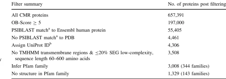
[image:3.595.159.544.581.715.2]We also developed bioinformatics protocols for large-scale target selection. A round of target selection was conducted to select tractable prokaryotic proteins expected to be rel-evant to human biology. The 657,391 proteins in the CMR (‘Omniome’) database [11] was the starting point for this work. The progress of these proteins through the target selection filters is summarised in Table1. Proteins pre-dicted to be amenable to producing diffraction-quality crystals (OB-ScoreC5) were searched against Ensembl human [12] with PSIBLAST [13], which identified 55,405 matches with expected structural similarity according to published thresholds [14]. In order to explore novel struc-ture space, proteins with a match to the PDB [15] were excluded, leaving 4,461 proteins. Uniprot [16] identifiers were inferred to provide a mechanism for obtaining pre-calculated annotations, particularly Pfam [17] and Gene Ontology (GO) [18] data. Proteins were excluded by the criteria of one or more predicted TMHMM2 transmem-brane regions, sequence length outwith 60–600 amino acids, and more than 20% predicted SEG low-complexity [19]. A total of 344 Pfam families were inferred for 3,008 proteins that passed the above filters. The total set of sequences from the 344 Pfam families were searched against the PDB (BLASTP, 1E-6, 90% identify, 90% query coverage) to exclude proteins where a homology modelling template was available. The final filtered set contained 1,329 proteins from 143 Pfam families. These proteins were then ranked according to OB-Score, and a custom scoring system ‘SOFA’ (see below).
Table 1 CMR target selection summary
a Matches defined by Rost curve [14]
b Uniprot identifiers inferred by perfect sequence match or BLASTP (1E-6, 90% query coverage, 90% identity)
Filter summary No. of proteins post filtering
All CMR proteins 657,391
OB-ScoreC5 197,000
PSIBLAST matchato Ensembl human protein 55,405
No PSIBLAST matchato PDB 4,461
Assign UniProt IDb 4,306
No TMHMM transmembrane regions &B20% SEG low-complexity, sequence length 60–600 amino acids
3,508
Infer Pfam family 3,008 (344 families)
A Gene Ontology-based scoring system (Specificity Of Functional Annotation, ‘SOFA’) was implemented to estimate the extent of available functional annotation for candidate targets, because functional annotation is often important for interpretation of new protein structures. The GO term with the least number of children was the starting point for calculating a protein’s SOFA score. From this term, the ratio of the parent to child terms was calculated; higher scores indicated more parents and therefore esti-mated more knowledge was available about the protein’s function. Where proteins had leaf-node GO term(s) the SOFA score was always higher than for proteins with no leaf-node GO term. Also, a greater number of leaf node terms corresponded to higher scores.
Further large-scale target selection was conducted with the aim of identifying tractable proteins relevant to Staphylococcus aureustherapeutics development. A set of 253 identifiers were taken from the literature [20, 21], indicating genes essential for infectivity in mouse models and/or essential for viability for growth in rich media. These were mapped to 1637 S. aureus proteins in CMR, corresponding to 1 MSSA (MSSA476) and 5 MRSA (MRSA252, Mu50, COL, MW2, and N315) strains. These proteins were subject to a series of filters similar to those described above. The filtering criteria were OB-ScoreC3, \2 TMHMM2 transmembrane regions, B20% SEG low-complexity, sequence length 60–600 amino acids. PSI-BLAST matches to PDB and Ensembl human proteins were excluded according to published thresholds. Matches to human proteins were excluded because these were considered to be less attractive therapeutic targets. Pfam matches were inferred, leaving 51 proteins from 36 Pfam families and 50 proteins without a Pfam match. SOFA scoring was applied to these 101 proteins and further analyses of these proteins including TarO and literature searching proposed a final set of 20 targets from 11 func-tional categories. This final set of 20 S. aureus proteins were submitted to the pipeline for production.
Cloning
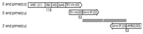
With the exception of purified proteins provided by col-laborators, open reading frames (ORFs) of all targets were cloned using a modified version of the Gateway cloning system. Each target was cloned with an N-terminal TEV protease cleavable Hisx6 tag, with the BP recombination site relocated out of the cloning sequence. Target genes were amplified using one common and two gene-specific primers. The common primer, encoding the attB1 recom-bination site, RBS, ATG start codon, six histidine residues and TEV protease site (Fig.1) was generated by PCR. The primers used for the above process were 50-GGGGACAA GTTTGTACAAAAAAGCAGGCTTCGAAGGAGATATAC
ATATG-30 (designated ATTF; attB1 site is in italics) and 50-GCCCTGAAAATACAGGTTTC-30(designated ATTR) with template of NdeI/NcoI-DNA fragments released from pEHISTEV plasmid DNA [22]. PCR products thus gener-ated were purified by ethanol precipitation and resuspended to a final concentration of 4lM. Target genes were amplified in PCR reactions consisting of 5 ll of each pair of gene-specific primers (10lM), 2.5ll of the common primers, 5ll of dNTP mixture (200lM each), 10ll of thermo polymerase buffer (59) containing 2 mM MgSO4
and 4% DMSO, 2 units ofPfuDNA polymerase and 10 ng of DNA template in a total volume of 50ll. Two-stage PCR amplifications were carried out in a 96-well formatted PCR plate. After denaturing the template at 95°C for 5 min, amplifications were carried out at 95°C for 1 min, Tm-5 for 1 min and 72°C for 4 min for 5 cycles and then followed by another 25 cycles using the same procedure except that an annealing temperature of 62°C was employed instead of Tm-5 to increase the specificity of the amplification. PCR products were cleaned using the PCR cleaning kit (Promega) and diluted to a concentration of 50 ng/ll.
BP recombination was carried out as described in the Gateway cloning instruction manual using pDONR221 as donor vector. The recombination reaction consisted of 100 ng ofattB-PCR products, 100 ng of pDONR221 vec-tor and 1ll of BP clonase II enzyme mix in TE buffer to a total volume of 10ll. The mixture was incubated at 25°C for 1 h and then further incubated at 37°C for 15 min following the addition of 2ll proteinase K.E. coli DH5a chemical competent cells were transformed with 2ll reaction mix and the transformed cells were spread onto L-agar plates containing 50 lg/ml kanamycin. Plasmid DNA was prepared by picking two colonies and cultivating in separate 10 ml L-broth media containing 50lg/ml kanamycin, prior to insert verification by agarose gel electrophoresis.
[image:4.595.305.544.56.114.2]LR recombination was carried out using pDEST14 as the destination vector with two verified pDONR221 clones. The recombination reaction contained 100 ng of entry clone pDONR221 DNA, 100 ng of pDEST14 vector, 1ll of LR clonase II enzyme mix in TE buffer to a total volume of 10ll. The reaction mixture was incubated for 1 h at 25°C and then incubated at 37°C for 15 min after adding 2ll of proteinase K. A total volume of 2ll of each BP reaction was transformed into 50ll of DH5a chemical competent cells and selected for ampicillin resistance on an L-agar plate. Two clones were picked and the plasmid DNA isolated for each LR reaction. The insertion sequence of each clone was verified and the prepared DNA was stored at-80°C for expression trials.
Small-scale expression
TheE. colicompetent cells BL21 (DE3), C43 (DE3), and BL21 (DE3)-CodonPlus (Strategene) were transformed with the pDEST14 expression vectors. Small-scale expression trials were carried out in Lysogeny Broth (LB; 10 g Tryptone, 5 g Yeast Extract, 10 g NaCl), Tryptone phosphate broth (TPB; 20 g Tryptone, 2 g K2HPO4, 2 g
KH2PO4, 5 g NaCl) and auto-induction media, prepared
in-house using the recipe from [23] or purchased as ‘Magic Media’ (Invitrogen). For LB and TPB cultures, starter cultures were prepared by inoculating LB supplemented with ampicillin (final concentration 100lg/ml) with freshly-transformed colonies on LB/ampicillin plates, and incubating overnight at 37°C, 200 rpm. Alternatively, the transformation mix was used directly as inoculum for starter cultures. Growth media (5 ml) in 50 ml Falcon tubes was inoculated with overnight starter culture (1:100 dilution factor) prior to incubation at 37°C, 200 rpm. At mid-log growth phase (OD600*0.6–0.8), protein
expression was induced with 0.4 mM IPTG. For LB cul-tures, incubation continued for a further 3 h at 37°C for one set of cultures, whilst another set was incubated at 25°C overnight. TPB cultures were incubated overnight at 25°C and 15°C post-induction. For protein expression in auto-induction media, freshly transformed colonies were used to inoculate 3-ml auto-induction media supplemented with 100lg/ml ampicillin (two colonies per target protein). Cultures were then incubated at 37°C, 300 rpm until cul-tures turned slightly turbid at which point one set was further incubated at 37°C and the other set at 25°C, both for 42 h. Cultures were harvested by centrifugation and pellets were stored at -20°C. Cell lysis was achieved either chemically using Bugbuster HT solution (Novagen) or mechanically by sonication. Whole cell lysates were ana-lysed for target protein expression by sodium dodecyl sulphate polyacrylamide gel electrophoresis (SDS–PAGE; [24] using pre-cast gels (Invitrogen). For target protein
localization, cell lysates were spun down and the super-natant was analysed for His6-tagged soluble protein
expression using the BioSprint 15 workstation according to manufacturer’s instructions (Qiagen). The resulting eluates were analysed by SDS-PAGE. Soluble protein expression was scored on the basis of whether protein bands com-mensurate with the estimated molecular weight were identified on SDS-PAGE gels. Band intensities were used to estimate the amount of soluble protein expressed. For uniformity, protein expression at\5 mg/l (low expression) was classified as 1S, 5–10 mg/l (medium expression) as 2S, and[10 mg/l (high expression) as 3S.
Large-scale expression of target proteins
Protein identities were verified by mass spectrometry prior to scale-up. Overnight starter cultures were used to inoc-ulate 1–6 l (depending on expression levels obtained from small-scale solubility screening experiments) LB, TPB or auto-induction media supplemented with ampicillin. Opti-mal growth conditions from sOpti-mall-scale expression trials were replicated for large scale expression. Prior to har-vesting (centrifugation at 2,4009g, for 30 min at 4°C), 1-ml aliquots were analyzed for protein expression and to estimate final yields. Cell pellets were stored at -80°C until required for purification.
Where appropriate, selenomethionine-labelled proteins were produced using the method of [25]. Essentially, freshly transformed BL21(DE3) or B834(DE3) cells served as inoculum for starter cultures. After overnight incubation, starter cultures were pelleted and washed 3 times with phosphate buffer (1 g NH4Cl, 3 g KH2PO4, 6 g Na2
H-PO47H2O/l) prior to inoculation (1:20) of
selenomethio-nine-incorporation media prepared as follows: 100 ml glucose solution (20% glucose, 0.3% MgSO4, 0.01%
Fe2(SO4)3, 0.01% thiamine) was added to 900 ml phosphate
buffer (same as above). The pH of the media was adjusted to 7.4 prior to addition of ampicillin and 50 mg/lL-(?) Sele-nomethionine. Cells were cultivated at 37°C until OD600was
between 0.8 and 1.0, at which point IPTG was added to a final concentration of 0.2 mM. Cultures were incubated at 25°C overnight, harvested and stored as described above.
Preparation of His6-tagged tobacco etch virus (TEV)
protease
uncleavable N-terminal His6 and C-terminal polyarginine
tags [26]. Following protein expression at 25°C, the His6
-TEV protease was purified by nickel-immobilized metal affinity chromatography (Ni2?-IMAC), followed by desalting chromatography. Aliquots (5 mg/ml) of TEV protease were prepared and stored at-80°C.
Purification
Cell pellets were resuspended in buffer A (see below for all buffer constituents) supplemented with Dnase1 (Sigma) and EDTA-free protease inhibitor cocktail tablets (Roche) prior to cell lysis on ice using the One Shot cell disruptor (Con-stant Systems Ltd), continuously cooled with cold tap water. Cell lysate was clarified by centrifugation at 39,0009gfor 1 h at 4°C and the supernatant was filtered using a 0.45lm filter (Millipore) prior to purification. Fully-automated purification was carried out on the AKTAxpress chroma-tography system using pre-packed columns (GE Health-care). The purification procedure comprised of (1) Ni2? -immobilized metal affinity chromatography (Ni2?-IMAC) using buffers B and C for the wash and elution steps respectively (2) Desalting chromatography (DS) in buffer D for buffer exchange and removal of imidazole, (3) incuba-tion with TEV protease for cleavage of the His6 tag, (4)
capture of the His6-tagged TEV protease on a second Ni2?
-IMAC column and (5) Gel filtration chromatography (GF) in buffer E to separate out contaminants and aggregates from the target proteins. A schematic representation of the fully-automated version is shown in Fig.2a. To facilitate on-line off-column cleavage of the His6-tag, a 50-ml superloop
preloaded with His6-TEV protease was incorporated into the
chromatography system. This arrangement facilitated the direct loading of the DS eluate into the TEV protease-loaded superloop. For manual purification, the Ni2?-IMAC and TEV cleavage steps were carried out on the bench, and the AKTAxpress system was used for DS and GF. All proteins were purified at room temperature and analysed by SDS-PAGE. Aliquots of purified proteins were transferred into thin-walled PCR tubes and flash frozen in liquid nitrogen prior to storage at-80°C.
Buffer A: 20 mM sodium phosphate pH 7.4, 500 mM NaCl, 10% glycerol, 10–30 mM imidazole
Buffer B: 20 mM sodium phosphate pH 7.4, 500 mM NaCl, 10% glycerol, 30–50 mM imidazole
Buffer C: 20 mM sodium phosphate pH 7.4, 500 mM NaCl, 10% glycerol, 300 mM imidazole
Buffer D: 20 mM Tris pH 7.5, 500 mM NaCl, 10% glycerol
Buffer E: 10 mM Tris pH 7.5, 150 mM or 500 mM NaCl, 10% glycerol (optional)
Crystallization
The pre-crystallization test (PCT) (Hampton Research) was used to determine the optimal protein concentration (OPC) for crystallization trials. All screening experiments were set up as sitting drops in 96-well crystallization plates at 2 or 3 protein concentrations using either the Cartesian Honeybee X8?1 in the Hamilton-Rhombix-Thermo integrated crys-tallization and imager system or an offline Cartesian Hon-eybee 963. Drop sizes consisting of 0.15ll protein ?
0.15ll precipitant (protein concentrations: 19OPC and 29
OPC) and 0.3ll protein?0.15ll precipitant (protein concentration: 19 OPC) were employed. Initially 4 com-mercial crystallization screens chosen from JCSG?, Clas-sics, Pegs, pHClear, Anions, Cations (Qiagen) and Wizard I & II (Emerald Biosystems) were used to screen all proteins. Eventually, this practice was phased out in favour of 3 sto-chastic screens [27] prepared in-house alongside the com-mercial JCSG? screen. All screening experiments were incubated at 20°C and imaged at regular intervals with a Rhombix-Thermo imager.
The traditional grid screen approach around the initial hit condition(s) was used primarily for crystal optimization purposes. If this failed, a stochastic approach to optimiza-tion was often able to generate suitable single crystals. In both cases, 24 and 96 well grid/stochastic screens based on the original mother liquor were designed and built using a Hamilton Microlab Star and crystallization screens were then set either as before on one of the robots using nanoliter volumes or manually using largerll drop sizes.
Data collection and structure solution
CCP4 REFMAC5 [40] was mainly used for refinement, although some structures were refined with PHENIX. The quality of all structures was checked with MOLPROBITY [41] and STAN (http://xray.bmc.uu.se/usf/www.html).
Protein information management system (PIMS)
Given the large volume of data produced by the project and shared between multiple researchers, a database system was required to store and process the information. A companion SPoRT funded project, Protein Information Management System (PIMS), was initiated at the same time as the SSPF to provide a laboratory information sys-tem for the project and other similar projects within the UK. The PIMS database was used to store and share experimental results within the project.
Results and discussion
Protein production
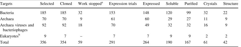
All targets have been divided into four groups on the basis of their origins. (1) Targets from Sulfolobus solfataricus andThermoproteus tenaxconstitute the Archaea group (2) ORFs from the archaeal Pyrobaculum spherical virus (PSV) and Sulfolobus islandicus rudivirus (SIRV), along with ORFs from Mycobacterium andStreptomyces riophages were classified as archaeal viruses and bacte-riophages (3) several pathogenic bacteria make up the bacterial group; and (4), the eukaryote group consisted of proteins of human, frog and trypanosome origins. Statistics for all target groups at various stages of the pipeline are presented in Table 2. Work was stopped on a few targets
[image:7.595.72.518.49.478.2]for two main reasons: structure of target or ortholog was determined in our laboratory or elsewhere, or bioinfor-matics analysis or publications suggested that the targets were either insoluble or membrane-associated.
The Gateway cloning system was adopted and modified by the SPoRT laboratory. This cloning strategy is inde-pendent of restriction enzymes, thus permitting standard-ized conditions for all of our cloning needs. We found our modified version to be very efficient for our experiments. Although PCR reactions using the double-stranded com-mon and gene-specific primers yielded sufficient PCR product with our standardized cycles, amplification of GC rich sequences predictably yielded less PCR product. The amplified PCR fragments were successfully used for BP recombination with the protocol described. Transformation of E. coli cells with 2ll of the recombination reaction produced more than 30 colonies on all plates with[99% of the isolated pDEST14 clones containing the right inserts. Intermittent sequencing of cloned genes showed that there was no significant increase in the rate of mutation during the cloning process. Overall, we found this strategy to be very robust as over 350 targets were transferred success-fully to the vector, irrespective of the origin of the gene.
Our expression strategy involved the use of three dif-ferent variables comprising expression strain, growth medium and temperature. At the outset of the project, three E. coli (DE3) expression strains (BL21, C43 and Codon Plus) were screened for expression. The rationale for includingE. coliBL21(DE3) Codon Plus was to overcome the anticipated codon bias of archaeal and eukaryotic genes. However, preliminary results from small scale expression trials using this strain showed that expression levels were not improved, and therefore further usage was discontinued. The C43(DE3) strain is particularly useful for expression of toxic proteins inE. coli[42, 43]. Initial expression trials comparing C43(DE3) and BL21(DE3) cells revealed that the former was particularly beneficial for expression of the archaeal viral proteins. Overall, 67% of targets scaled-up were expressed in C43(DE3) and the rest in BL21(DE3). Growth media employed in our facility consisted of LB, TPB and auto-induction media. We found
the auto-induction media to be particularly useful for small-scale expression screening as it facilitated parallel screening by eliminating the need to measure cell densities prior to induction. However, since most of the targets were equally soluble using either TPB or LB, we opted for these relatively inexpensive and simpler media at the scale-up stage. Consequently, only 4% of proteins were scaled-up using auto-induction media, with the rest shared equally between LB and TPB. Targets scaled-up using auto-induction media and TPB were those that either expressed at the 1S level or a minority that did not express at all in LB media. Overall 72% (190/264) of all expressed targets were soluble (Table2), a success rate attributable to the different expression systems and conditions utilized by the SPoRT laboratory. Although protein targets of prokaryotic origins constitute most of the soluble proteins (63%; 120/190), it is not certain if this is due to the larger number of prokaryotic targets compared to the others or the use of E. coli for expression. The majority of soluble targets expressed suf-ficiently: 27% at the 3S level, 60% at the 2S level, and 13% at the 1S level. It was tempting to rescue insoluble proteins using fusion proteins such as MBP and thioredoxin, but we were not convinced that this would significantly increase the overall number of soluble targets. Moreover, this would require more manpower and resources. All soluble proteins scaled-up successfully, although the volume of media had to be adjusted depending on the expression level. Typically 2 l of culture was grown for 3S proteins, 4 l for 2S proteins and 6 l for 1S proteins.
[image:8.595.51.550.73.170.2]Initially, small-scale cell lysis was carried out using Bugbuster for convenience. Eventually, this was replaced by sonication for two main reasons. Firstly, we observed that higher protein yields of poorly-expressing proteins were recovered by sonication compared to Bugbuster. Secondly, we observed that in certain cases, protein yields were inconsistent between estimated and final yields from large-scale cultures, presumably due to the detergents in Bugbuster. Although not convenient for a high-throughput approach, we adopted sonication in order to eliminate any ambiguities. A consistent and practical plan was adopted for the expression process in order to feed the pipeline
Table 2 SPoRT laboratory pipeline scoreboard
Targets Selected Cloned Work stoppeda Expression trials Expressed Soluble Purified Crystals Structure
Bacteria 185 185 32 153 148 120 99 32 22
Archaea 70 70 9 61 60 29 27 11 9
Archaea viruses and bacteriophages
92 92 18 70 49 32 32 16 9
Eukaryotesb 9 7 – 7 7 9 9 2 2
Total 356 354 59 291 264 190 167 61 42
a Number of targets that were cloned but were not submitted for expression trials
continuously with proteins for processing. Typically, up to twenty targets were chosen per week for expression trials. Concurrently, proteins judged to be soluble from the pre-vious week and confirmed by mass spectrometry were prepared in large quantities. The BioSprint 15 workstation, which can perform 15 nickel pull-down experiments in about 30 min, expedited sample processing times. Addi-tionally, the Nanodrop Spectrophotometer was very bene-ficial as it provided a quick, easy and efficient means for not only measuring optical densities of cultures, but also spectra of DNA and proteins. As it was never the case that all twenty proteins were soluble, up to ten proteins were scaled up per week and passed on for purification. All protein expression experiments were routinely carried out on a weekly basis by one technician, with one PDRA trouble-shooting problems such as conflicting expression and mass spectrometry results. Such problems were usually resolved by repeating the trials and/or gene sequencing.
Affinity tags are ideal for HTP protein purification because a single chromatography method can be adopted. The advantages of using the His6-tag are well-documented.
Using either manual or fully-automated purification meth-ods, we obtained[90% protein purity in sufficient amounts for about 88% (167/190) of all soluble proteins. A typical elution profile for proteins purified using the fully-auto-mated version is shown in Fig.2b, with clearly identifiable Ni2?-IMAC, DS and GF peaks. The fully-automated method, incorporating on-line off-column TEV cleavage, was made possible by the inclusion of the 50-ml superloop in-series, as described in Materials and Methods. This method required no user-intervention once the sample was applied to the system. Also, the fully-automated method was less time-consuming, eliminating all manual handling steps such as sample loading and pooling between each chromatographic step. TEV cleavage using the fully-auto-mated method was just as efficient as the manual method. Up to ten proteins were routinely purified per week by one technician. However, in several cases, it was difficult to control the final volume of eluates from all columns lead-ing up to the GF column, which is limited to a maximum permissible volume of 5 ml. Elution volumes from chro-matography steps prior to GF were typically larger than this volume. The outcome of this was that only 50% of the purified protein eventually made it to GF. In the latter stages of the project, after several unsuccessful attempts were made to rectify the situation, we simply purified all our targets manually. Consequently, the number of proteins purified weekly reduced to about six.
Crystallization and crystal structures
All purified protein samples were screened successfully using the nanolitre-scale Rhombix-Hamilton-Thermo
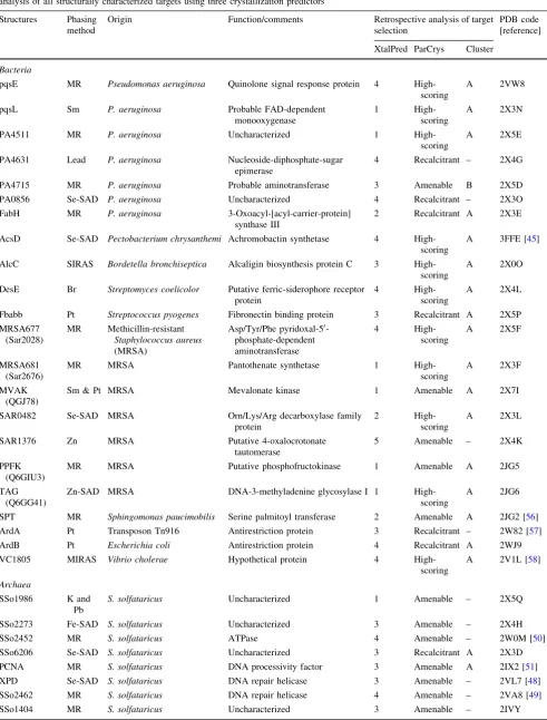
integrated system. This technology not only reduced protein production costs (final protein yield was not always important), but allowed for immediate setting up of crystal trials soon after purification. As shown in Table2, 61 of the 167 purified proteins (37%) formed diffraction-quality crystals. Despite the nanolitre-scale size of the crystalli-zation drops, most of the crystals obtained were robust enough to provide complete datasets suitable for structural determination. As shown in Table2, 42 structures have been solved thus far, representing 25% (42/167) of the targets that entered crystallization trials. Different phasing methods were used for structure determination with two-thirds of the structures using methods other than molecular replacement (see Table3). This outcome was anticipated since most of the SPoRT target sequences did not display significant homology ([20%) with protein sequences in all databases at the outset of the project.
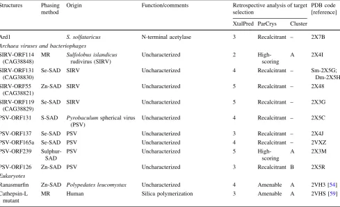
Retrospective analysis of target selection
Table 3 Summary of all SPoRT laboratory crystal structures showing phasing methods, origins and functions of proteins, and retrospective analysis of all structurally characterized targets using three crystallization predictors
Structures Phasing method
Origin Function/comments Retrospective analysis of target selection
PDB code [reference]
XtalPred ParCrys Cluster
Bacteria
pqsE MR Pseudomonas aeruginosa Quinolone signal response protein 4
High-scoring
A 2VW8
pqsL Sm P. aeruginosa Probable FAD-dependent
monooxygenase
1
High-scoring
A 2X3N
PA4511 MR P. aeruginosa Uncharacterized 1
High-scoring
A 2X5E
PA4631 Lead P. aeruginosa Nucleoside-diphosphate-sugar epimerase
4 Recalcitrant – 2X4G
PA4715 MR P. aeruginosa Probable aminotransferase 3 Amenable B 2X5D
PA0856 Se-SAD P. aeruginosa Uncharacterized 4 Recalcitrant – 2X3O
FabH MR P. aeruginosa 3-Oxoacyl-[acyl-carrier-protein]
synthase III
2 Recalcitrant A 2X3E
AcsD Se-SAD Pectobacterium chrysanthemi Achromobactin synthetase 4 High-scoring
A 3FFE [45]
AlcC SIRAS Bordetella bronchiseptica Alcaligin biosynthesis protein C 3 High-scoring
A 2X0O
DesE Br Streptomyces coelicolor Putative ferric-siderophore receptor
protein
4
High-scoring
A 2X4L
Fbabb Pt Streptococcus pyogenes Fibronectin binding protein 3 Recalcitrant A 2X5P
MRSA677 (Sar2028)
MR Methicillin-resistant
Staphylococcus aureus
(MRSA)
Asp/Tyr/Phe pyridoxal-50 -phosphate-dependent aminotransferase
4
High-scoring
A 2X5F
MRSA681 (Sar2676)
MR MRSA Pantothenate synthetase 1
High-scoring
A 2X3F
MVAK (QGJ78)
Sm & Pt MRSA Mevalonate kinase 1 Amenable A 2X7I
SAR0482 Se-SAD MRSA Orn/Lys/Arg decarboxylase family protein
2
High-scoring
A 2X3L
SAR1376 Zn MRSA Putative 4-oxalocrotonate tautomerase
5 Amenable – 2X4K
PPFK (Q6GIU3)
MR MRSA Putative phosphofructokinase 1 Amenable A 2JG5
TAG (Q6GG41)
Zn-SAD MRSA DNA-3-methyladenine glycosylase I 1 High-scoring
A 2JG6
SPT MR Sphingomonas paucimobilis Serine palmitoyl transferase 2 Amenable A 2JG2 [56]
ArdA Pt Transposon Tn916 Antirestriction protein 3 Recalcitrant – 2W82 [57]
ArdB Pt Escherichia coli Antirestriction protein 4 Recalcitrant A 2WJ9
VC1805 MIRAS Vibrio cholerae Hypothetical protein 4 High-scoring
A 2V1L [58]
Archaea
SSo1986 K and Pb
S. solfataricus Uncharacterized 1 Amenable – 2X5Q
SSo2273 Fe-SAD S. solfataricus Uncharacterized 3 Amenable – 2X4H
SSo2452 MR S. solfataricus ATPase 4 Amenable – 2W0M [50]
SSo6206 Se-SAD S. solfataricus Uncharacterized 3 Recalcitrant A 2X3D
PCNA MR S. solfataricus DNA processivity factor 3 Amenable A 2IX2 [51]
XPD Se-SAD S. solfataricus DNA repair helicase 3 Amenable – 2VL7 [48]
SSo2462 MR S. solfataricus DNA repair helicase 4 Amenable – 2VA8 [49]
and 11 proteins by ParCrys versus pI/GRAVY plot. This discrepancy may be due to different parameters utilized by the predictors namely; size, homologs in PDB, GRAVY index and pI. Therefore, it is reasonable to suggest that at least two predictors should be utilized in order to provide a wider coverage of crystallization space.
Scientific highlights
Reports describing the crystal structures and biochemical analyses of 10 SPoRT-generated targets have been pub-lished in peer-reviewed journals (see Table3 for refer-ences). Experimental details of crystal structures yet to be published are presented in Supplementary information. Below, we summarize a few examples to highlight the scientific impact of a few of our accomplishments.
Iron is an essential nutrient and microorganisms syn-thesize low molecular weight high affinity iron chelators (siderophores) in order to sequester environmental or host iron. The synthesis of these compounds is both a potential antibacterial target and of interest biochemically. We have reported the crystal structure and biochemical analysis of AcsD, an enzyme involved in the biosynthesis of
achromobactin from the Gram negative plant pathogen Pectobacterium chrysanthemi [45] and the structure of AlcC, a new member of the superfamily, will be reported in due course. Other antimicrobial targets included those selected from Pseudomonas aeruginosa, an opportunistic antibiotic-resistant human pathogen. The Pseudomonas quinolone signal (PQS) is implicated in both pathogenesis and formation of biofilms and was of particular interest. In total eightP. aeruginosastructures have been determined. Methicillin-resistantStaphylococcus aureus(MRSA) is the cause of considerable concern in hospitals. We employed comparative two-dimensional gel analysis and mass spec-trometry [46, 47] to identify proteins that were either upregulated or downregulated in MRSA compared to methicillin-sensitiveS. aureus(MSSA), and therefore con-sidered to be essential with potentially novel folds. Seven structures have been determined from this target.
[image:11.595.53.543.73.371.2]Archaeal proteins are frequently used as tools to understand equivalent proteins in eukaryotes, most partic-ularly those involved in DNA replication and repair. Of the 61 archaeal targets selected, 60 expressed, about half were soluble and nine structures were determined. These inclu-ded crystal structures of two helicases implicated in DNA
Table 3continued Structures Phasing
method
Origin Function/comments Retrospective analysis of target selection
PDB code [reference]
XtalPred ParCrys Cluster
Ard1 S. solfataricus N-terminal acetylase 3 Recalcitrant – 2X7B
Archaea viruses and bacteriophages
SIRV-ORF114 (CAG38848)
MR Sulfolobus islandicus
rudivirus (SIRV)
Uncharacterized 2
High-scoring
A 2X4I
SIRV-ORF131 (CAG38830)
Se-SAD SIRV Uncharacterized 4 Recalcitrant – Sm-2X5G;
Dm-2X5H
SIRV-ORF55 (CAG38821)
Zn-SAD SIRV Uncharacterized 5 Recalcitrant – 2X48
SIRV-ORF119 (CAG38829)
Se-SAD SIRV Uncharacterized 5 Recalcitrant – 2X3G
PSV-ORF131 S-SAD Pyrobaculumspherical virus (PSV)
Uncharacterized 4 Recalcitrant – 2X5C
PSV-ORF137 Se-SAD PSV Uncharacterized 3 Recalcitrant – 2X4J
PSV-ORF165a Se-SAD PSV Uncharacterized 4 Recalcitrant – 2VXZ
PSV-ORF239 Sulphur-SAD
PSV Uncharacterized 5
High-scoring
A 2X3M
PSV-ORF126 Zn-SAD PSV Uncharacterized 3 Recalcitrant B 2X5R
Eukaryotes
Ranasmurfin Zn-SAD Polypedates leucomystax Uncharacterized 4 Amenable A 2VH3 [54]
Cathepsin-L mutant
MR Human Silica polymerization 3 Amenable A 2VHS [59]
repair: XPD from S. tokodaii [48] and hel308 from S. solfataricus[49], a RadA paralog [50] and a heterotrimeric PCNA sliding clamp [51].
Sulfolobus islandicus rudivirus (SIRV) and Pyrobacu-lum spherical virus (PSV) are dsDNA viruses that infect archaea and their proteins show little or no detectable homology to any proteins of known function [52,53]. We reasoned that structural data from these viruses could help elucidate viral protein function and improve our under-standing of viral evolution. The virus genomes (48 ORFs from SIRV and 45 ORFs from PSV) were of a suitable size for us to attempt complete coverage. We were able to determine eight structures having cloned and attempted to express all genes.
Biological research is of course very broad but we were able to contribute to a number of local projects by viding structural data. The most striking was a novel pro-tein harvested from the foam nests of Polypedates leucomystax, a tropical frog species from Malaysia. The crystal structure of the protein, named ranasmurfin, revealed intra- and inter-molecular cross-links featuring the lysine-tyrosine quinone (LTQ) co-factor. At 1.16 A˚ reso-lution, the amino acid sequence was identified directly from the electron density map and verified by mass spec-trometry to be a novel protein [54].
Conclusions
It is clear from many studies that structure determination can be largely automated and the efficiency greatly improved. What is more, our effort demonstrates that this can be carried out on a relatively small scale and be focused to deliver results to the scientific community. The ability to run such a pipeline appears to be a skill in itself. Our own efficiency improved year on year as new working practices and approaches developed with experience. Despite this success, we have not been able to reach the point of guaranteeing a structure for every target nor in being able a priori to identify which targets will give structures. Target selection is improving and new tools are emerging that should continue to drive up the target to structure ratio (reviewed in [55]), although this is not the same as a structure for every biological target. A small group can gain efficiency by being able to adjust and alter procedures. For example, we found the use of heavy atoms a very efficient first attempt to solve structures rather than always proceeding to selenomethionine.
Our experience is that a structural biology laboratory within a collaborative centre, bringing the techniques of modern structural biology to bear on novel problems, has some advantages. The pipeline has the capacity to move very quickly to respond to a scientific need. Personnel who
run the pipeline improved in efficiency over the time of the project. In a collaboration, the expertise of those carrying out the structural biology can be leveraged in the analysis of the structure, design and interpretation of subsequent experiments.
Efficiency and automation require significant intellec-tual resources. We observe that in this lies an issue for the future. Even in our relatively small scale effort, we were unable to match the productivity of the pipeline at gener-ating structures with publication. Only in cases where targets were carefully chosen in advance or the structure was immediately scientifically significant, were we able to follow this more traditional path to publications. With collaborations, we were able to perform significant bio-chemical analysis for some proteins. However, as we report, many structures have been determined with very limited or no analysis. By fully reporting the experimental detail and depositing the data, we aim to prompt others to perform this analysis.
Acknowledgments THE SSPF was supported by grants from The Scottish Funding Council (references SSPF and SULSA), The Biotechnology and Biological Sciences Research Council (reference BB/S/B14450), European Union under framework 7 (reference Aeropath)
Open Access This article is distributed under the terms of the Creative Commons Attribution Noncommercial License which per-mits any noncommercial use, distribution, and reproduction in any medium, provided the original author(s) and source are credited..
References
1. Banci L, Bertini I, Cusack S, de Jong RN, Heinemann U, Jones EY, Kozielski F, Maskos K, Messerschmidt A, Owens R, Per-rakis A, Poterszman A, Schneider G, Siebold C, Silman I, Sixma T, Stewart-Jones G, Sussman JL, Thierry JC, Moras D (2006) First steps towards effective methods in exploiting high-throughput technologies for the determination of human protein structures of high biomedical value. Acta Crystallogr D Biol Crystallogr 62(Pt 10):1208–1217
2. Lee WH, Atienza-Herrero J, Abagyan R, Marsden BD (2009) SGC—structural biology and human health: a new approach to publishing structural biology results. PLoS One 4(10):e7675 3. Bendtsen JD, Nielsen H, von Heijne G, Brunak S (2004)
Improved prediction of signal peptides: SignalP 3.0. J Mol Biol 340(4):783–795
4. Krogh A, Larsson B, von Heijne G, Sonnhammer EL (2001) Predicting transmembrane protein topology with a hidden Mar-kov model: application to complete genomes. J Mol Biol 305(3): 567–580
5. Li X, Romero P, Rani M, Dunker AK, Obradovic Z (1999) Predicting protein disorder for N-, C-, and internal regions. Genome Inform Ser Workshop Genome Inform 10:30–40 6. Yang ZR, Thomson R, McNeil P, Esnouf RM (2005) RONN: the
detection of natively disordered regions in proteins. Bioinfor-matics 21(16):3369–3376
7. Canaves JM, Page R, Wilson IA, Stevens RC (2004) Protein biophysical properties that correlate with crystallization success in Thermotoga maritima: maximum clustering strategy for structural genomics. J Mol Biol 344(4):977–991
8. Overton IM, van Niekerk CA, Carter LG, Dawson A, Martin DM, Cameron S, McMahon SA, White MF, Hunter WN, Naismith JH, Barton GJ (2008) TarO: a target optimisation system for struc-tural biology. Nucleic Acids Res 36(Web Server issue):W190– W196
9. Overton IM, Barton GJ (2006) A normalised scale for structural genomics target ranking: the OB-score. FEBS Lett 580(16):4005– 4009
10. Overton IM, Padovani G, Girolami MA, Barton GJ (2008) Par-Crys: a Parzen window density estimation approach to protein crystallization propensity prediction. Bioinformatics 24(7):901– 907
11. Peterson JD, Umayam LA, Dickinson T, Hickey EK, White O (2001) The comprehensive microbial resource. Nucleic Acids Res 29(1):123–125
12. Hubbard TJ, Aken BL, Beal K, Ballester B, Caccamo M, Chen Y, Clarke L, Coates G, Cunningham F, Cutts T, Down T, Dyer SC, Fitzgerald S, Fernandez-Banet J, Graf S, Haider S, Hammond M, Herrero J, Holland R, Howe K, Johnson N, Kahari A, Keefe D, Kokocinski F, Kulesha E, Lawson D, Longden I, Melsopp C, Megy K, Meidl P, Ouverdin B, Parker A, Prlic A, Rice S, Rios D, Schuster M, Sealy I, Severin J, Slater G, Smedley D, Spudich G, Trevanion S, Vilella A, Vogel J, White S, Wood M, Cox T, Curwen V, Durbin R, Fernandez-Suarez XM, Flicek P, Kasprzyk A, Proctor G, Searle S, Smith J, Ureta-Vidal A, Birney E (2007) Ensembl 2007. Nucleic Acids Res 35(Database issue):D610– D6177
13. Altschul SF, Madden TL, Schaffer AA, Zhang J, Zhang Z, Miller W, Lipman DJ (1997) Gapped BLAST and PSI-BLAST: a new generation of protein database search programs. Nucleic Acids Res 25(17):3389–3402
14. Rost B (1999) Twilight zone of protein sequence alignments. Protein Eng 12(2):85–94
15. Berman H, Henrick K, Nakamura H, Markley JL (2007) The worldwide protein data bank (wwPDB): ensuring a single, uni-form archive of PDB data. Nucleic Acids Res 35(Database issue):D301–D303
16. Apweiler R, Bairoch A, Wu CH, Barker WC, Boeckmann B, Ferro S, Gasteiger E, Huang H, Lopez R, Magrane M, Martin MJ, Natale DA, O’Donovan C, Redaschi N, Yeh LS (2004) UniProt: the universal protein knowledgebase. Nucleic Acids Res 32(Database issue):D115–D119
17. Bateman A, Coin L, Durbin R, Finn RD, Hollich V, Griffiths-Jones S, Khanna A, Marshall M, Moxon S, Sonnhammer EL, Studholme DJ, Yeats C, Eddy SR (2004) The Pfam protein families database. Nucleic Acids Res 32(Database issue):D138– D141
18. Harris MA, Clark J, Ireland A, Lomax J, Ashburner M, Foulger R, Eilbeck K, Lewis S, Marshall B, Mungall C, Richter J, Rubin GM, Blake JA, Bult C, Dolan M, Drabkin H, Eppig JT, Hill DP, Ni L, Ringwald M, Balakrishnan R, Cherry JM, Christie KR, Costanzo MC, Dwight SS, Engel S, Fisk DG, Hirschman JE, Hong EL, Nash RS, Sethuraman A, Theesfeld CL, Botstein D, Dolinski K, Feierbach B, Berardini T, Mundodi S, Rhee SY, Apweiler R, Barrell D, Camon E, Dimmer E, Lee V, Chisholm R, Gaudet P, Kibbe W, Kishore R, Schwarz EM, Sternberg P, Gwinn M, Hannick L, Wortman J, Berriman M, Wood V, de la Cruz N, Tonellato P, Jaiswal P, Seigfried T, White R (2004) The gene ontology (GO) database and informatics resource. Nucleic Acids Res 32(Database issue):D258–D261
19. Wootton JC, Federhen S (1996) Analysis of compositionally biased regions in sequence databases. Methods Enzymol 266:554–571
20. Forsyth RA, Haselbeck RJ, Ohlsen KL, Yamamoto RT, Xu H, Trawick JD, Wall D, Wang L, Brown-Driver V, Froelich JM, Kedar GC, King K, McCarthy M, Malone C, Misiner B, Robbins D, Tan Z, Zhu ZY, Carr G, Mosca DA, Zamudio C, Foulkes JG, Zyskind JW (2002) A genome-wide strategy for the identification of essential genes in Staphylococcus aureus. Mol Microbiol 43(6):1387–1400
21. Ji Y, Zhang B, Van SF, Horn WP, Woodnutt G, Burnham MK, Rosenberg M (2001) Identification of critical staphylococcal genes using conditional phenotypes generated by antisense RNA. Science 293(5538):2266–2269
22. Liu H, Naismith JH (2009) A simple and efficient expression and purification system using two newly constructed vectors. Protein Expr Purif 63(2):102–111
23. Studier FW (2005) Protein production by auto-induction in high density shaking cultures. Protein Expr Purif 41(1):207– 234
24. Laemmli UK (1970) Cleavage of structural proteins during the assembly of the head of bacteriophage T4. Nature 227(5259): 680–685
25. Guerrero SA, Hecht HJ, Hofmann B, Biebl H, Singh M (2001) Production of selenomethionine-labelled proteins using simpli-fied culture conditions and generally applicable host/vector sys-tems. Appl Microbiol Biotechnol 56(5–6):718–723
26. Kapust RB, Tozser J, Fox JD, Anderson DE, Cherry S, Copeland TD, Waugh DS (2001) Tobacco etch virus protease: mechanism of autolysis and rational design of stable mutants with wild-type catalytic proficiency. Protein Eng 14(12):993–1000
27. Rupp B (2003) Maximum-likelihood crystallization. J Struct Biol 142(1):162–169
28. Otwinowski Z, Minor W (1997) Processing of X-ray diffraction data collected in oscillation mode. In: Methods in enzymology. Academic Press, New York, pp 307–326
29. Leslie AGW (1992) Recent changes to the MOSFLM package for processing film and image plate data. Joint CCP4? ESF-EA-MCB newsletter on protein crystallography, no. 26
30. Evans P (2006) Scaling and assessment of data quality. Acta Crystallogr D Biol Crystallogr 62(Pt 1):72–82
31. Kabsch W (1993) Automatic processing of rotation diffraction data from crystals of initially unknown symmetry and cell con-tants. J Appl Crystallogr 26:795–800
32. Schneider TR, Sheldrick GM (2002) Substructure solution with SHELXD. Acta Crystallogr D Biol Crystallogr 58(Pt 10 Pt 2):1772–1779
33. Terwilliger TC, Berendzen J (1999) Automated MAD and MIR structure solution. Acta Crystallogr D Biol Crystallogr 55(Pt 4):849–861
34. Terwilliger TC (2000) Maximum-likelihood density modifica-tion. Acta Crystallogr D Biol Crystallogr 56(Pt 8):965–972 35. Adams PD, Grosse-Kunstleve RW, Hung LW, Ioerger TR,
McCoy AJ, Moriarty NW, Read RJ, Sacchettini JC, Sauter NK, Terwilliger TC (2002) PHENIX: building new software for automated crystallographic structure determination. Acta Crys-tallogr D Biol CrysCrys-tallogr 58(Pt 11):1948–1954
36. Brunger AT, Adams PD, Clore GM, DeLano WL, Gros P, Grosse-Kunstleve RW, Jiang JS, Kuszewski J, Nilges M, Pannu NS, Read RJ, Rice LM, Simonson T, Warren GL (1998) Crystallography & NMR system: a new software suite for macromolecular structure determination. Acta Crystallogr D Biol Crystallogr 54(Pt 5): 905–921
38. Emsley P, Cowtan K (2004) Coot: model-building tools for molecular graphics. Acta Crystallogr D Biol Crystallogr 60(Pt 12 Pt 1):2126–2132
39. McRee DE (1999) XtalView/Xfit—a versatile program for manipulating atomic coordinates and electron density. J Struct Biol 125(2–3):156–165
40. Murshudov GN, Vagin AA, Dodson EJ (1997) Refinement of macromolecular structures by the maximum-likelihood method. Acta Crystallogr D Biol Crystallogr 53(Pt 3):240–255
41. Davis IW, Leaver-Fay A, Chen VB, Block JN, Kapral GJ, Wang X, Murray LW, Arendall WB, 3rd, Snoeyink J, Richardson JS, Richardson DC (2007) MolProbity: all-atom contacts and struc-ture validation for proteins and nucleic acids. Nucleic Acids Res 35(Web Server issue):W375–W383
42. Dumon-Seignovert L, Cariot G, Vuillard L (2004) The toxicity of recombinant proteins in Escherichia coli: a comparison of over-expression in BL21(DE3), C41(DE3), and C43(DE3). Protein Expr Purif 37(1):203–206
43. Miroux B, Walker JE (1996) Over-production of proteins in Escherichia coli: mutant hosts that allow synthesis of some membrane proteins and globular proteins at high levels. J Mol Biol 260(3):289–298
44. Slabinski L, Jaroszewski L, Rychlewski L, Wilson IA, Lesley SA, Godzik A (2007) XtalPred: a web server for prediction of protein crystallizability. Bioinformatics 23(24):3403–3405
45. Schmelz S, Kadi N, McMahon SA, Song L, Oves-Costales D, Oke M, Liu H, Johnson KA, Carter LG, Botting CH, White MF, Challis GL, Naismith JH (2009) AcsD catalyzes enantioselective citrate desymmetrization in siderophore biosynthesis. Nat Chem Biol 5(3):174–182
46. Seetharamappa J, Oke M, Liu H, McMahon SA, Johnson KA, Carter L, Dorward M, Zawadzki M, Overton IM, van Niekirk CA, Graham S, Botting CH, Taylor GL, White MF, Barton GJ, Coote PJ, Naismith JH (2007) Expression, purification, crystallization, data collection and preliminary biochemical characterization of methicillin-resistant Staphylococcus aureus Sar2028, an aspar-tate/tyrosine/phenylalanine pyridoxal-50-phosphate-dependent aminotransferase. Acta Crystallogr Sect F Struct Biol Cryst Commun 63(Pt 5):452–456
47. Seetharamappa J, Oke M, Liu H, McMahon SA, Johnson KA, Carter L, Dorward M, Zawadzki M, Overton IM, van Niekirk CA, Graham S, Botting CH, Taylor GL, White MF, Barton GJ, Coote PJ, Naismith JH (2007) Purification, crystallization and data collection of methicillin-resistant Staphylococcus aureus Sar2676, a pantothenate synthetase. Acta Crystallogr Sect F Struct Biol Cryst Commun 63(Pt 6):488–491
48. Liu H, Rudolf J, Johnson KA, McMahon SA, Oke M, Carter L, McRobbie AM, Brown SE, Naismith JH, White MF (2008) Structure of the DNA repair helicase XPD. Cell 133(5):801–812
49. Richards JD, Johnson KA, Liu H, McRobbie AM, McMahon S, Oke M, Carter L, Naismith JH, White MF (2008) Structure of the DNA repair helicase hel308 reveals DNA binding and autoin-hibitory domains. J Biol Chem 283(8):5118–5126
50. McRobbie AM, Carter LG, Kerou M, Liu H, McMahon SA, Johnson KA, Oke M, Naismith JH, White MF (2009) Structural and functional characterisation of a conserved archaeal RadA paralog with antirecombinase activity. J Mol Biol 389(4):661–673 51. Williams GJ, Johnson K, Rudolf J, McMahon SA, Carter L, Oke M, Liu H, Taylor GL, White MF, Naismith JH (2006) Structure of the heterotrimeric PCNA from Sulfolobus solfataricus. Acta Crystallogr Sect F Struct Biol Cryst Commun 62(Pt 10):944–948 52. Haring M, Peng X, Brugger K, Rachel R, Stetter KO, Garrett RA, Prangishvili D (2004) Morphology and genome organization of the virus PSV of the hyperthermophilic archaeal genera Pyro-baculum and Thermoproteus: a novel virus family, the Globulo-viridae. Virology 323(2):233–242
53. Peng X, Blum H, She Q, Mallok S, Brugger K, Garrett RA, Zillig W, Prangishvili D (2001) Sequences and replication of genomes of the archaeal rudiviruses SIRV1 and SIRV2: relationships to the archaeal lipothrixvirus SIFV and some eukaryal viruses. Virology 291(2):226–234
54. Oke M, Ching RT, Carter LG, Johnson KA, Liu H, McMahon SA, White MF, Bloch C Jr, Botting CH, Walsh MA, Latiff AA, Kennedy MW, Cooper A, Naismith JH (2008) Unusual chro-mophore and cross-links in ranasmurfin: a blue protein from the foam nests of a tropical frog. Angew Chem Int Ed Engl 47(41):7853–7856
55. Marsden RL, Orengo CA (2008) Target selection for structural genomics: an overview. Methods Mol Biol 426:3–25
56. Yard BA, Carter LG, Johnson KA, Overton IM, Dorward M, Liu H, McMahon SA, Oke M, Puech D, Barton GJ, Naismith JH, Campopiano DJ (2007) The structure of serine palmitoyltrans-ferase; gateway to sphingolipid biosynthesis. J Mol Biol 370(5): 870–886
57. McMahon SA, Roberts GA, Johnson KA, Cooper LP, Liu H, White JH, Carter LG, Sanghvi B, Oke M, Walkinshaw MD, Blakely GW, Naismith JH, Dryden DT (2009) Extensive DNA mimicry by the ArdA anti-restriction protein and its role in the spread of antibiotic resistance. Nucleic Acids Res 37(15):4887– 4897
58. Sheikh MA, Potter JA, Johnson KA, Sim RB, Boyd EF, Taylor GL (2008) Crystal structure of VC1805, a conserved hypothetical protein from a Vibrio cholerae pathogenicity island, reveals homology to human p32. Proteins 71(3):1563–1571