ISSN Online: 2162-2191 ISSN Print: 2162-2183
DOI: 10.4236/abc.2019.96014 Nov. 29, 2019 179 Advances in Biological Chemistry
Effects of Sonication Processing on the
Behavior of the Synthesis Human Serum
Albumin-SPIONs Loaded PLGA Nanoparticles
S. Vidawati
1, S. Barbosa
2, P. Taboada
2, V. Mosquera
21Faculty of Post Graduate, National Institute of Science and Technology, Jakarta, Indonesia
2Grupo de Física de Coloides y Polímeros, Departamento de Física de la Materia Condensada, Universidad de Santiago de
Compostela, Santiago de Compostela, Spain
Abstract
This paper reports the most prominent contributions in the field of biode-gradable polymeric nanoparticles from poly (lactic-co-glycolic acid) (PLGA) used as a protein/drug delivery. We use a combination of Human Serum Al-bumin (HSA)-superparamagnetic iron oxide nanoparticles (SPIONs) loaded PLGA nanoparticles. To obtain protein stabilization, the optimization of each step of synthesis nanoparticle is required. One of the most common problems in encapsulating protein to PLGA nanoparticles is the presence of several challenges as a problem of instability. We explained how the effect of the various sonication processing on the synthesis HSA-SPIONs loaded PLGA nanoparticles would be one of the crucial parameters for stability.
Keywords
Nanoparticles, PLGA, SPIONs, HSA, Sonication
1. Introduction
Nanoparticles are dense and spherical structures range from 100 nm - 200 nm in size and are made from natural or synthetic polymers. Various medications can be delivered using nanoparticles, such as hydrophilic small drug, hydrophobic small drug, vaccines, and biological macromolecules. Nanoparticles also allow the administration of specific organs or cells or controlled drug delivery.
In connections with the safety of the polymers used for encapsulation, Poly (lactic-co-glycolic acid) (PLGA) is one of the most successfully used biodegrada-ble polymers, because its hydrolysis leads to metabolite monomers, lactic acid
How to cite this paper: Vidawati, S., Bar-bosa, S., Taboada, P. and Mosquera, V. (2019) Effects of Sonication Processing on the Behavior of the Synthesis Human Serum Albumin-SPIONs Loaded PLGA Nano- par-ticles. Advances in Biological Chemistry, 9, 179-188.
https://doi.org/10.4236/abc.2019.96014
Received: October 17, 2019 Accepted: November 26, 2019 Published: November 29, 2019
Copyright © 2019 by author(s) and Scientific Research Publishing Inc. This work is licensed under the Creative Commons Attribution International License (CC BY 4.0).
http://creativecommons.org/licenses/by/4.0/
DOI: 10.4236/abc.2019.96014 180 Advances in Biological Chemistry
and glycolic acid. PLGA has been selected to design nanoparticles as the drug de-livery systems in variety of biomedical applications, such as vaccination, cancer, inflammation and other diseases. PLGA is approved by the US FDA and European Medicine Agency (EMA) in a variety of drug delivery systems in humans.
The development of nanotechnology is explained in medical sciences, e.g. SPIONs (Superparamagnetic Iron Oxide Nanoparticles). SPIONs appear with significant potential application in Magnetic Resonance Imaging (MRI), drug delivery, magnetic hyperthermia, tissue repair, detoxification of biological fluids, and in cell separation, etc. The development of nanoparticles for the delivery of contrast agents has emerged in recent years because of the possibility of produc-ing multifunctional nanoparticles that can specifically target tumors [1]. PLGA is used to formulate nanoparticles that encapsulate superparamagnetic iron oxide for MRI. This system enhances the imaging effect along with increasing the half-life of nanoparticles in the bloodstream, thereby reducing side effects [2].
This encapsulation of these therapeutic proteins in PLGA nanoparticles has emerged as a promising alternative to overcome all these problems as well as to contribute with certain additional benefits. Combining proteins into the polymer matrix provides protection against enzymatic and hydrolytic degradation in vi-vo, maintains their integrity and activity, can increase their bioavailability and in some cases can target therapeutic protein to the target area.
Biodegradable nanoparticles production contains stable therapeutic proteins, mostly in terms of technical barriers. The precise assessment of the stability and quantifying of protein encapsulation remains difficult for major tasks and bar-rier prior to analysis [3] [4] [5] [6]. To enable protein stabilization, the optimiza-tion of each step of nanoparticles producoptimiza-tion is required. One of the most com-mon techniques for encapsulating proteins into PLGA nanoparticles presents several challenges as a matter of instability [7]. Often protein instability is closely related to the presence of water or interfaces during particle preparation and some new techniques. Proteins from therapeutic should be studied on a case-by-case basis, so as to bring to the stage of future processing and stress factors that dam-age them.
To address this problem, many studies have focused on optimizing the for-mulation process in order to improve protein stability during the processing of procedures. The purpose of this study is investigated the effect of sonication processing on the behavior of the encapsulated PLGA nanoparticles for pro-tein/drug delivery. For this study, we used HSA as a protein model. We use en-capsuled PLGA loaded combinations of SPIONs and HSA. These nanoparticles are characterized for their physicochemical properties.
DOI: 10.4236/abc.2019.96014 181 Advances in Biological Chemistry
processes. Sonication with high-powered pulses is used to increase the disper-sion of nanoparticles in the preparation of nanofluids. High-intensity sonication is used for the processing of liquids such as mixing, emulsifying, dispersing and de-agglomeration, or milling. When liquids are sonicated with high intensity, the sound waves that spread to the liquid media produce high-pressure (com-pression) and low-pressure (rarefaction) cycles, with rates depending on the frequency. Variations of the sonication intensity probe are studied to determine its effect on the characteristics of nanoparticles, such as average agglomerate size, polydispersity of the solution, and surface charge. The processing condi-tions have an important effect on the morphology, particle size and the forma-tion of a stable nanoparticles phase.
2. Materials and Methods
2.1. Materials
PLGA of 38 - 54 kDa with 50:50 lactide-glycolide ratio, Pluronic F127, FeCl2,
FeCl3, and Human Serum Albumin (HSA), obtained from Sigma–Aldrich (St.
Louis, MO, USA). Oleic acid with purty 90% obtained from Alfa Aesar (Karlshrue, Germany). All other chemicals and solvents obtained from Sigma–Aldrich. Pure water of Milli-Q quality is used in all preparations.
2.2. Synthesis of SPIONs
Oleic acid-stabilized Fe3O4 SPIONs are synthesized with the method
co-precipitation. In summary, an aqueous solutions of 0.1 M of FeCl3 (30 mL)
and FeCl2 (15 mL) prepared with N2 purged-water were mixed; then, 3 mL of 5
M solution of ammonia was added in small aliquots of 0.6 mL while stirring. A black precipitate is made indicating the formation of SPIONs. After 20 min of stirring under N2 atmosphere, 56.4 mg of oleic acid was added to the SPIONs
and the temperature increased to 80˚C and kept for 30 min while stirring to evaporate the ammonia. The magnetic nanoparticles were washed twice by cen-trifugation at 9000 rpm for 20 min and the precipitate was lyophilized and stored at 4˚C.
2.3. Synthesis of HSA-SPIONs-PLGA Nanoparticles
DOI: 10.4236/abc.2019.96014 182 Advances in Biological Chemistry
that the difference in the power and timing of sonication parameters are very small could yield significantly very different result from each synthesis of HSA-SPIONs-PLGA nanoparticles.
Then, this organic solution added a wise drop with a syringe pump (0.166 mL/min) to an aqueous solution (50 mL) containing Pluronic F127 (typically 1
wt% if not otherwise stated) while stirring at 10˚C. After sonication with power 100 W for 15 minutes from this experiment to homogenize the resulting disper-sion, the organic solvent completely evaporated under mechanical stirring over-night, the dispersion subsequently centrifuged twice at 9000 rpm for 20 min and 20˚C. Subsequently, the supernatant was removed and the final precipitates are stored in the freezer.
2.4. Characterization of Nanoparticles
In this study, all of nanoparticles were characterized using TEM image, Zeta Po-tential, and UV-Vis spectroscopy measurements.
The Transmission electron microscopy (TEM) is used for described particle size and morphology of nanoparticles. Samples are prepared for analysis by evaporation a nanoparticles dispersion on a carbon-coated cooper grid without staining (TEM). TEM images of nanoparticles are obtained by a Philips CM-12 (Philips, Netherlands) microscope operating at 120 kV. HR-TEM images and selected area electron diffraction (SAED) patterns obtained with the transmis-sion electron microscope (Carl-Zeiss Libra 200 FE-EFTEM, Germany) operating at 200 kV.
UV-Vis measurements are used to describe Human Serum Albumin (HSA) released. UV-Vis spectroscopy measurements were performed in a CARY 100 Bio UV-Visible (Agilent Technologies, Santa Clara, USA) spectrophotometer.
The zeta potentials of nanoparticles are obtained by triplicate with a Zetasizer Nano ZS (Malvern, UK), using disposable folded capillary cells. Each experiment is repeated at least three times.
3. Result and Discussion
3.1. SPIONs
In this study, oleic acid-stabilized SPIONs were obtained with a co-precipitation method [3]. Without the coating, SPIONs tend to be aggregated, they are also hydrophobic and, when injected into the bloodstream, are coated by plasma proteins (called opsonization). The hydrophilic coating may prevent or signifi-cantly reduse opsonization, and through electrostatic interactions or steric hin-drance, it decreases the aggregation of the SPIONs.
DOI: 10.4236/abc.2019.96014 183 Advances in Biological Chemistry
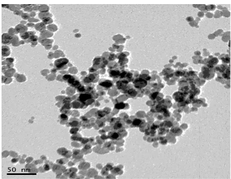
Figure 1. TEM image of superparamagnetic iron oxide nanoparticles.
the size and shape. The magnetic microspheres of SPIONs (with diameter size of 5 - 20 nm) in biocompatible, non-toxic (FDA approved) and biodegradable po-lymeric microspheres, such as popo-lymeric PLGA nanoparticles are recognized as desirable promising for application in spintronics and biomedicine.
The Zeta potential of SPIONs in this study around +45 mV, the zeta potential representing a surface charge of the particles in a colloidal suspension, is one of the most important factors defining their stability, a tendency to aggregate (thus defines them effective size), as well as their ability to bind serum proteins. In spite of the fact that most of them are charged negatively, the more positive the charge of a SPION is, the stronger its ability to bind serum proteins [8].
3.2. HSA-SPIONs Loaded PLGA Nanoparticles
Many studies have been studied further on the combination of PLGA nanopar-ticles with SPIONs and protein or vaccines or amongst others [3] [9]. Emulsifi-cation by soniEmulsifi-cation a preparation method to the drug-loaded systems of biode-gradable PLGA nano-carriers contains SPIONs and HSA. Parameter preparation for the formation of PLGA nanoparticles for protein delivery
In this study, we were informed about the effect of sonications on the beha-vior of synthesis biodegradable PLGA nanoparticles for protein delivery using a combination of SPIONs and HSA. We used various of sonication parameters on the each synthesis encapsulated processing.
pa-DOI: 10.4236/abc.2019.96014 184 Advances in Biological Chemistry
rameters and the power sonication accurately are crucial to produce the poly-meric HSA-SPIONs loaded PLGA nanoparticles.
The ultrasonic emulsification has been studied for decades and has recently garnered increased interest [10] [11]. The study compares the ultrasonic emulsi-fication with the dispersing rotors [11] [12] finding ultrasound to be competitive or even superior in terms of droplet size and energetic efficiency. Oil-in-water emulsions, the system is also found useful for biodegradable nanoparticles prep-arations using the solvent extraction/evaporation method. The sonication power is controlled by the transfer oscillation amplitude. To measure the power con-sumed for emulsification, the power intake of a high frequency generator was recorded using a standard household power monitor. For 100%, 80% and 60% of the maximum amplitude, the power intake is of 32 W, 25 W and 17 W, respec-tively. The actual power assessment transferred to the emulsion is usually done by measuring the heat taken by the emulsion, which for current ultrasonic flow-through cell will be difficult to do with reasonable accuracy. However, it makes sense to assume that the power consumption by the generator should be comparable to that delivered to the emulsion [13] [14]. PLGA nanoparticles, forming emulsion in ultrasonic flow-cell via rapid, oil-in-water emulsions, the particle sizes is increased with less sonication power, although the difference is less pronounced than observed for the oil emulsions. By increasing the concen-tration of the polymer solution, and hence its viscosity, larger particles are pro-duced. Obviously, viscosity is not the only physicochemical. Set the emulsifica-tion parameter, as it has been noted for the oil-in-water emulsion. Factors such as the tension of the interface and surfactants conformity are equally important, especially with regard to droplet coalescence.
The size and morphology of HSA-SPIONs loaded PLGA nanoparticles of this experiment is characterized by TEM. TEM image are used to obtain important information about the main size and morphology of nanoparticles. TEM is an important technique whose unique ability to probing the internal structure of individual nanoparticles.
The results TEM images of HSA-SPIONs loaded PLGA nanoparticles are dis-played interesting phenomenon information in the synthesis of HSA-SPIONs loaded PLGA nanoparticles processing with the sonication variation parameters.
Figure 2 to number 5 has provided information about TEM image 0.05 g/ml
HSA-SPIONs loaded PLGA nanoparticles which multple parameters of sonica-tion processing combinasonica-tions for mixture the PLGA + SPIONs + HSA solusonica-tions and in vitro release of HSA from HSA-SPIONs loaded PLGA nanoparticles. In this study, HSA-SPIONs loaded PLGA nanoparticles had a zeta potential be-tween −4.2 mV until −10.15 mV.
DOI: 10.4236/abc.2019.96014 185 Advances in Biological Chemistry
Figure 2. TEM images of HSA-SPIONs loaded PLGA nanoparticles with sonication step
in the first power of 20 W for 10 min, than the second power of 60 W for 4 min.
but from UV-Vis measurement results are notified that HSA does not contain in the polymeric PLGA nanoparticles.
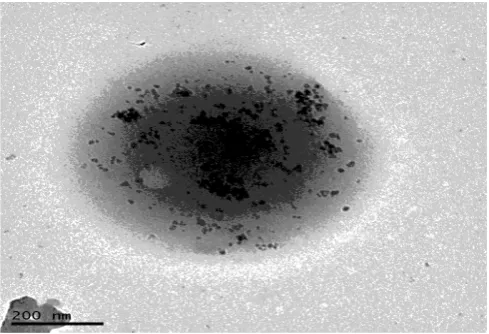
Another parameter of the sonication variation for PLGA + SPIONs + HSA solutions displayed TEM image Figure 3. PLGA + SPIONs + HSA is a mixture with sonication parameters: the first power of 60 W for 2 minutes and a second power of 20 W for 10 minutes in an ice bath. TEM image is performed polymer-ic 0.05 g/ml HSA-SPIONs-PLGA nanopartpolymer-icles on this parameter as a donut structure (Figure 3). PLGA + SPIONs + HSA solutions is mix with parameters similar to previous sonication parameters as in Figure 3, but the results are sig-nificant different obtained from UV-Vis measurement. Unfortunately, from the UV-Vis spectrometry, it does not contain HSA in the polymeric PLGA nanopar-ticles.
Figure 4 shows the result of sonication variation using the first power of 60 W
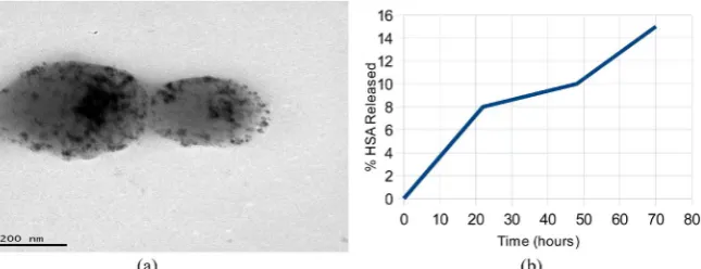
for 1 minute, than the second power of 20 W for 10 minutes in an ice bath. The TEM morphology image of nanoparticles in Figure 4(a) is displayed spherical. They have a heterogeneous structure. Polymeric HSA-SPIONs loaded PLGA nanoparticles have a donut-shaped structure, and another one has perfect round nanoparticle. UV-Vis measurement results indicate that the HSA in the PLGA nanoparticles only 50 percent. In vitro the release of HSA profile was observed, and all result of this study according to Gopferich et al. [14] about the release of protein. From biodegradable nanoparticles in Figure 4(a), HSA release around 5.5% to 120 hours (see Figure 4(b)).
Figure 5 performed TEM image of 0.05 g/ml HSA-SPIONs loaded PLGA
DOI: 10.4236/abc.2019.96014 186 Advances in Biological Chemistry
Figure 3. TEM images of HSA-SPIONs loaded PLGA nanoparticles with sonication step
[image:8.595.209.536.275.429.2]in the first power of 60 W for 2 min, than the second power of 20 W for 10 min.
Figure 4. TEM images of HSA-SPIONs loaded PLGA nanoparticles with sonication step
in the first power of 60 W for 1 min, than the second power of 20 W for 10 min (4a) and in vitro release of HSA from HSA-SPIONs loaded PLGA nanoparticles (4b).
Figure 5. TEM images of HSA-SPIONs loaded PLGA nanoparticles with sonication step
in the power of 20 W for 12 min (5a) and in vitro release of HSA from HSA-SPIONs loaded PLGA nanoparticles (5b).
4. Conclusion
[image:8.595.214.537.490.614.2]DOI: 10.4236/abc.2019.96014 187 Advances in Biological Chemistry
tion of sonication power in the processing of synthesis nanoparticles into a crucial-parameter obtains excellent results and stability in polymeric HSA-SPIONs loaded PLGA nanoparticles. These results have important implications for their poten-tial applications such as protein/drug delivery.
Acknowledgements
This study was supported by the Erasmus Mundus II EXPERTS III (SV).
Conflicts of Interest
The authors declare no conflicts of interest regarding the publication of this pa-per.
References
[1] Acharya, S. and Sahoo, S.K. (2011) PLGA Nanoparticles Containing Various Anti-cancer Agents and Tumour Delivery by EPR Effect. Advanced Drug Delivery Re-views, 63, 170-183.https://doi.org/10.1016/j.addr.2010.10.008
[2] Wang, Y., Ng, Y.G., Chen, Y., Shuter, B., Yi, J., Ding, J., Wang, S. and Feng, S.G. (2008) Formulation of Superparamagnetic Iron Oxides by Nanoparticles of Biode-gradable Polymers for Magnetic Resonance Imaging. Advanced Functional Mate-rials, 18, 308-318.https://doi.org/10.1002/adfm.200700456
[3] Vidawati, S., Barbosa, S., Taboada, P., Topete, A., Villar, E. and Mosquera, V. (2018) Study of Human Serum Albumin-SPIONs Loaded PLGA Nanoparticles for Protein Delivery. Advances in Biological Chemistry, 8, 91-100.
https://doi.org/10.4236/abc.2018.85008
[4] Wolf, M., Wirth, M., Pittner, F. and Gabor, F. (2003) Stabilisation and Determination of the Biological Activity of L-asparaginase in Poly(D,L-lactide-co-glycolide) Na-nospheres. International Journal of Pharmaceutics, 256, 141-152.
https://doi.org/10.1016/S0378-5173(03)00071-1
[5] Park, T.G., Lu, W. and Crotts, G. (1995) Importance of in Vitro Experimental Con-ditions on Protein Release Kinetics, Stability and Polymer Degradation in Protein Encapsulated Poly(D,L-lactic acid-co-glycolic acid) Microspheres. Journal of Con-trolled Release, 33, 211-222.https://doi.org/10.1016/0168-3659(94)00084-8
[6] Crotts, G. and Park, T.G. (1998) Protein Delivery from Poly(lactic-co-glycolic acid) Biodegradable Microspheres: Release Kinetics and Stability Issues. Journal of Mi-croencapsulation, 15, 699-713.https://doi.org/10.3109/02652049809008253
[7] Van de Weert, M., Hennink, W.E. and Jiskoot, W. (2000) Protein Instability in Poly(Lactic-Coglycolic Acid) Microparticles. Pharmaceutical Research, 17, 1159-1167. https://doi.org/10.1023/A:1026498209874
[8] Bravo-Osuna, I., Ponchel, G. and Vauthier, C. (2007) Tuning of Shell and Core Characteristics of Chitosan-Decorated Acrylic Nanoparticles. European Journal of Pharmaceutical Sciences, 30, 143-154.https://doi.org/10.1016/j.ejps.2006.10.007 [9] Topete, A., Melgar, D., Alatorre-Meda, M., Iglesias, P., Argibay, B., Vidawati, S.,
Barbosa, S., Costoya, J.A., Taboada, P. and Mosquera, V. (2014) NIR Light Active Hybrid Nanoparticles for Combined Imaging and Bimodal Therapy of Cancerous Cells. Journal of Materials Chemistry B, 2, 6967-6977.
https://doi.org/10.1039/C4TB01273A
DOI: 10.4236/abc.2019.96014 188 Advances in Biological Chemistry Emulsions of Lorazepam for Intravenous Injection. International Journal of Phar-maceutics, 216, 1-8.https://doi.org/10.1016/S0378-5173(00)00664-5
[11] Maa, Y.F. and Hsu, C.C. (2001) Performance of Sonication and Microfluidization for Liquid-Liquid Emulsification. Pharmaceutical Development and Technology, 4, 233-240.https://doi.org/10.1081/PDT-100101357
[12] Abismaı, B., Canselier, J.P., Wilhelm, A.M., Delmas, H. and Gourdon, C. (1999) Emulsification by Ultrasound: Drop Size Distribution and Stability. Ultrasonics Sonochemistry, 6, 75-83.https://doi.org/10.1016/S1350-4177(98)00027-3
[13] Ratoarinoro, F., Contamine, A.M., Wilhelm, J., et al. (1995) Power Measurement in Sonochemistry. Ultrasonics Sonochemistry, 2, S43-S47.
https://doi.org/10.1016/1350-4177(94)00010-P
[14] Loning, J.M., Horst, C. and Hoffmann, U. (2002) Investigations on the Energy Conversion in Sonochemical Processes. Ultrasonics Sonochemistry, 9, 169-179.