University of Warwick institutional repository:http://go.warwick.ac.uk/wrap
A Thesis Submitted for the Degree of PhD at the University of Warwick
http://go.warwick.ac.uk/wrap/73509
This thesis is made available online and is protected by original copyright. Please scroll down to view the document itself.
The effects of factors influencing human oocyte maturation upon
fertilization and
pre implantation
embryo development.
Jennifer Louise Cavilla BSc (Hons)
A thesis submitted for the degree of Doctor of Philosophy
Department of Biological Sciences University of Warwick
Contents
Contents List of Figures List of Tables Abbreviations Acknowledgements Dedication
Declaration Summary
Chapter 1 Introduction 1.1 Oocyte development
1.1.1 Overview
1.1.1.1 Summary of follicular growth 1.1.1.2 Follicle selection
1.1.1.3 Maturation of the dominant follicle 1.1.2 Meiosis
1.1.2.1 Meiosis I 1.1.2.2 Meiosis II
1.1.3 Key stages of developmental competence 1.1.3.1 Nuclear maturation
1.1.3.2 Cytoplasmic maturation 1.2 In vitro growth of follicles
1.3 The current state of IVM of human oocytes 1.3.1 Background
1.3.2 Content of media used for IVM 1.3.3 ICSI in human IVM programmes
1.3.4 Clinical pregnancies resulting from IVM 1.3.5 Role of prior stimulation in IVM
1.3.6 Different stimulation 1.3.7 Importance of PCO
1.3.8 The expectations and limitations of IVM 29
compared to conventional IVF
1.3 .8.1 Potential benefits of NM 29
1.3.8.2 Limitations of IVM 31
1.4 Polycystic Ovaries 32
1.4.1 Definition and diagnosis 32
1.4.2 Incidence 33
1.4.3 Pathogenesis of
peo
341.4.4 Complications with fertility treatment 36
1.5 Epidermal growth factor (EGF) 39
1.5.1 Discovery 39
1.5.2 Structure and synthesis 39
1.6 Meiosis activating sterol (MAS) 42
1.6.1 Discovery 42
1.6.2 Structure and synthesis 43
1.7 Project aims and objectives 48
Chapter 2 Materials and Methods 51
2.1 Media preparation 51
2.1.1 5% w/v Human serum albumin (hSA) 51
2.1.2 Earle's balanced salt solution (EBSS) 51
2.1.3 Phosphate buffered saline (PBS) 51
2.1.4 Follicle flushing medium 52
2.1.4.1 Patients undergoing transvaginal oocyte retrieval 52
for ICSI
2.1.4.2 Patients undergoing laparoscopic oocyte retrieval 52
2.1.5 In vitro maturation (IVM) medium 52
2.1.6 Sperm storage medium 53
2.1.7 Hyaluronidase 53
2.1.8 Minimal essential medium (MEM) 53
2.1.9 Embryo culture medium 54
2.1.9.1 PI medium with gentamycin 54
2.1.9.3 Gl and G2 medium 55
2.1.10 Pre-equilibration of mineral oil used in ICSI procedure 55
2.2 Oocyte maturation in vitro 55
2.2.1 EGF 55
2.2.1.1 EGF preparation and culture system 55
2.2.2 FF-MAS 56
2.2.2.1 FF-MAS preparation and culture system 56
2.3 Source and collection of oocytes 57
2.3.1 PCO patients 57
2.3.1.1 Recruitment of patients 57
2.3.1.2 Theatre procedure 57
2.3.1.3 Oocyte identification 58
2.3.1.4 Cumulus grading 58
2.3.1.5 Oocyte viability and maturity assessment 58
2.3.1.6 Oocyte allocation 59
2.3.2 ICSI patients 59
2.3.2.1 Recruitment 59
2.3.2.2 Theatre procedure 59
2.3.2.3 Oocyte identification and randomisation 59
2.3.2.4 Oocyte maturity assessment for ICSI 59
2.3.3 Collection of mouse oocytes 60
2.4 ICSI 61
2.4.1 Sperm preparation 61
2.4.2 Removal of cumulus cells 62
2.4.3 ICSI reagents and set up 62
2.5 Embryo culture 64
2.5.1 Embryo culture media 64
2.5.2. Morphological evaluation of embryo quality 64
2.5.3 Fluorescent staining of chromatin 65
2.6 Chromosome spreading 65
2.7 Immunocytochemistry 66
2.7.1 Fluorescent labelling of tubulin and chromatin 66
2.8.1 Set up and measurements 67
2.8.2 Oocyte diameter 67
2.8.3 Oocyte + zona diameter 67
2.8.4 Zona pellucida thickness 68
2.8.5 Perivitelline space (PVS) 68
2.9 Statistics 68
Chapter 3 Patient data 71
3.1 Introduction 71
3.2
pea
patients72
3.2.1 Clinical details of patients 72
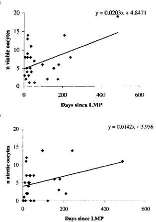
3.2.2 Relationship between age and number of oocytes collected. 74 3.2.3 Relationship between days since last menstrual period 75
(LMP) and the number of viable and oocytes collected.
3.2.4 Relationship between patient weight and the number of 76 viable and atretic oocytes collected.
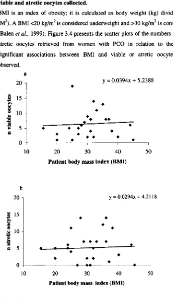
3.2.5 Relationship between patient body mass index (BMI) and 78 the number of viable and atretic oocytes collected.
3.3 ICSI patients 79
3.3.1 Clinical details of patients 79
3.4 Discussion 82
Chapter 4 Addition of EGF to culture of oocytes from PCO patients 87
4.1 Introduction 87
4.2 Aims 90
4.3 Patients and oocytes 4.3.1 Patients 4.3.2 Oocytes
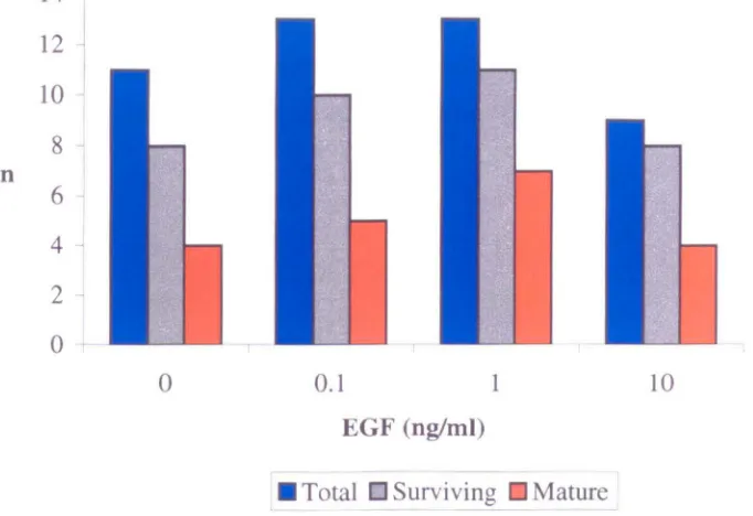
4.4 Effect of EGF on survival and maturation
4.5 Effects of EGF on fertilization and embryo development 4.6 Effect of cumulus cells on oocyte development
4.7 Discussion
Chapter 5 Effects of FF -MAS during in vitro maturation culture 10 1
5.1 Introduction 101
5.2 Aims 104
5.3 Patients and oocytes 104
5.3.1 Patients 104
5.3.20ocytes 104
5.4 Effect of FF-MAS on survival and maturation 104
5.5 Effects of FF-MAS on fertilization and embryo development 112
5.6 Effect of cumulus cells on oocyte development 116
5.7 Discussion 118
Chapter 6 Assessment of normality of maturation in vitro: 127
metaphase spindles and chromosome assessments
6.1 Introduction 127
6.2 Aims 134
6.3 Chromosome assessments of MI oocytes from patients 134
undergoing a cycle of ICSI treatment
6.3.1 Patients 134
6.3.20ocytes 134
6.3.3 Results from chromosome spreading 134
6.4 Effect of FF-MAS on spindle formation in in-vitro maturing 136
oocytes from patients undergoing a cycle of ICSI treatment
6.4.1 Patients 136
6.4.2 Oocytes 136
6.4.3 Spindle analysis 136
6.4.3.1 Mouse oocytes 136
6.4.3.2 Human oocytes 137
6.5 Discussion 145
Chapter 7 Image analysis 151
7.1 Introduction 151
7.2 Oocytes from pca patients, cultured with or without FF-MAS 154
7.2.2 Oocyte + zona diameter 7.2.3 Zona pellucida thickness
7.2.4 Perivitelline space (PVS) thickness
7.3 Oocytes from patients undergoing ICSI treatment, cultured with or without FF-MAS
7.3.1 Oocyte diameter
7.3.2 Oocyte
+
zona diameter 7.3.3 Zona pellucida thickness7.3.4 Perivitelline space (PVS) thickness 7.4 Discussion
Chapter 8 General Conclusions
Chapter 9 References
Appendices
166 171 176
180
180
188
192198
202209
217
Appendix 1- Figure Al Meiosis and follicular growth in the female a1
Appendix 11- Tables of media compositions a2-a7
Appendix III- Figure A2 Representative images of oocytes a8 demonstrating the variable extent of cumulus cover
Appendix IV-Figure A3 Image of oocyte demonstrating the various a9 parameters measured using the image analysis package
List of Figures
Figure 1.1 Figure 1.2 Figure 1.3 Figure 1.4 Figure 1.5 Figure 1.6 Figure 1.7 Figure 2.1 Figure 3.1 Figure 3.2 Figure 3.3 Figure 3.4Stages of folliculogenesis in the adult human ovary and level of atresia in the eight classes of growing follicles.
Diagrammatic illustration of the morphological features of nuclear maturation.
Images of the stages of human oocyte maturation in vitro.
Diagrammatic illustration of the activation of maturation promoting factor (MPF).
Linear view of the EGF receptor protein. Structural diagrams of FF-MAS and T -MAS.
MAS intermediates in the biosynthesis of cholesterol from lanosterol.
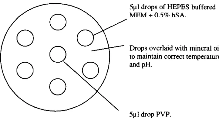
Diagram of microinjection dish as seen from above, used for IeSI procedure.
Scatter plots of the number of (a) viable and (b) atretic oocytes retrieved from unstimulated polycystic ovaries according to the age of the patient.
Scatter plots of the number of (a) viable and (b) atretic oocytes according to the number of days since the last menstrual period (LMP).
Scatter plots of the number of (a) viable oocytes and (b) atretic oocytes retrieved according to the weight of patients with peo.
Figure 4.1
Figure 4.2
Figure 4.3
Figure 4.4
Figure 5.1
Figure 5.2
Figure 5.3
In-vitro maturation of immature oocytes (n=46) collected from patients (n=8) with polycystic ovaries and cultured with or without epidermal growth factor (EGF).
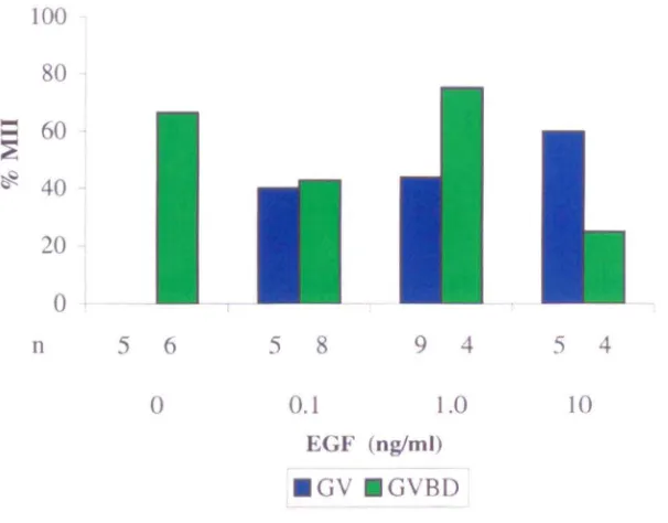
Maturation of germinal vesicle (GV) and germinal vesicle breakdown (GVBD) oocytes from patients with polycystic ovaries (PCO) in the presence or absence of epidermal growth factor (EGF).
Proportions of germinal vesicle (GV) and germinal vesicle breakdown (GVBD) oocytes becoming atretic in vitro after collection from polycystic ovaries (PCO) patients.
Levels of cumulus cover on oocytes on day-2 recovered from polycystic ovaries (PCO) patients and cultured in a) vehicle, b) O.lng/ml EGF, c) 1.Ong/ml EGF, d) IOng/ml EGF.
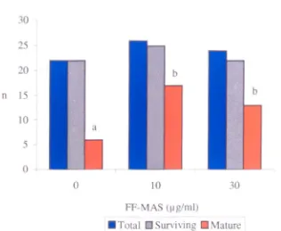
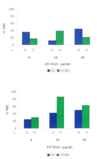
In-vitro maturation of immature oocytes (n= 128) collected from patients (n= 19) with polycystic ovaries and cultured with or without meiosis activating sterol (FF-MAS).
In-vitro maturation of immature oocytes (n=72) donated by patients undergoing intracytoplasmic sperm injection (ICSn (n=28) and cultured with or without meiosis activating sterol (FF-MAS).
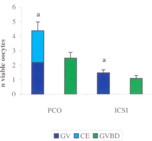
Maturity at collection of oocytes recovered from polycystic ovaries (PCO) and intracytoplasmic sperm injection (ICSn patients.
91
92
93
96
105
106
Figure 4.1
Figure 4.2
Figure 4.3
Figure 4.4
Figure 5.1
Figure 5.2
Figure 5.3
In-vitro maturation of immature oocytes (n=46) collected from patients (n=8) with polycystic ovaries and cultured with or without epidermal growth factor (EOP).
Maturation of germinal vesicle (OV) and germinal vesicle breakdown (OVBD) oocytes from patients with polycystic ovaries (PCO) in the presence or absence of epidermal growth factor (EOP).
Proportions of germinal vesicle (GV) and germinal vesicle breakdown (GVBD) oocytes becoming atretic in vitro after collection from polycystic ovaries (PCO) patients.
Levels of cumulus cover on oocytes on day-2 recovered from polycystic ovaries (PCO) patients and cultured in a) vehicle, b) O.lng/ml EOF, c)
I.Ong/ml EOF, d) IOng/ml EOP.
In-vitro maturation of immature oocytes (n=128) collected from patients (n=19) with polycystic ovaries and cultured with or without meiosis activating sterol (FF-MAS).
In-vitro maturation of immature oocytes (n=72) donated by patients undergoing intracytoplasmic sperm injection (lCSn (n=28) and cultured with or without meiosis activating sterol (FF-MAS).
Maturity at collection of oocytes recovered from polycystic ovaries (PCO) and intracytoplasmic sperm injection (lCSn patients.
91
92
93
96
105
106
Figure 5.4 Figure 5.5 Figure 5.6 Figure 6.1 Figure 6.2 Figure 6.3 Figure 6.4 Figure 6.5 Figure 6.6
Maturation of germinal vesicle (GV) and germinal vesicle breakdown (GVBD) O()Cytes from (a) patients with polycystic ovaries (PCO) and (b) patients undergoing intracytoplasmic sperm injection (lCSI) in the presence or absence of meiosis activating sterol (FF-MAS).
Proportions of germinal vesicle (GV) and germinal vesicle breakdown (GVBD) oocytes becoming atretic in vitro after collection from polycystic ovaries (PCO) or intracytoplasmic sperm injection (ICSI) patients.
Cumulative time course of maturation in vitro for maturing oocytes from polycystic ovaries (PCO) and intracytoplasmic sperm injection (lCSI) patients. Spread of chromosomes from a human oocyte at first meiotic metaphase, during maturation in vitro. Mouse oocytes attained with FITC-Iabelled anti-tubulin antibodies (green) and DAPI (blue).
Nuclear staining with Hoechst 33258 of human oocytes that arrested at the GV stage after culture in control conditions.
Combined DAPI and FITC fluorescence in a mature human oocyte cultured in 30~g/ml FF-MAS, at anaphase of MIl.
Examples of abnormal spindles in three human oocytes matured in vitro in the presence of 30~glml FF-MAS.
Example of a normal shaped spindle in an oocyte that failed to fertilize.
Figure 7.1
Figure 7.2
Figure 7.3
Figure 7.4
Frequency histogram showing mean oocyte diameters on day of collection (day-2) of oocytes from unstimulated patients with PCO.
a) Viable oocytes
b) Oocytes becoming mature in vitro
c) Oocytes that progressed to maturity expressed as a percentage of viable oocytes on day-2
Frequency histogram showing oocyte diameters for patients having unstimulated polycystic ovaries on day 0 (day of ICSI for mature oocytes)
a) Immature or atretic oocytes b) Mature oocytes
c) Oocytes that progressed to maturity expressed as a percentage of viable oocytes on day O.
Diameters of in vitro matured oocytes on day 0 obtained from unstimulated patients with PCO in relation to their competence to fertilize and cleave. a) Frequency histogram of diameters of in vitro matured oocytes on day O.
b) Proportion of mature oocytes fertilizing (2pn) according to their diameter on day O.
c) Frequency histogram of fertilized oocytes undergoing cleavage or arresting at the 2pn stage according to the oocyte diameter on day O.
Relationship between time and oocyte diameter during in vitro culture in a) control, b) lOJJg!ml FF-MAS and c) 30,..g!ml FF-FF-MAS.
158
160
163
Figure 7.5 Figure 7.6 Figure 7.7 Figure 7.8 Figure 7.9 Figure 7.10 Figure 7.11
Frequency histogram showing mean oocyte diameters (including zona) on day of collection (day-2) of oocytes from unstimulated patients having PCO
a) Viable oocytes
b) Oocytes becoming mature in vitro
Frequency histogram showing mean oocyte diameters (including zona) on day of ICSI (day 0) of oocytes from unstimulated patients having PCO. a) Immature or atretic oocytes
b) Mature oocytes
Frequency histogram of fertilized oocytes obtained from unstimulated patients with PCO. undergoing cleavage or arresting at the 2pn stage according to the oocyte
+
zona diameter on day O.Relationship between time and oocyte + zona diameter during in vitro culture in a) control, b) 10J.lg/ml FF-MAS and c) 30jJg/ml FF-MAS.
Frequency histogram showing mean zona pellucida thickness on day of collection (day-2) of oocytes from unstimulated patients having PCO
a) Viable oocytes
b) Oocytes becoming mature in vitro
Frequency histogram showing mean zona pellucida thickness on day 0 (day of ICSI for mature oocytes) of oocytes from unstimulated patients having PCO a) Immature or atretic oocytes
b) Mature oocytes
Relationship between time and zona pellucida thickness during in vitro culture in a) control, b)
10jJg/ml FF-MAS and c) 30J.lg/ml FF-MAS.
Figure 7.12
Figure 7.13
Figure 7.14
Figure 7.15
Figure 7.16
Frequency histogram showing mean perivitelline space on day of collection (day-2) of oocytes from unstimulated patients with PCO.
a) Viable oocytes
b) Oocytes becoming mature in vitro
Frequency histogram showing mean perivitelline space on day 0 (day of ICSI for mature oocytes) of oocytes from unstimulated patients with PCO.
a) Immature or atretic oocytes b) Mature oocytes
Relationship between time and mean perivitelline space (PVS) during in vitro culture in a) control, b) lOJ! glml FF-MAS and c) 301-1 glml FF-MAS collected on day-2 from patients with PCO.
Frequency histogram showing mean oocyte diameters on day of collection of immature oocytes from patients undergoing ICSI treatment.
a) Viable oocytes
b) Oocytes becoming mature in vitro
c) Oocytes that progressed to maturity expressed as a percentage of viable oocytes on day of collection. Frequency histogram showing mean oocyte diameters for patients undergoing ICSI treatment on day 0 (day of ICSI for mature oocytes)
a) Immature or atretic oocytes b) Mature oocytes
c) Oocytes that progressed to maturity expressed as a percentage of viable oocytes on day O.
177
178
179
181
Figure 7.17
Figure 7.18
Figure 7.19
Figure 7.20
Figure 7.21
Diameters of in vitro matured oocytes on day 0 obtained from patients undergoing IeSI in relation to their competence to fertilize and cleave.
a) Frequency histogram of diameters of in vitro matured oocytes on day O.
b) Proportion of mature oocytes fertilizing (2pn) according to their diameter on day O.
c) Frequency histogram of fertilized oocytes undergoing cleavage or arresting at the 2pn stage according to the oocyte diameter on day
O.
Numbers of oocytes donated by patients undergoing IeSI treatment, fertilizing and cleaving after maturation in vitro according to oocyte diameter a) Oocytes that matured in 23-24 hr
b) Oocytes that matured in 46-48 hr
Relationship between time and oocyte diameter during in vitro culture in a) control, b) 1OJ.1g/ml FF-MAS and c) 30J.1g/ml FF-FF-MAS.
Frequency histogram showing mean oocyte diameters (including zona) on day of collection of oocytes from patients undergoing IeSI treatment. a) Viable oocytes
b) Oocytes becoming mature in vitro
Frequency histogram showing mean oocyte diameters (including zona) on day of IeSI (day 0) of oocytes from patients undergoing IeSI treatment. a) Immature or atretic oocytes
b) Mature oocytes
184
185
187
189
Figure 7.22
Figure 7.23
Figure 7.24
Figure 7.25
Figure 7.26
Frequency histogram of fertilized o<>cytes obtained from patients undergoing ICSI treatment, undergoing cleavage or arresting at the 2pn stage according to the oocyte + zona diameter on day O. a) Oocytes that matured in 23-24 hr
b) Oocytes that matured in 46-48 hr
c) Oocytes that fertilized or fertilized and cleaved, irrespective of the time taken to mature in vitro Frequency histogram showing mean zona pellucida thickness on day of collection of oocytes from patients undergoing ICSI treatment
a) Viable oocytes
b) Oocytes becoming mature in vitro
c) Oocytes that progressed to maturity expressed as a percentage of all viable o<>cytes on day of collection
Frequency histogram showing mean zona pellucida thickness on day 0 (day of ICSI for mature o<>cytes) of oocytes from patients undergoing ICSI treatment. a) Immature or atretic oocytes
b) Mature oocytes
Relationship between time and zona pellucida thickness during in vitro culture in a) control, b) 101Jg/ml FF-MAS and c) 30IJg/ml FF-MAS. These oocytes were collected from patients undergoing ICSI treatment.
Frequency histogram showing mean perivitelline space on day of collection of oocytes from patients undergoing ICSI treatment.
a) Viable o<>cytes
b) Oocytes becoming mature in vitro
191
194
195
196
Figure 7.27
Figure 7.28
Figure Al Figure A2
Figure A3
Frequency histogram showing mean perivitelline space on day 0 (day of ICSI for mature oocytes) of oocytes from patients undergoing ICSI treatment. a) Immature or atretic oocytes
b) Mature oocytes
Relationship between time and mean perivitelline space (PVS) during in vitro culture in a) control, b) lOpglml FF-MAS and c) 30pglml FF-MAS collected from patients undergoing ICSI treatment.
Meiosis and follicular growth in the female
Representative images of oocytes demonstrating the variable extent of cumulus cover
Image of oocyte demonstrating the various parameters measured using the image analysis package
200
201
al a8
List of Tables
Table 3.1 Table 3.2 Table 3.3 Table 3.4 Table 4.4 Table 5.1 Table 5.2 Table 5.3Clinical details of patients with polycystic ovaries (PCO) who donated immature oocytes for culture with or without EGF.
Clinical details of patients with polycystic ovaries (PCO) who donated immature oocytes for culture with or without FF-MAS.
Clinical details of patients undergoing intracytoplasmic sperm injection (ICSI) treatment who donated immature oocytes for culture with or without FF-MAS, chromosome assessment or for culture with or without FF-MAS for spindle analysis. Aetiology of infertility for patients (n=57) undergoing ICSI treatment who donated immature oocytes for this study.
Fertilization and cleavage rates of in-vitro matured oocytes arising from polycystic ovaries (PCO) patients.
Fertilization rates of in-vitro matured oocytes arising from patients with polycystic ovaries (PCO) or those being treated with intracytoplasmic sperm injection (ICSI).
Cleavage rates of in-vitro matured oocytes arising from patients with polycystic ovaries (PCO) or those being treated with intracytoplasmic sperm injection (lCSI).
Stage and grade of embryos arising from fertilization of in vitro matured oocytes retrieved from patients with polycystic ovaries (PCO) or those being treated with intracytoplasmic sperm injection (lCSI).
Table 5.4 Table 5.5 Table 5.6 Table 6.1 Table 6.2 Table 7.1 Table 7.2 Table 7.3 Table 7.4 Table 7.5
Polar body types observed in mature oocytes obtained from patients with PCO, cultured with or without FF-MAS
Polar body types observed in mature oocytes donated by patients undergoing ICSI treatment, cultured with or without FF-MAS
Effects of cumulus presence at collection (day-2) of oocytes recovered from patients with polycystic ovaries (PCO) and cultured in 30).1g1ml FF-MAS,
10).1g1ml FF- MAS or control.
Outcome of in vitro culture with or without FF-MAS of oocytes donated by patients undergoing ICSI treatment.
Chromatin staging of non-matured oocytes donated by patients undergoing ICSI treatment and cultured with or without FF-MAS.
Control oocyte measurements.
Total number of oocyte measurements available for each parameter measured on the day of oocyte collection and on day 0 (day of insemination of mature oocytes) for both patient groups.
Effects of the levels of cumulus cover on oocytes at collection (day-2) recovered from patients with PCO on outcome of in vitro culture.
Mean diameters of oocytes from patients with PCO in relation to the outcome of in vitro culture according to cumulus grade at collection (day-2) Zona pellucida thickness on day+ 1 and day+2 of oocytes collected from patients with PCO that fertilized after ICSI insemination on day+ I and those that failed to fertilize or became atretic.
Table 7.6
Table Al
Table A2 Table A3 Table A4
Table A5
Table A6
Table A7
Zona pellucida thickness on day+ 1 and day+2 of oocytes collected from patients undergoing ICSI treatment according to whether they fertilized or failed to fertilize/became atretic.
Composition ofEBSS lOx (Gibco) Composition of PBS (Gibco)
Composition of Hams FlO (ICN Biomedicals) Composition ofMl99 2x (Gibco)
Composition of PI medium with gentamycin (Irvine Scientific).
Composition of S 1 and S2 media (Scandinavian IVF Science)
Composition of Gardner's G 1.2 and G2.2 sequential media (Scandinavian IVF Science)
197
a2
a2 a2-3
a4 as
a6
Abbreviations
ART Assisted reproductive technology
BMI Body mass index
bp cAMP CG CMT d DMAP EBSS EGF ERK FF-MAS FGF FSH GnRHa GV GVBD g hCG hr hSA ICSI IGF-l IV IVF IVM KB kDa Base pairs
Cyclic adenosine monophosphate Degrees centigrade
Cortical granule
Cytoplasmic microtubule day
6-Dimeth y laminopurine Earles balanced salt solution Epidermal growth factor Extracellular regulated kinase Meiosis activating sterol Fibroblast growth factor Follicle stimulating hormone
Gonadotrophin releasing hormone agonist Germinal vesicle
Germinal vesicle breakdown Gram
Human chorionic gonadotrophin Hour
Human serum albumin
Intracytoplasmic sperm injection Insulin-like growth factor-type I International units
In vitro fertilization In vitro maturation Kilobase
Kilodalton Litre
MAP kinase Mitogen activated protein kinase MIS Meiosis-inducing substance
MPF Maturation (or M-phase) promoting factor
MIl Metaphase II
~g Microgram
~I ~m ~M mg mI min M MT MTOC ng nM OHSS PI % pb PBS PCO
pcos
PDGF PN PVS RNA mRNA RT TGF-a VEGF v/v w/v Microlitre Micrometre Micromolar Milligram Millilitre Minute Molar MicrotubuleMicrotubule organizing center Nanogram
Nanomolar
Ovarian hyperstimulation syndrome Prophase I
Percentage polar body
Phosphate buffered saline Polycystic ovaries
Polycystic ovarian syndrome Platelet-derived growth factor Pronuclei
Perivitelline space Ribonucleic acid Messenger RNA Room temperature
Transforming growth factor- a Vascular endothelial growth factor Volume/volume
Acknowledgements
I would like to express my sincere thanks to my supervisor Geraldine Hartshorne. I
am
most grateful for her patience, supervision and unfailing support throughout my PhD studies and during the preparation of this thesis.The considerable help, support & encouragement for the entirety of my PhD from all my colleagues at the CRM, particularly the lab staff are gratefully acknowledged.
Hazel Baker performed the mouse '2 cell to blastocyst' biotoxicity assay and Animal House Personnel performed the mice intraperitoneal injections of 6-12 IU hCG. Anne-Grete Byskov & Mogens Baltsen generously prepared and supplied the FF-MAS. Special thanks to Mr. Richard Kennedy and Mr. Lucas Klentzeris for performing the laparoscopic oocyte collections. My thanks are also due to Charles Tees for his help with the chromosomal and spindle analysis. From the Statistics department, I
am
grateful to Fotios Siannis for his advice and guidance.I would like to express my sincere thanks and gratitude to Mr. Richard Kennedy and the R&D Committee for the financial support of these studies. For invaluable computer assistance, my personal thanks go to Mike Swann. I would also like to thank my "little sister" Sarah for her speedy and helpful retrieval of last minute references.
Dedication
This thesis is dedicated to my late father, without his love, faith & support I would never have achieved the things that I feel privileged to have been able to.
Declaration
All the results in this thesis were obtained as a result of original work by the author, under the supervision of Dr. Geraldine Hartshorne, unless otherwise stated in the text. All sources of information have been acknowledged by means of reference. None of the work contained in this thesis has been submitted for any previous degree. A paper published as a result of this work is presented in Appendix V.
Summary
The competence of oocytes to mature and undergo fertilization and embryonic development is known to be influenced by the conditions under which their maturation occurs. Suboptimal maturation in vitro currently limits the use of immature oocytes for embryo creation. This project examines the relationship between the conditions of in vitro maturation of human oocytes and aspects of their subsequent developmental competence through the in vitro creation and analysis of research embryos. This work is essential in defining effective and safe conditions for the use of human immature oocytes in programmes of clinical treatment to alleviate infertility.
Human immature oocytes were exposed in vitro to various concentrations of meiosis activating sterol (FF-MAS), an endogenous mediator of follicle and oocyte function, or epidermal growth factor (EOP), in the absence of other hormonal support. Their survival and further development relative to controls were measured by assessing the proportions maturing, fertilizing by sperm injection (ICSI), and cleaving in vitro. Image analysis was used to measure various dimensions of oocytes and embryos daily. A pilot study of chromosome and spindle configurations at meiotic metaphase was also conducted.
The major findings of this project are that FF-MAS supplementation of maturation medium has different positive effects upon immature oocytes arising from patient groups having different endocrine profiles and yielding differing oocyte populations. FF-MAS at 30J.lg/ml promotes survival of oocytes from unstimulated patients with polycystic ovaries (p<O.02S) and promotes maturation of oocytes from gonadotrophin-stimulated patients undergoing ICSI treatment (p<O.OS). A response to FF-MAS in terms of oocyte growth was evident in immature oocytes from both types of patients (p<O.05). Mature, immature and atretic oocytes from patients with
pca
became significantly different in terms of oocyte diameter when cultured in FF-MAS (10 or 30J.lg/ml), contrasting with those cultured in control conditions, or obtained from patients undergoing ICSI treatment. However, significant (p<O.OS) enlargement of oocyte diameter between oocyte recovery and the day of insemination occurred when oocytes from patients undergoing ICSI were cultured with FF-MAS.The proportion of mature oocytes fertilized by ICSI was not affected by FF-MAS in the maturation culture but there was a non-significant tendency towards an improved chance of cleavage when FF-MAS was present. Embryonic development beyond early cleavage was not observed, despite conditions supportive of blastocyst production from in vivo matured oocytes, suggesting that developmental competence remained compromised even in the presence of FF-MAS. A pilot study to ascertain whether the incidence of spindle defects or chromosome irregularities in in vitro matured metaphase oocytes was influenced by the presence of FF-MAS gave equivocal results.
Chapter 1
Introduction
''The shortage of human immature oocytes for research is the limiting factor in developing methods for using them in clinical practice" (Coskun et al., 1998).
This thesis examines the maturation of human oocytes in vitro under a variety of conditions. This topic is important because little is known about human oocytes despite their widespread collection and use in infertility treatments. Oocytes are key cells, which embody the ovarian reserve of female gametes. They are laid down in early development and their quality is critical for embryo formation and the successful continuation of the human species.
In 1935, Pincus and Enzmann for the first time observed in mammals that intact oocytes, isolated from antral follicles, proceed through meiotic maturation spontaneously. In 1939, Pincus and Saunders liberated rabbit and human oocytes from their follicles in vitro and demonstrated how both matured spontaneously in less than 12 hours. This was originally considered as potentially the most appropriate way to obtain mature oocytes for in vitro fertilization (IVF) techniques (Edwards, 1965a). In retrospect, the time allowed for maturation of oocytes in vitro, based on the data of Pincus and Saunders (1939), was probably inadequate, judging from the work of Edwards (1965b).
1.1 Oocyte development
1.1.1 Overview
1.1.1.1 Summary of follicular growth
The development of oocytes that are competent to become functional embryos is a prolonged process, starting in fetal life, when a stock of primordial follicles is formed. The oocytes are stored arrested at an immature stage of meiotic prophase, and are incompetent to develop further unless the follicle grows and provides an environment conducive to support full development. Ovulation does not occur until after puberty. In the human female, approximately seven million primordial follicles are initially formed; at birth this number has declined to 1-2 million primordial follicles, of these approximately 400 ovulate throughout the reproductive life, the rest undergoing atresia (Baker, 1963; Salha et al., 1998; Hardy et al., 2000). In one menstrual cycle of a mature woman, normally only a single dominant follicle achieves ovulation.
There are three successive steps in the production of a dominant follicle (Salha et al., 1998)
• Growth of a daily cohort of primordial follicles.
• Recruitment of several small antral follicles by waves of pituitary gonadotrophins.
• Selection and maturation of the preovulatory follicle.
Figure 1.1 provides a diagrammatic illustration of follicular growth in the human. Although follicle growth is a continuous process occurring from early infancy until the end of the reproductive phase, follicles do not attain ovulatory sizes or produce significant quantities of oestrogen during childhood due to the absence of FSH and LH stimulation. At puberty, coordinated cyclic gonadotrophin stimulation initiates the menses and results in oocyte maturation and ovulation at midcycle (Salha et al., 1998).
secreted by the oocyte; it separates the oocyte from the surrounding granulosa cells and is penetrated by cytoplasmic processes from the granulosa cells, which interact with the gap junctions. The gap junctions form at the points where the cytoplasmic processes contact the oocyte surface, transferring nutrients and regulatory molecules into the oocyte and also between adjacent granulosa cells, providing the basis for intercellular communication and potential syncytial coordination of the follicle (Downs, 1995).
At the primordial follicle stage, the oocyte measures approximately 30flm diameter (Gosden et ai., 1987). Oocyte growth and increased RNA polymerase activity is indicative of follicle growth initiation (Gosden et ai., 1993), and is followed by clonal and radial expansion of pregranulosa cells. At the preantral stage, the enlarged oocyte is encapsulated by >2 granulosa cell layers in close proximity without an antrum.
Figure 1.1 Stages of folliculogenesis in the adult human ovary and level of atresia in the eight classes of growing follicles.
The granulosa cell (gc) numbers and their corresponding estimated follicle diameter indicate the limits of each class.
1.1.1.2 Follicle selection
The follicle selected to ovulate (dominant follicle) is normally the largest healthy follicle with a diameter of 5.5-8.2mm at the start of the follicular phase (Gougeon and Lefevre, 1983). Its granulosa cells are most responsive to FSH, and modulation of FSH sensitivity may be achieved by local growth factor involvement, for example, a timely and selective activation of insulin-like growth factor-l (lGF-1) in the chosen follicle, might promote cytodifferentation and proliferation of granulosa cells (Adashi, 1993).
The maturing follicle that achieves dominance does so via feedback inhibition of FSH secretion from the pituitary gland and increased sensitivity to FSH. The latter, combined with the fact that the dominant follicle requires less FSH to maintain preovulatory growth than is required to stimulate follicular growth at an earlier stage, enables the dominant follicle to inhibit the development of less mature follicles, without inhibiting its own growth (Salha et al., 1998).
1.1.1.3 Maturation of the dominant follicle
During the follicular phase, the preovulatory follicle increases in size by both cellular multiplication and fluid accumulation in the antrum until the ovulatory LH surge. The dominant follicle begins to synthesize systemically detectable oestradiol from the mid-follicular phase. As follicle maturation occurs, the granulosa cells acquire LH receptors in response to FSH stimulation, conferring responsiveness of large follicles but not smaller ones to LH. As preovulatory follicular maturation continues, the vascularity of the follicle increases, thereby increasing the deli very of gonadotrophin to the maturing follicle as well as the systemic availability of its products, notably steroids (Zeleznik et at., 1981).
1998) and peripheral migration of cortical granules (Hartshorne, 1999). Cumulus expansion is a result of hyaluronic acid-rich proteoglycan matrix secretion by the cumulus cells, causing the dispersal of cumulus cells within the matrix, which are ovulated approximately 37 hr after the LH surge along with a mature MIl-arrested oocyte. This expanded matrix is a critical factor for reproductive function, since it holds the oocyte in position together with its cumulus cells, facilitates follicular extrusion and oviductal fimbria capture, facilitates sperm penetration and fertilization (Salustri et al., 1996; Matzuk, 2000). Potential roles in communication with the oviductal wall via sloughed cumulus cells may also be envisaged.
1.1.2 Meiosis
Meiosis is termed cell reduction; it is a form of cell division occurring only in the formation of gametes, where the number of chromosomes is reduced to a haploid number (in humans, n=23). In addition to halving the chromosome number, meiosis serves to promote genetic variation, through the mixing of maternal and paternal chromosomes and genes.
Following several mitotic divisions in the developing fetus, oogonia enter meiosis during the early second trimester of pregnancy. Meiosis is comprised of two stages: meiosis I and II, as illustrated in Figure Al (Appendix I).
1.1.2.1 Meiosis I
Meiosis I begins in the fetal ovary and has a complex extended prophase stage, which is divided into four sub-stages (Briggs et al., 1999).
Prophase I
• Leptotene- chromosomes condense, and are attached to nuclear membrane. • Zygotene- homologous chromosomes align and homologous loci synapse. The
synaptonemal complex forms, allowing sites for the exchange of genetic material (recombination). Bivalents (sets of synapsed homologues) result. • Pachytene- shortening and thickening of bivalents, genetic recombination
occurs between chromatids of paired homologous chromosomes.
to ovulation or degeneration. A stable conformation of the nucleus known as a germinal vesicle is present in the oocyte.
All immature oocytes are diploid (2n) with a 4C DNA content. Progress beyond diplotene of meiotic prophase I occurs only in maturing oocytes or those in the stages of atresia where control of the oocyte by surrounding somatic cells is lost.
Metaphase I
A spindle forms at the periphery; chromosomes are randomly aligned on the equatorial plate prior to centromere repulsion and chromosome separation.
Anaphase I
The chromosomes move to opposite ends of the spindle. Telophase I
Cell division results in the formation of daughter cells. Formation of the secondary oocyte with expulsion of first polar body (pb) (containing a set of chromosomes). The division is asymmetric due to the peripheral position of the spindle, resulting in a minimal reduction in volume of the oocyte.
1.1.2.2 Meiosis II
There is no replication of DNA or formation of a nucleus after completion of meiosis I, the cell progresses directly to MIl (Veeck, 1991).
Metaphase II
A spindle reforms, the 23 chromosomes (each two chromatids) align on the equatorial plate. There is a diploid amount of DNA, but a haploid set of chromosomes, as the strands have not yet separated. The second meiotic arrest occurs and the oocyte is ovulated in this stage. Meiosis II is resumed only upon sperm penetration or parthenogenetic activation.
Anaphase II
The chromatids split at the centromeres, moving to the opposite ends of the spindle. Telophase II
1.1.3 Key stages of developmental competence
"It is only by considering all aspects of maturation that the production in vitro of mammalian eggs with a developmental potential equivalent to that of in-vivo
counterparts will be achieved" (Fulka et al., 1998).
Human oocyte maturation is defined as the reinitiation and completion of the first
meiotic division from the germinal vesicle (GV) stage (prophase I) to MIl, with accompanying cytoplasmic maturation for fertilization and early embryonic
development (Cha and Chian, 1998). Although the nuclear and cytoplasmic
programmes can proceed as independent processes, the acquisition of full
developmental competence is conferred only when the two processes are closely
integrated (Moor et al.,
1998;
Fulka et al.,1998).
According to Barnesand
Sirard (2000), the acquisition of developmental competence via reinitiation of meiosis isprobably a common signaling or differentiation pathway that occurs in the oocyte
and/or associated granulosa, regardless of whether the oocyte is destined to ovulate or
degenerate.
1.1.3.1 Nuclear maturation
Nuclear maturation refers to the resumption of meiosis from prophase I arrest (PI) and the progression of meiosis to its next physiological arrest point (Mm.
Morphological evidence of resumption of meiosis is the disappearance of the oocyte's
nucleolus and nuclear (GV) envelope, a process termed germinal vesicle breakdown
(GVBD) (Eppig, 1996).
Nuclear maturation can be morphologically evaluated by observation of the extrusion
of the first pb (Smitz and Cortvrindt, 1999) as illustrated in Figure 1.2.
Immature oocytes: GVBD MI Mature oocyte:
Prophase I MIl
Figure 1.2 Diagrammatic illustration of the morphological features of nuclear maturation.
Figure 1.3 shows images of oocytes at the various stages of maturation during in vitro culture.
a b
GV
d
Many of the proteins that regulate mitosis also regulate meiosis. The G2 to M-phase
transition in fully-grown oocytes is driven by maturation (or M-phase) promoting factor (MPF)- the primary molecule involved in meiotic cell cycle progression (Eppig, 1996; Fulka et al., 1998; Briggs et al., 1999; Trounson et al., 2(01).
Maturation (or M-phase) promoting factor (MPF)
MPF is a serine-threonine kinase protein heterodimer composed of a regulatory subunit, cyclin B and a catalytic subunit p34cdC2; see Figure 1.4 for diagrammatic
representation of the molecular structure.
The two molecules cyclin B and p34cdC2 become associated, and dephosphorylation of p34cdC2 occurs on residues tyrosine-15 and threonine-14, catalysed by a phosphatase encoded by the cdc25 gene (Gautier et al., 1991; Gabrielli et al., 1992; Izumi and Maller, 1993; Eppig, 1996; Fulka et al., 1998; Trounson, 2(01). Active MPF is required for the initiation of nuclear maturation and the condensation of metaphase I chromosomes, MPF activity is detected before or coincident with GVBD. MPF is activated in vivo by the endogenous LH surge at the onset of oocyte maturation. Trounson and colleagues (2001) demonstrated the activation of MPF activity in IVM studies using human oocytes from unstimulated ovaries, leading them to conclude that the activation of MPF in vitro occurs in a manner similar to that for oocytes in vivo.
Entry into anaphase I correlates with the inactivation of MPF, probably caused by proteolytic degradation of the cyclin unit of MPF (Murray et al., 1989). Entry into Mil requires a second increase in active MPF activity, c-mos maintains a high level of MPF, thereby arresting cell cycle progression until fertilization. Sperm penetration and the resultant increase in intracellular Ca2+ levels induce cyclin degradation and
completion of meiosis (Fulka et al., 1998; Trounson, 2(01).
To summarize, oocytes acquire competence to complete nuclear maturation in at least two sequential steps (Eppig, 1996).
Thr-16
o
Inacti velPre-MPF
p34Cdc2 dephosphorylation
I
Cdc250
Active MPF
Figure 1.4. Diagrammatic illustration of the activation of maturation promoting factor (MPF).
Adapted from Eppig (1996) and Trounson et al. (2001).
Additional factors, which may affect meiotic progression, include MAP kinase and
cAMP.
Mitogen activated protein kinase (MAP) kinase
MAP kinase is a serine-threonine kinase, alternatively known as extra-cellular
regulated kinase (ERK) (Trounson, 2(01); that is activated via a protein kinase cascade at the onset of oocyte maturation in mouse (Verlhac et aI., 1993), pig (Inoue et al., 1995) and Xenopus (Haccard et aI., 1990). MAP kinase, although activated at
the onset of oocyte maturation in mice (Trounson, 2(01) is not necessarily required for OVBD in mice (Sun et aI., 1999b; Trounson, 2(01).
In human oocytes, p42ERK2 is the main form of MAP kinase (Sun et aI., 1999a; Trounson, 2(01). MAP kinase has not been widely studied in human oocytes, although it is known to be inactive in immature oocytes, active in mature oocytes and
there is a decrease in activity after the formation of pronuclei following fertilization
Cyclic adenosine monophosphate (cAMP)
cAMP participates in meiotic arrest and may prevent GVBD by the inhibition of MPF activation, or by the down regulation of p34cdC2 dephosphorylation. Once cAMP decreases below a threshold level, meiosis can resume.
Two other purines, hypoxanthine and adenosine, have also been shown to prevent GVBO in vitro by promoting elevated cAMP levels in the oocyte. It is thought that these purines enter the oocyte via the gap junctions. Hypoxanthine prevents the degradation of cAMP and adenosine promotes the generation of cAMP by stimulating adenylate cyclase, acting as a substrate for cAMP production (Epigg, 1996; Briggs et al., 1999).
In response to the LH surge at ovulation, gap junctions are lost following mucification of the granulosa cells, resulting from hyaluronic acid production, this causes a decrease in the levels of cAMP, below the threshold required to maintain GV status (Briggs et al., 1999).
1.1.3.2 Cytoplasmic maturation
Cytoplasmic maturation refers to the processes that prepare the oocyte for fertilization, activation, formation of PN and preimplantation development. Similarly to nuclear maturation, competence to undergo cytoplasmic maturation is acquired in sequential steps by GV-stage oocytes. Although nuclear and cytoplasmic maturation occur in synchrony, the acquisition by GV-stage oocytes of competence to undergo cytoplasmic maturation is independent of competence to complete nuclear maturation. Most deficiencies in oocytes during maturation are believed to be associated with cytoplasmic reprogramming rather than with meiotic progression; and the effects of cytoplasmic aberrations are normally associated with cleavage, blastulation and the peri-implantation stages, rather than early development (Moor et ai., 1998).
Protein synthesis
during oocyte maturation is stored in a stable, dormant form until after GVBD (Bachvarova, 1985). Moor et al. (1998) suggested that the reduced developmental potential in human oocytes matured in vitro might be attributable to abnormal cytoplasmic maturation, as well as sub-optimal culture conditions and incomplete growth. Trounson (2001) studied the protein content of in vitro matured MIl human oocytes, obtained from unstimulated ovaries. A reduced protein content was observed compared to in vivo matured MIl oocytes retrieved from stimulated ovaries. The proteins not detected in oocytes matured in vitro probably include molecules essential for cell cycle regulation and normal embryo development.
Inositol lipids and calcium
Sperm binding and fusion promote Ca2+-dependent changes that result in oocyte activation. Ca2+ is released from the intracellular stores via an IP3-dependent signal transduction mechanism (Homa, 1995). The oocyte's capacity to release intracellular Ca2+ is relatively low during the early stages of maturation, but reaches maximum sensitivity at MIl (Fujiwara et aI., 1993). The release of Ca2+, essential for oocyte activation and pronuclear formation, normally coincides with nuclear maturation (Eppig, 1996).
Glutathione production
The levels of glutathione in oocytes rise during maturation (Perreault, 1988). Glutathione is a reducing agent, which aids sperm decondensation and may playa role in the formation of the male pronucleus, as well as potentially protecting from pathological oxidation processes and free radicals (Yoshida et aI., 1993; De Matos et al., 1996).
Competence for the release of cortical granules
Follicle size
The size of the follicle from which the oocyte originates influences its developmental capacity, as aspects of developmental competence are acquired during follicular growth (Tsuji et al., 1985).
A significantly higher percentage of human oocytes retrieved during the follicular phase of the menstrual cycle from follicles of 9-15mm diameter underwent GVBD, than did oocytes collected during the luteal phase from follicles of 3-4mm in diameter (60% and 48% respectively). The percentage of oocytes reaching the Mll stage did not differ significantly between the two groups (Whitacre et al., 1998). Conversely, a minimum follicle diameter of 5mm was reported by Wynn et al. (1998) to be required for oocytes capable of in vitro maturation.
It is possible that follicles of <tOmm in diameter contain developmentally less competent oocytes than larger follicles (Dubey, 1995; Trounson et al., 1998). The ability of gonadotrophin priming to influence developmental competence is dependent on the stage and size of follicle development (Barnes, 2(00).
Oocyte size
The human oocyte has a size-dependent ability to resume meiosis and complete maturation, with an oocyte diameter of >115/-lm reported to be necessary for the progression from GVBD to MIl (Durinzi et at., 1995).
A better understanding of the minimal requirements for oocyte growth and maturation
will enable the development of consistent techniques of in vitro culture of follicles and in vitro maturation of oocytes. In vitro maturation of human oocytes is currently undertaken with limited success, due to our incomplete knowledge of the factors controlling developmental competence.
assessments of oocyte/embryo quality, which do not provide a reliable indication of the prospect of pregnancy (Briggs et aI., 1999). With the continuation of ongoing research into these areas worldwide, the resulting knowledge gained and consequently increased pregnancy rates; IVM is expected to become a clinically viable technique for infertility treatment.
1.2 In vitro growth of follicles
Methods for the long-term culture of whole human follicles are at present in their infancy; therefore oocytes retrieved from enlarged antral follicles are usually used in IVM studies. However, in future, it is likely that the technology for early follicle growth in vitro will be combined with that for in vitro maturation of oocytes to extend the period of oocyte culture. Hence a brief description of follicle techniques is provided here.
Prospective applications for oocytes obtained from follicular culture include oocyte donation, oocyte storage, IVF, animal production technology, support of endangered species and in the experimental study of follicular and oocyte development (Gosden et
al., 1993; Salha et al., 1998; Cortvrindt and Smitz, 2001).
complexes, which were cultured for a further 14 days to complete oocyte development.
The total development period of -three weeks corresponds to the estimated time required for full growth in vivo (Pedersen, 1970). According to Gougeon (1986), a period in excess of six months is required for the equivalent in vivo with human primordial follicles, due to the differing growth rates between small and large mammals, however it is possible that artificial manipulation in vitro might enable the schedule to be hastened.
Murine follicles are ideal for developing a culture system, since they are small, readily available and grow to full size within a short time-span. The formation of primordial follicles, which have an oocyte in first prophase of meiosis and a single layer of flattened granulosa cells, occurs synchronously within a few days after birth in rats and mice (Fortune et al., 2(00). Activation of a few primordial follicles begins almost immediately thereafter, yielding the first group of antral follicles around day 15 postpartum (Hirshfield, 1991). Thus, the timing of follicle formation and the initiation of primordial follicle activation in rodents, coupled with an ovarian size that allows organ culture of whole ovaries. make them very useful models.
larger size of ovaries precludes the whole-ovary culture system used with rodents. There are also interspecies differences in regulation and timing of maturation; mouse oocytes acquire GVBD competence at an earlier stage of follicular development than MIl competence, which is attained only when the oocyte has reached maximum size soon after follicular antrum formation (Erickson and Sorensen, 1974; Sorensen and Wassarman, 1976). In contrast, at antrum formation, ungulate oocytes (bovine and porcine) have not reached maximum size and exhibit low meiotic competence (McGaughey et ai., 1979; Motlik et ai., 1984; Motlik and Fulka, 1986). Even after the acquisition of maximum oocyte size during antral growth, MIT competence is low and increases only with increasing follicular size (Gilchrist et ai., 1995).
Despite difficulties encountered with for example, the duration of the follicular growth span from the primordial to Graafian stage, changes in the nutritional requirements of the cells, cellular interactions, morphogenesis and the sheer increase in volume associated with antrum formation, human follicle culture is making progress. Picton and Gosden (2000) suggest that at present, the best strategy is likely to be initiation of follicle growth in situ in slices of ovarian cortex, isolation of the follicles or granulosa-oocyte complexes once they have progressed to preantral stages for individual culture. Followed by IVM of the oocytes within their cumulus cells once full oocyte growth has been achieved. The preservation of cellular interactions and the phenotype of follicle cells is likely to increase the chances of succeeding at each stage, as these provide the physiological environment in which oocytes develop. Culture of intact follicles beyond -500-1 OOOJ.lm in diameter is unlikely to succeed due to failure of oxygen diffusion (Gosden et al., 1986). Therefore, beyond this point, oocyte isolation or a non-intact follicle culture system, as in mice is likely to be essential (Cortvrindt et ai., 1996).
cryopreserved human primordial and primary follicles can be achieved by homologous transplantation of the frozen-thawed ovarian tissue (Nugent et al., 1997; Smitz and Cortvrindt, 1999), although there was variable survival of the follicles within the transplants. No human pregnancies have resulted from these auto transplants to date
The technique of in vitro growth of immature follicles in combination with in vitro maturation and cryopreservation, promises to be a powerful technology in assisted reproduction. However, over the past decade there has been no real progress in applying these techniques for use in animals and humans with follicles that undergo a long growth period (Telfer et al., 2(00). Although it is possible to grow primordial follicles to pre-antral stages in slices of ovarian tissue, and support antrum formation in isolated pre-antral follicles, we are still some considerable way from growing and maturing pre-antral follicles to the pre-ovulatory stage in vitro (Hardy et al., 2(00). However, Gosden (2000) predicts that eventually it will be possible to produce viable embryos from oocytes that have been grown and matured entirely in vitro. This achievement will provide the foundations of a technology that not only has important practical application but also provides the most valuable model for investigating oogenesis.
1.3 The current state of IVM of human oocytes 1.3.1 Background
The final 36 hr of human oocyte formation are critical for the normal functioning of the resulting gamete. During this time, oocytes resume meiosis from their prenatal arrest in diplotene of meiotic prophase I, and progress to metaphase II. They also undergo cytoplasmic maturation (section 1.1.3.2) in preparation for fertilization and early embryo development, which is largely dependent upon oocyte constituents until after embryonic genome activation, which occurs principally between the 4-8 cell stages of embryonic growth (Braude, 1988).
(Veeck et al., 1983; Barnes et al., 1996; Coskun et al., 1998), probably due to disruption of the normal follicular control mechanisms regulating this important stage of development. When oocytes are aspirated from antral follicles early in the follicular phase, follicular and oocyte growth are incomplete at the time of oocyte retrieval, and some follicles may already have initiated the process of atresia. Collected oocytes are usually matured in the 48 hr subsequent to retrieval, a shortened period of time compared with the natural cycle, in which nuclear maturation follows the luteinization signal and cytoplasmic maturation is a progressive phenomenon (Cheung et al., 2000; BFS Policy and Practice Sub-Committee, 2(01). It is possible that the maturational and developmental anomalies observed in in vitro matured oocytes are attributable to their truncated growth phase and thus the inability to complete all the necessary transcriptional and translational changes required for complete maturation and developmental competence.
Following the work of Lonergan et al. (1997), Anderiesz et al. (2000) artificially extended the pre-maturation growth phase of human and mice oocytes in vitro using 6-Dimethylaminopurine (DMAP) which reversibly inhibits GVBD. DMAP blocks GVBD and cell cycle progression in immature oocytes by inhibiting the post-translational dephosphorylation of p34cdC2 that triggers MPF activity (Jessus et al., 1991), but does not interfere with protein synthesis (Rime et al., 1989; Fulka et al., 1991; Trounson et al., 2001). It was hypothesized that DMAP treatment may synchronize nuclear maturational events in oocytes, but there was no evidence of this effect in either mouse or human oocytes. DMAP treatment of human oocytes had no effect on fertilization or development to the blastocyst stage, but increased the developmental capacity of mouse embryos. Anderiesz et al. (2000) concluded that lengthening the prematuration growth phase, by temporarily inhibiting kinase activity with DMAP, does not directly improve oocyte developmental competence.
this is not without problems, as a proportion of the animals thus produced are abnormal (Young et al., 1998; Sinclair et al., 2(00).
The challenge with human Q()Cytes is to provide optimal conditions for IVM by mimicking the microendocrine environment of the developing follicle to enable the immature oocyte to achieve nuclear and cytoplasmic maturation. When utilising in vitro matured oocytes, conditions for fertilization and culture as well as uterine receptivity must also be optimal if implantation is to occur (Russell, 1998). IVM is particularly challenging in the human because folliculogenesis is a lengthy process encompassing many complex changes in the oocyte and its surrounding follicle cells (Hardy et al., 2(00) which are known to have a bearing upon later developmental competence. Moreover, disruption of these processes may potentially have devastating effects upon development.
The production of embryos in vitro exposes them to hazards not normally encountered in vivo, and as a result, there have been unforeseen consequences including the large offspring syndrome (LOS), also termed the fetal oversize syndrome (Walker et al., 1992; Behboodi et al., 1995; Farin and Farin, 1995; Walker et al., 1996; Thompson, 1997; Young et al., 1998; McEvoy et al., 2000) which has not however been evident in human IVF offspring. The birth of grossly enlarged offspring was the first marked and unexpected adverse consequence of culturing cattle or sheep zygotes ex utero (McEvoy et al., 2000). However, it is also associated with an increased abortion rate, increased gestation lengths, physical abnormalities and increased mortality and morbidity (Maxfield et al., 1998; Ranilla et al., 1998; Sinclair et al., 1999; Telfer et al., 2(00). Altered genomic imprinting is a likely cause of this syndrome (Sinclair et
al., 2(00); imprinted genes (Le. genes which express only the maternal or paternal
allele) play key roles in the control of fetal growth (Khosla et al., 2(01) and are possible candidates for involvement in livestock LOS (Young and Fairburn, 2000).
threatened in many ways. Experience from the culture of cattle and sheep embryos to the blastocyst stage of development may help to avoid some of these dangers (McEvoy et al., 2(00).
1.3.2 Content of media used for IVM
The content of the medium used for NM may affect the outcome. A key experiment in the 1990s, which re-awakened interest in human NM, used human follicular fluid to supplement the culture medium (Cha et al., 1991) but, with the growing preference for defined media, the roles of individual factors are now being assessed and the inclusion of biological fluids is declining.
Various hormones included in the culture medium have promoted oocyte maturation and subsequent embryo development, for example, epidermal growth factor (EGF, G6mez et al., 1993a,b; Goud et al., 1998) or follicle stimulating hormone (FSH, Barnes et al., 1996; Durinzi et al., 1997) with or without human chorionic gonadotrophin (hCG, Jaroudi et al., 1997; Cha et al., 2000; Liu et aI., 1997).
Granulosa cells contain mRNA for FSH receptors from very early in follicle development (Oktay et al., 1997), whereas LH receptors are induced much later, in response to FSH stimulation, and are principally located at the periphery of large follicles (Amsterdam et al., 1975). Receptors for the gonadotrophins are lacking on oocytes, so any effects observed must be mediated via the attached cumulus cells (Hartshorne, 1999). Cumulus cells are highly metabolically active and their steroid production may be influenced by hormonal supplementation in vitro (Durinzi et al.,
1997).
1.3.3 Intracytoplasmic sperm injection (ICSI) in human IVM programmes
programmes (BFS Policy and Practice Sub-Committee, 2(01). ICSI of in vitro matured oocytes has increased the likelihood of normal fertilization; overcoming problems with zona hardening due to extended culture (Barnes et a/., 1995, 1996; Nagy et a/., 1996; Cha and Chian, 1998; Hwang, et aI., 2(00), and failure of the cortical reaction probably due to the inadequate peripheral migration of cortical granules during maturation.
1.3.4 Clinical pregnancies resulting from IVM
The full developmental competence of an oocyte and an embryo can ultimately be evaluated only by live births. In 1983, Veeck et al. reported the first live birth resulting from successful NM as part of an NF programme in which patients received ovarian stimulation drugs. Subsequently, Cha et al. (1991) reported IVM with unstimulated oocytes in a donor oocyte programme.
Following this there have been a number of reports of the successful use of IVM resulting in clinical pregnancies (Veeck et al., 1983; Trounson et aI., 1994; Barnes et a/., 1995; Nagy et ai., 1996; Edirisinghe et aI., 1997; Jaroudi et ai., 1997; Liu et ai.,
1997; Cha and Chian, 1998; Russell et at., 1998; Thornton et at., 1998; Tucker et at., 1998; De Vos et a/., 1999; Chian et ai., 1999a,b; Jaroudi et aI., 1999; Mikkelsen et ai., 1999; Cha et a/., 2000; Chian et ai., 2000; Mikkelsen et aI., 2000; Smith et aI., 2000; Abdul-Jalil et a/., 2001;Chian et a/., 2001; Wu et a/., 2(01) however the live birth rate associated with NM remains low. Pregnancy rates of approximately 2% result from the transfer of up to three embryos, derived from oocytes matured in vitro. The simultaneous transfer of many more embryos derived from oocytes matured in vitro may result in higher pregnancy rates (Cha et ai., 2(00).
1.3.5 Role of prior ovarian stimulation in IVM
Various protocols including partial or minimal exogenous hormonal stimulation are used in preparation for IVM of human oocytes. In preliminary studies of the treatment of women for one or three days with recombinant human FSH (rFSH) early in the follicular phase, no difference in the recovery rate of oocytes, maturation, fertilization or development in culture was demonstrated (Trounson et al., 1998). Mikkelsen et al. (1999) confirmed this finding. After the treatment of women with rFSH for three days, on days 3-5 of the cycle, no benefit was observed from extending the rFSH pre-treatment from three to six days to produce follicles >10mm in diameter.
However, Wynn et al. (1998) administered a truncated course of 600IU rFSH over five days (300 IU on day two, 150IU on day four and six). A mean of 7.5 oocytes was recovered after rFSH treatment compared with 5.2 oocytes from untreated oocytes. After 48 hr in culture, supplemented with FSH and hCG, significantly more oocytes completed maturation to MIl following FSH stimulation than in untreated women (71.1% and 43.5% respectively). Significantly fewer degenerating oocytes were seen both at the time of collection and after 48 hr of culture from patients treated with rFSH.