Original Article
MicroRNA-503 attenuates hypoxia-induced pulmonary
artery smooth muscle cell proliferation
through directly targeting Bcl-2
Zhi Zheng1, Xiaofei Zheng2
1Sichuan Province People’s Hospital, Chengdu, China; 2Chengfei Hospital, Chengdu, China
Received February 15, 2017; Accepted April 30, 2018; Epub March 15, 2019; Published March 30, 2019
Abstract:Emerging evidence has identified the vital role of microRNAs (miRNAs) in the etiology of pulmonary arterial hypertension (PAH). However, little is well-understood about the role of miR-503 in PAH. Herein, we aimed to determine the expression profile of miR-503 and explore its modulatory mechanism in PAH. Serum samples were collected from patients with idiopathic pulmonary arterial hypertension (IPAH) (n=31) and healthy partici-pants (n=30), and qRT-PCR was performed to study the expression levels of miR-503. Cell proliferation and migra-tion were assessed by MTT assay and wound healing assay, respectively. Cell apoptosis and cell cycle distribumigra-tion were analyzed by flow cytometry. We found that the expression levels of miR-503 in IPAH patients’ sera were evidently decreased. After transfection with miR-503 mimics, overexpression of miR-503 significantly inhibited the hypoxia-induced excessive proliferation, migration and cell cycle progression of pulmonary arterial smooth muscle cells (hPASMCs). Further, Bcl-2 was identified as a direct target of miR-503 in hPASMCs through bioinformatics and luciferase reporter analysis. Taken together, our findings provide the first clues that miR-503 serves a critical role in the pathogenesis of PAH and may serve as a potential therapeutic strategy for this disease.
Keywords: Pulmonary arterial hypertension, hPASMC, miR-503, Bcl-2, cell cycle
Introduction
Pulmonary arterial hypertension (PAH), a severe pathophysiological and hemodynamic condi-tion of the pulmonary circulacondi-tion, is defined by a mean pulmonary artery pressure (mPAP) ≥25 mmHg based on hemodynamic criteria [1]. The disease is characterized by excessive pulmo-nary vascular remodeling, small vessel occlu-sion and loss, and increased pulmonary vascu-lar resistance which could eventually lead to right heart failure and death [2, 3]. Recent reg-istries have shown that the prevalence of PAH in USA varies from 4.5 to 12.3 per 100,000 people [1]. Although enormous efforts have been made, PAH still confers an unacceptable high mortality rate of 10-15% per annum with a median survival of 6-7 years [4]. Accordingly, it is of great clinical significance to identify spe -cific molecular targets and develop more effec -tive therapies for this fatal disease.
[9]. However, up to now, there are limited reports about the expression pattern and bio-logical function of miR-503 in PAH.
In the present study, we took advantage of an
in vitro model of PAH to investigate whether miR-503 attenuates the proliferation and migration of pulmonary arterial smooth muscle cells (PASMCs) under hypoxia and to further explore the molecular mechanisms regulating this process. Our findings revealed that miR-503 might be a potential target in PAH ther- apy.
Materials and methods
Clinical samples
The present study enrolled 31 therapy-naïve patients who were diagnosed with idiopathic pulmonary arterial hypertension (IPAH) by right heart catheterization (RHC) at Chengfei Hos-
Cell culture, transfection and hypoxia treat-ment
Human pulmonary arterial smooth muscle cells (hPASMCs), purchased from the Cell Bank of the Chinese Academy of Sciences (Shanghai, China), were cultured in SmGM-2 smooth mus-cle cell growth medium (Lonza Japan, Tokyo, Japan) supplemented with 10% fetal bovine serum (FBS; HyClone, Logan, UT, USA). Cells were used for experiments between passages 5 and 8.
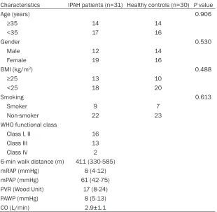
[image:2.612.92.402.97.400.2]MiR-503 mimics and corresponding negative control miR (miR-NC) were purchased from RiboBio (Guangzhou, China). Transient trans-fection of cells was achieved with Lipofectamine 2000 (Invitrogen, Life Technologies, Carlsbad, CA, USA) following the manufacturer’s protocol. Cell culture media was changed after 6 hours to remove the transfection reagent. 48 hours after transfection, transfection efficacy was Table 1. Demographic and clinical characteristics of IPAH patients and
healthy controls
Characteristics IPAH patients (n=31) Healthy controls (n=30) P value
Age (years) 0.906
≥35 14 14
<35 17 16
Gender 0.530
Male 12 14
Female 19 16
BMI (kg/m2) 0.488
≥25 13 10
<25 18 20
Smoking 0.613
Smoker 9 7
Non-smoker 22 23
WHO functional class
Class I, II 16
Class III 13
Class IV 2
6-min walk distance (m) 411 (330-585)
mRAP (mmHg) 8 (4-12)
mPAP (mmHg) 61 (42-75)
PVR (Wood Unit) 17 (8-24)
PAWP (mmHg) 8 (5-13)
CO (L/min) 2.9±1.1
Values are presented as mean ± SD or median (interquartile range). BMI: body mass
index; WHO: World Health Organization; mRAP: mean right atrial pressure; mPAP: mean pulmonary arterial pressure; PVR: pulmonary vascular resistance; PAWP: pulmonary artery
wedge pressure; CO: cardiac output.
WHO clinical classifica -tion [10], inclusion crite-ria including mean pul-monary artery pressu- re (mPAP) ≥25 mmHg, pulmonary artery wedge pressure (PAWP) ≤15 mmHg. 30 healthy par-ticipants without PAH or significant cardiorespi -ratory disease were also recruited. The character-istics of IPAH patients and healthy participants are listed in Table 1. This study was approved by the Ethics Committee of Chengfei Hospital and the written informed co- nsents were obtained from all subjects prior to participation.
For hypoxia experiments, hPASMCs were pla- ced in a humidified airtight incubator that was constantly infused with a hypoxic gas mixture (3% O2, 5% CO2, and 92% N2). The oxygen con-centration was monitored continuously (Forma 3130; Thermo Scientific, Rockford, IL, USA). At the same time, normoxic hPASMCs were placed in an incubator infused with air (21% O2, 5% CO2 and 74% N2).
RNA extraction and quantitative reverse tran-scription-PCR (qRT-PCR)
Total RNA was isolated from serum samples and cells using mirVanaTM PARIS miRNA isola-tion kit (Ambion, Foster City, CA, USA) according to manufacturer’s instructions. The quantity and quality of the total RNA extracted were determined spectrophotometrically (Shanghai Spectrum Instruments Co., Ltd., Shanghai, China) and RNA integrity was determined by gel electrophoresis.
Each miRNA was specifically reverse tran -scribed to cDNA using a TaqMan MicroRNA Reverse Transcription Kit, and the relative lev-els of miR-503 to the control U6 were detected by PCR using the All-in one TM miRNA qRT-PCR reagent kit (GeneCopoeia, Rockville, USA). The primers were as follows: miR-503, RT: 5’-GTCGTATCCAGTGCAGGGTCCGAGGTATTCG-CACTGGATACGACCTGCAG-3’, forward primer: 5’-TAGCAGCGGGAACAGTT-3’ and reverse pri-mer: 5’-GTGCAGGGTCCGAGGT-3’; U6, RT: 5’-A-ACGCTTCACGAATTTGCGT-3’, forward primer: 5’-CTCGCTTCGGCAGCACA-3’ and reverse prim -er: 5’-AACGCTTCACGAATTTGCGT-3’. The data were analyzed using 2-ΔΔCt method. All reactions
were performed in triplicate on a 7500 Fast Real-Time PCR system (Applied Biosystems, USA).
Western blot
Proteins were isolated from cells using RIPA lysis buffer (Beyotime, Beijing, China), and the protein concentration was measured by a BCA protein assay kit (Pierce, Rockford, IL, USA). Equal quantities of proteins were loaded and separated by SDS-PAGE and transferred to PVDF membranes (Millipore, Billerica, MA, USA). The membrane was then plotted with pri-mary antibodies against p27 (1:1000; Cell Signaling Technology, Boston, MA, USA), Cyclin D1 (1:1000; Santa Cruz Biotechnology, Santa
Cruz, CA, USA), Bax (1:500; Abcam, Cambridge, UK) and Bcl-2 (1:500; Abcam) overnight at 4°C on a shaker. Then the membranes were incu-bated with HRP-conjugated secondary antibod-ies for 1 h at room temperature, and visualized using enhanced chemiluminescence assay (ECL, Thermo, Rockford, USA). β-actin was used as a internal control.
Luciferase activity assay
For the binding of miR-503 to Bcl-2 3’UTR, the 3’UTR segment of the Bcl-2 gene was amplified by PCR and inserted into the pGL3-luciferase reporter plasmid (Promega, Madison, WI, USA). A mutant construct in miR-503 binding sites of Bcl-2 3’UTR region also was generated us-ing Quick Change Site-Directed Mutagen- esis Kit (Agilent, Roseville City, CA, USA). Co-transfections of luciferase reporter plasmid and pRL-TK vector expressing the Renilla lucif-erase (Promega) into the miR-503 overexpre- ssing or control hPASMCs were accomplish- ed by using Lipofectamine 2000. Luciferase activity was measured 48 hours after transfe- ction by the Dual-Luciferase Reporter Assay System (Promega).
MTT assay
Cell proliferation was determined by 3-(4, 5-dimethylthiazol-2-yl)-2, 5-diphenyltetrazolium bromide (MTT) assay. hPASMCs were seeded in 96-well plates (5000 cells/well) and transfect-ed with either miR-NC or miR-503 before expo-sure to hypoxia. After incubation for 48 h, 10 µl of MTT solution (5 mg/ml) was added to each well for 4 h. The 570 nm absorbance was inves-tigated using a microplate reader (Bio-Rad, Hercules, CA, USA).
Wound healing assay
Flow cytometry analysis
For cell cycle assay, hPASMCs were fixed in 70% ethanol at 4°C overnight. Then, the cells were analyzed by using a CycletestTM Plus DNA Reagent Kit (BD Biosciences, Bedford, MA, USA). The cell cycle was determined with a FACS Calibur flow cytometer (BD Biosciences) and data were analyzed with ModFit 3.0 soft-ware (BD Biosciences).
The apoptosis rate of hPASMCs was tested using an Annexin V-FITC/PI Apoptosis Detection Kit (BD Biosciences) in a FACS Calibur flow cytometer, and the data were analyzed using the Cell Quest Pro software (BD Bioscience).
Statistical analysis
Statistical analyses were conducted with GraphPad Prism 6 (GraphPad Software, La Jolla, California, USA). All in vitro experiments were carried out at least 3 times and results were presented as means ± standard deviation (SD). A receiver operating characteristic (ROC) curve analysis was performed to evaluate the diagnostic value of serum miR-503 for discrimi-nating between IPAH patients and healthy indi-viduals. Differences between groups were determined using Student’s t-test, Chi-square test or one-way analysis of variance (ANOVA) test followed by post-hoc test when appropri-ate. All the differences were regarded as statis-tically significant when P<0.05.
Results
Serum miR-503 level is markedly reduced in IPAH patients
To study the role of miR-503 in PAH, we exam-ined the miR-503 levels in sera from 31 IPAH patients and 30 healthy participants. As indi-cated in Figure 1A, the serum level of miR-503 was significantly decreased in IPAH patients than controls (P<0.05). Then, we examined the diagnostic efficiency of serum miR-503 in IPAH through ROC curve analysis. As shown in Figure 1B, the area under the curve (AUC) of the ROC curve was 0.890 (95% CI: 0.808-0.973). When the level of serum miR-503 was at the optimum cutoff point (0.0434), sensitivity and specificity were 90.0% and 77.4%, respectively. These results indicated that serum miR-503 level possessed a strong diagnostic efficiency for discriminating IPAH patients from healthy individuals.
MiR-503 overexpression attenuates hPASMC proliferation and migration induced by hypoxia
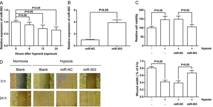
[image:4.612.93.520.73.226.2]Hypoxia is a critical factor in the pathogenesis of PAH. As shown in Figure 2A, miR-503 expres-sion was gradually decreased in hPASMCs after exposure to hypoxia in a time-dependent man-ner. To further study the biological functions of 503, we induced overexpression of miR-503 by miR-miR-503 mimics in hPASMCs, and the efficiencies were confirmed by qRT-PCR. The
Figure 1. Serum miR-503 level is markedly reduced in IPAH patients. A. Levels of serum miR-503 in healthy controls and IPAH patients were measured. The results were expressed as 2-ΔCt. ΔCt = Ct
miR-503-CtU6. The data are
Figure 2. MiR-503 overexpression attenuates hPASMC proliferation and migration induced by hypoxia. A. miR-503 expression in PASMCs after hypoxia stimulation for 0, 6, 12 and 24 h was detected by qRT-PCR analysis. B. miR-503 mimics and NC mimics were transfected into hPASMCs, and the transfection efficiency was detected by qRT-PCR analysis. C. Cell proliferation was detected by MTT assay in hPASMCs. D. Cell migration was detected by wound heal-ing assay in hPASMCs. The data are expressed as mean ± SD.
expression of miR-503 in hPASMCs was signifi -cantly enhanced after transfection of miR-503 mimics (Figure 2B).
The effect of miR-503 up-regulation on cell pro-liferation of hPASMCs was examined by MTT assay. The results showed that overexpression of miR-503 rescued the excessive proliferation of hypoxia-treated hPASMCs after 48 h of incu-bation (Figure 2C). The effect of miR-503 on cell migration of hPASMCs was determined by wound healing assay. As demonstrated in Figure 2D, the migratory ability of hPASMCs was dramatically increased under hypoxia treatment, whereas miR-503 up-regulation reserved the hypoxia-induced promotion of migration of hPASMCs.
MiR-503 overexpression arrests hPASMC cell cycle progression induced by hypoxia
Above findings showed miR-503 could inhibit the proliferation of hPASMCs, and then impact of miR-503 overexpression on cell cycle was further assessed by flow cytometry. As shown in Figure 3A, hypoxia treatment reduced the proportion of hPASMCs in G0/G1-phase, and the proportion of hPASMCs in S and G2/M phases markedly increased as compared to under normoxia. The deregulated cell cycle
pro-gression was obviously restored by transfec-tion of miR-503 mimics.
Next, western blotting was used to investigate the expression of cell cycle relative proteins. As shown in Figure 3B, p27 expression was signific-antly decreased and Cyclin D1 was significant-ly increased in hypoxia-treated hPASMCs, and transfection of miR-503 mimics obviously re- versed these effects.
MiR-503 overexpression restores hPASMC apoptosis inhibited by hypoxia
Flow cytometric analysis showed that the pro-portion of the cell population undergoing ap- optosis was decreased in hPASMCs under hypoxia treatment, but this was reversed by transfection of miR-503 mimics in hPASMCs (Figure 4A).
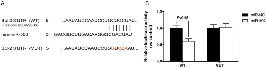
MiR-503 directly targets 3’-UTR of Bcl-2 in hPASMCs
Given that the biological effects of altered miRNA expression rely on the significance of their target genes, we explored targets of miR-503 using the TargetScan bioinformatics algo-rithm (http://www.targetscan.org/). Our analy-sis revealed that Bcl-2 is a potential target of miR-503 based on putative target sequences of the Bcl-2 3’-UTR (Figure 5A). To confirm Bcl-2 as a direct target of miR-503, a dual-luciferase reporter assay was performed in hPASMCs. The two luciferase constructs containing the WT or MUT 3’-UTR of the Bcl-2 gene were then separately transfected into hPASMCs, together with either NC or miR-503. As a result, miR-503 dramatically reduced the luciferase activity of the WT-Bcl-2 3’UTR but not of the mutant in hPASMCs (Figure 5B). Thus, the complementa-ry sequences on Bcl-2 3’-UTR were then a direct target of miR-503.
Discussion
The pathogenesis of PAH is a critical topic, but it is not yet fully understand. The present study is the first, to our knowledge, to identify decreased miR-503 expression in serum sam-ples of treatment-naive IPAH patients and hypoxia-treated hPASMCs. Overexpression of miR-503 alleviated disease phenotypes in hypoxia-treated hPASMCs through regulating Bcl-2 expression.
MiRNAs have been investigated as a diagnostic and prognostic biomarker in numerous diseas-es. The extreme stability of circulating miRNAs in biological fluids and their resistance to vari -ous storage conditions make them good
candi-dates for the development of minimally invasive biomarkers [11, 12]. Several studies examined circulating miRNAs and have demonstrated that they are correlated with PAH development and progression. For example, plasma miR-150 levels are decreased and correlate with surviv-al in PAH patients [13]. In the present study, we found markedly decreased expression of miR-503 in IPAH patients compared to controls. Consistent with our findings, previous animal experiments showed that miR-503 was reduced in the lung and microdissected pulmonary artery of monocrotaline-induced PAH rats [14]. MiR-503 was also identified as an onco-sup -pressor in human tumorigenesis, including prostate cancer [15], osteosarcoma [16] and hepatocellular carcinoma [17]. In cardiovascu-lar system, a recent study reported that overex-pression of miR-503 increased the cellular pro-liferation and collagen production in mice neo-natal cardiac fibroblasts [18]. The clinical sig -nificance of miR-503 promoted us to explore its underlying mechanisms in PAH.
[image:8.612.91.520.73.184.2]The pulmonary vascular wall is made up of three resident cell types, including the endothe-lial (intima), smooth muscle (media) and fibro -blast (adventitia) cells [19]. It has been recog-nized that hypoxia is a stimulus to enhance PASMC proliferation and migration capabilities, which play key roles in promoting vascular remodeling during the development of PAH [20]. Some miRNAs, such as miR-206 [21] and miR-322 [22], are involved in the hypoxia-induced excessive PASMC proliferation and migration. Our results showed that miR-503 expression was reduced in hypoxia-treated hPASMCs, and excessive hPASMC proliferation and migration caused by hypoxia was signifi -cantly attenuated by miR-503 overexpression.
Deregulated cell proliferation is mainly attrib-uted to aberrant cell cycle regulation. Cyclin D1 is an important cell cycle gene that induces G1-to-S phase progression, leading to the pro-motion of cell proliferation [23]. Deregulated Cyclin D1 has been reported in PAH and has been implicated in PASMC proliferation [24, 25]. As a cyclin-dependent kinase inhibitor, p27 can cause cells in G1 arrest which contributes to the inhibition of PASMC proliferation [26, 27]. In this study, we found that miR-503 over-expression led to cell cycle arrest at G1 phase through increasing p27 expression and decreasing Cyclin D1 expression in hPASMCs. The B-cell lymphoma protein-2 (Bcl-2) protein family, which included both pro-apoptotic (e.g., Bax) and anti-apoptotic (e.g., Bcl-2) members, play a critical role in the development of apop-tosis [28]. Bcl-2 silencing can obviously attenu-ate hypoxia-triggered apoptosis resistance in pulmonary microvascular endothelial cells [29]. Several target genes regulated by miR-503 have been reported. For example, miR-503 reg-ulates cisplatin resistance of human non-small cell lung cancer cells and gastric cancer cells through targeting Bcl-2 [30, 31]. In this article, we also found that ectopic expression of miR-503 caused a remarkable reduction in Bcl-2 protein level and decreased luciferase activity of the Bcl-2 promoter, indicating that Bcl-2 is a direct target of miR-503 in hPASMCs.
Taken together, the present study presents evi-dences that miR-503 plays a critical suppres-sive role in PAH by directly binding the Bcl-2 3’UTR that leads to down-regulation of Bcl-2 expression level. Our observation may provide clues that enhancing miR-503 expression might be a potential therapeutic target for PAH in the near future.
Disclosure of conflict of interest
None.
Address correspondence to: Xiaofei Zheng, Cheng- fei Hospital, No 105 Jingyi Road, Qingyang District, Chengdu 610073, China. E-mail: zhengfei1230@ sina.com
References
[1] Galie N, Humbert M, Vachiery JL, Gibbs S, Lang I, Torbicki A, Simonneau G, Peacock A, Vonk Noordegraaf A, Beghetti M, Ghofrani A,
Gomez Sanchez MA, Hansmann G, Klepetko W, Lancellotti P, Matucci M, McDonagh T, Pierard LA, Trindade PT, Zompatori M and Hoeper M. 2015 ESC/ERS guidelines for the diagnosis and treatment of pulmonary hyper-tension: the joint task force for the diagnosis and treatment of pulmonary hypertension of the european society of cardiology (ESC) and the european respiratory society (ERS): en-dorsed by: association for european paediat-ric and congenital cardiology (AEPC), interna-tional society for heart and lung transplanta-tion (ISHLT). Eur Respir J 2015; 46: 903-975. [2] Handoko ML, de Man FS, Allaart CP, Paulus
WJ, Westerhof N and Vonk-Noordegraaf A. Perspectives on novel therapeutic strategies for right heart failure in pulmonary arterial hypertension: lessons from the left heart. Eur Respir Rev 2010; 19: 72-82.
[3] Tuder RM, Archer SL, Dorfmuller P, Erzurum SC, Guignabert C, Michelakis E, Rabinovitch M, Schermuly R, Stenmark KR and Morrell NW. Relevant issues in the pathology and pathobiology of pulmonary hypertension. J Am Coll Cardiol 2013; 62: D4-12.
[4] Benza RL, Miller DP, Barst RJ, Badesch DB, Frost AE and McGoon MD. An evaluation of long-term survival from time of diagnosis in pulmonary arterial hypertension from the REVEAL registry. Chest 2012; 142: 448-456. [5] Lee RC, Feinbaum RL and Ambros V. The C.
elegans heterochronic gene lin-4 encodes small RNAs with antisense complementarity to lin-14. Cell 1993; 75: 843-854.
[6] Bartel DP. MicroRNAs: genomics, biogenesis, mechanism, and function. Cell 2004; 116: 281-297.
[7] Potus F, Ruffenach G, Dahou A, Thebault C, Breuils-Bonnet S, Tremblay E, Nadeau V, Paradis R, Graydon C, Wong R, Johnson I, Paulin R, Lajoie AC, Perron J, Charbonneau E, Joubert P, Pibarot P, Michelakis ED, Provencher S and Bonnet S. Downregulation of microR-NA-126 contributes to the failing right ventri-cle in pulmonary arterial hypertension. Cir- culation 2015; 132: 932-943.
[8] Deng L, Blanco FJ, Stevens H, Lu R, Caudrillier A, McBride M, McClure JD, Grant J, Thomas M, Frid M, Stenmark K, White K, Seto AG, Morrell NW, Bradshaw AC, MacLean MR and Baker AH. MicroRNA-143 activation regulates sm- ooth muscle and endothelial cell crosstalk in pulmonary arterial hypertension. Circ Res 2015; 117: 870-883.
[10] Simonneau G, Gatzoulis MA, Adatia I, Celer- majer D, Denton C, Ghofrani A, Gomez San- chez MA, Krishna Kumar R, Landzberg M, Machado RF, Olschewski H, Robbins IM and Souza R. Updated clinical classification of pul-monary hypertension. J Am Coll Cardiol 2013; 62: D34-41.
[11] Creemers EE, Tijsen AJ and Pinto YM. Cir- culating microRNAs: novel biomarkers and extracellular communicators in cardiovascular disease? Circ Res 2012; 110: 483-495. [12] Wang F, Chen C and Wang D. Circulating
mi-croRNAs in cardiovascular diseases: from bio-markers to therapeutic targets. Front Med 2014; 8: 404-418.
[13] Rhodes CJ, Wharton J, Boon RA, Roexe T, Tsang H, Wojciak-Stothard B, Chakrabarti A, Howard LS, Gibbs JS, Lawrie A, Condliffe R, Elliot CA, Kiely DG, Huson L, Ghofrani HA, Tiede H, Schermuly R, Zeiher AM, Dimmeler S and Wilkins MR. Reduced microRNA-150 is associated with poor survival in pulmonary ar-terial hypertension. Am J Respir Crit Care Med 2013; 187: 294-302.
[14] Gubrij IB, Pangle AK, Pang L and Johnson LG. Reversal of microRNA dysregulation in an ani-mal model of pulmonary hypertension. PLoS One 2016; 11: e0147827.
[15] Guo J, Liu X and Wang M. miR-503 suppress- es tumor cell proliferation and metastasis by directly targeting RNF31 in prostate cancer. Biochem Biophys Res Commun 2015; 464: 1302-1308.
[16] Guo X, Zhang J, Pang J, He S, Li G, Chong Y, Li C, Jiao Z, Zhang S and Shao M. MicroRNA-503 represses epithelial-mesenchymal transition and inhibits metastasis of osteosarcoma by targeting c-myb. Tumour Biol 2016; 37: 9181-9187.
[17] Xiao Y, Tian Q, He J, Huang M, Yang C and Gong L. MiR-503 inhibits hepatocellular carcinoma cell growth via inhibition of insulin-like growth factor 1 receptor. Onco Targets Ther 2016; 9: 3535-3544.
[18] Zhou Y, Deng L, Zhao D, Chen L, Yao Z, Guo X, Liu X, Lv L, Leng B, Xu W, Qiao G and Shan H. MicroRNA-503 promotes angiotensin II-indu- ced cardiac fibrosis by targeting apelin-13. J Cell Mol Med 2016; 20: 495-505.
[19] Welsh DJ and Peacock AJ. Cellular responses to hypoxia in the pulmonary circulation. High Alt Med Biol 2013; 14: 111-116.
[20] Maxova H. [Experimental models and new ap-proaches to the treatment of pulmonary hy-pertension]. Cesk Fysiol 2012; 61: 24-29.
[21] Yue J, Guan J, Wang X, Zhang L, Yang Z, Ao Q, Deng Y, Zhu P and Wang G. MicroRNA-206 is involved in hypoxia-induced pulmonary hyper-tension through targeting of the HIF-1alpha/ Fhl-1 pathway. Lab Invest 2013; 93: 748-759. [22] Zeng Y, Liu H, Kang K, Wang Z, Hui G, Zhang X,
Zhong J, Peng W, Ramchandran R, Raj JU and Gou D. Hypoxia inducible factor-1 mediates expression of miR-322: potential role in proli- feration and migration of pulmonary arterial smooth muscle cells. Sci Rep 2015; 5: 12098. [23] Stacey DW. Cyclin D1 serves as a cell cycle
regulatory switch in actively proliferating cells. Curr Opin Cell Biol 2003; 15: 158-163. [24] Guo H, Zhang X, Cui Y, Deng W, Xu D, Han H,
Wang H, Chen Y, Li Y and Wu D. Isorhynch- ophylline protects against pulmonary arterial hypertension and suppresses PASMCs prolif-eration. Biochem Biophys Res Commun 2014; 450: 729-734.
[25] Zeng DX, Xu GP, Lei W, Wang R, Wang CG and Huang JA. Suppression of cyclin D1 by plas-mid-based short hairpin RNA ameliorated ex-perimental pulmonary vascular remodeling. Microvasc Res 2013; 90: 144-149.
[26] Sandal T. Molecular aspects of the mammali-an cell cycle mammali-and cmammali-ancer. Oncologist 2002; 7: 73-81.
[27] Yu L, Quinn DA, Garg HG and Hales CA. Gene expression of cyclin-dependent kinase inhibi-tors and effect of heparin on their expression in mice with hypoxia-induced pulmonary hy-pertension. Biochem Biophys Res Commun 2006; 345: 1565-1572.
[28] Danial NN and Korsmeyer SJ. Cell death: criti-cal control points. Cell 2004; 116: 205-219. [29] Cao Y, Jiang Z, Zeng Z, Liu Y, Gu Y, Ji Y, Zhao Y
and Li Y. Bcl-2 silencing attenuates hypoxia-induced apoptosis resistance in pulmonary microvascular endothelial cells. Apoptosis 2016; 21: 69-84.
[30] Qiu T, Zhou L, Wang T, Xu J, Wang J, Chen W, Zhou X, Huang Z, Zhu W, Shu Y and Liu P. miR-503 regulates the resistance of non-small cell lung cancer cells to cisplatin by targeting Bcl-2. Int J Mol Med 2013; 32: 593-598.