Supported by the Internal Grant Agency of the Mendel University in Brno, Czech Republic (Project No. TP5/2015).
Tissue Fatty Acid Deposition, Plasma Lipid
and Cytokine Profile in Pigs Fed a Diet with Fish Oil
or Palm Oil
Tomáš Komprda
1*, Veronika Rozíková
1, Monika Vícenová
2,
Nikola Procházková
1, Petra Ondráčková
2, Petra Pešková
1, Martin Faldyna
2 1Department of Food Technology, Mendel University in Brno, Brno, Czech Republic2Veterinary Research Institute, Brno, Czech Republic
*Corresponding author: komprda@mendelu.cz
ABSTRACT
KomprdaT., RozíkováV., VícenováM., ProcházkováN., OndráčkováP., PeškováP., Faldyna M.(2017): Tissue
fatty acid deposition, plasma lipid and cytokine profile in pigs fed a diet with fish oil or palm oil. Czech J. Anim. Sci., 62, 482–490.
The present study tested a hypothesis concerning a favourable effect of dietary fish oil on the tissue polyun-saturated fatty acid (PUFA) deposition, and on plasma lipid and cytokine profile. Thirty-two pigs divided into two groups of 16 animals each were fed for 70 days a diet with 2.5% of fish oil (F) and palm oil (P), respectively. The content of PUFA n-3 in the liver, muscle (m. quadriceps femoris), and visceral adipose tissue (VAT) of the F- and P-pigs was 530 and 129, 84 and 19, 1365 and 191 mg/100 g of the fresh tissue, respectively (differences between dietary groups were significant at P < 0.01 in all tissues). Dietary fish oil in comparison with palm oil decreased (P < 0.05) total plasma cholesterol, but also desirable high-density lipoprotein cholesterol, and had no effect (P > 0.05) both on low-density lipoprotein cholesterol and triacylglycerols. Moreover, dietary fish oil increased (P < 0.05) expression of the genes coding for not only anti-inflammatory cytokines IL-10 (P < 0.01) and TGF-β1 (P < 0.05), but also for pro-inflammatory cytokines IL-6 (P < 0.01), IL-12, and TNF-α (P < 0.05) in the VAT, and increased (P < 0.05) the expression of the IL6 gene in the liver. On the other hand, no significant differences (P > 0.05) between the F- and P-pigs in plasma levels of any tested cytokine were found out. It was concluded that an effect of dietary fish oil on tissue fatty acid deposition is undeniable, but its effects on plasma markers related to the risk of chronic degenerative diseases require further research.
Keywords: EPA; DHA; cholesterol; IL-10; TNF-α; cardiovascular diseases; feed ration; pig; muscle
Cardiovascular diseases (CVD) are currently a leading cause of mortality in economically de-veloped countries. Eating habits significantly af-fect risk of CVD via food composition, especially from the viewpoint of lipid fractions of ingested foods. As far as lipids are concerned, the so-called Western-type eating habit is characterized by a relatively high intake of saturated fatty acids
(SFA) and lower consumption of monounsaturated (MUFA) and polyunsaturated fatty acids (PUFA). Within PUFA, intake of PUFA n-6 highly prevails over PUFA n-3, which further increases the risk of CVD (Bragt and Mensink 2012).
to enrich pork with PUFA n-3. Possibilities of such an enrichment in foods in general were reviewed e.g. by Komprda (2012). Sobol et al. (2016) re-ported that loin and shoulder of pigs (with high intramuscular fat content) fed a diet enriched with the mixture of linseed, rapeseed, and fish oils meet the European Union recommendations for human nutrition for products considered as either PUFA n-3 sources or products with high PUFA n-3 content. Alvarez-Rodriguez et al. (2016) found out that total PUFA n-3 content (mainly α-linolenic acid, ALA) was greater in organic than in conventional pork, probably due to ALA content from dietary vegetable oils.
However, the conversion rate of dietary ALA (a precursor of the PUFA n-3 group) to higher func-tional metabolites eicosapentaenoic (EPA) and docosahexaenoic acid (DHA) in animal tissues is very low (Komprda et al. 2013). Therefore it is more suitable to use directly dietary EPA + DHA, a rich source of which is fish oil. From the viewpoint of a healthy nutrition, palm oil (with its high content of SFA), which is currently present in many foods, stays on the opposite pole to fish oil (PUFA n-3).
Therefore, one of the objectives of the present study was to compare fish oil and palm oil as the components of the pig feed ration from the view-point of a deposition of dietary fatty acids in the tissues relevant to human consumption: liver, muscle, and adipose tissue.
Moreover, regarding the above-mentioned CVD, their hallmarks are, among others, dislipidemia and low-level chronic inflammation in an organ-ism (especially in the vascular wall) (Calder 2013). Dislipidemia is characterized by a high plasma level of triacylglycerols (TAG), total cholesterol (TC), and low-density lipoprotein cholesterol (LDLC), and low level of high-density lipoprotein choles-terol (HDLC). As far as pigs are concerned, Sopkova et al. (2017) evaluated an effect of combining a dietary source of PUFA n-3 (flaxseed) with pro-biotics on the lipid metabolism of piglets after weaning, and reported a decrease of HDLC, but no change in TAG, TC, and LDLC, respectively.
Low-level chronic inflammation manifests itself (among others) by an increased plasma level of pro-inflammatory cytokines (Bragt and Mensink 2012). The effect of dietary fish oil on cytokine production in pigs is only scarcely described in available literature. Liu et al. (2003) tested the fish oil (7%) effect on cytokine production in
not-challenged and lipopolysaccharide-not-challenged crossbred pigs; in comparison with corn oil, fish oil only tended to reduce IL-1β and IL-2 produc-tion, respectively.
The second objective of the present study there-fore was to evaluate the lipid and cytokine levels in plasma of pigs fed a diet enriched with fish oil and palm oil, respectively. An intention was to use such a level of these oils (2.5% of the feed ration), which would be relevant to humans. In this case, pigs were considered a useful model of human nutrition.
Therefore, the present experiment tested a hy-pothesis that pigs fed a diet with fish oil, in com-parison with dietary palm oil, will deposit higher amounts of nutritionally favourable fatty acids in the food-processing relevant tissues, and will have a more favourable plasma lipid profile and a less inflammation-prone cytokine profile.
MATERIAL AND METHODS
Animals and dietary interventions. Thirty-two
pigs of both sexes (16 males, 16 females – from technical reasons it was not possible to obtain a weight-and-age-homogeneous same-sex group; Large White × Landrace; Bioprodukt Knapovec a.s., Czech Republic) at the age of eight weeks with the mean live weight of 25.5 ± 1.15 kg were used. The pigs were housed in an experimental stable in floored indoor pens (10 m2) of four animals each.
The experiment was performed in compliance with the Act No. 246/1992 Coll. of the Czech National Council on the protection of animals against cruelty, and with its amendment, the Act No. 162/1993 Coll., and was approved by the “Com-mission to protect animals against cruelty” of the Mendel University in Brno and of the Ministry of Agriculture of the Czech Republic.
using a KD-310-A-1015 KjelROC Analyzer; Opsis AB, Sweden), 56 g of fat (quantified as hexane/ 2-propanol extract), 48 g of crude fibre (determined using an ANKOM 220 Fiber Analyzer; ANKOM Technology, USA), and 758 g of nitrogen-free extractives (calculated as a remainder to 100%). Metabolizable energy content (calculated from nutrient content) was 13.6 MJ/kg. Basic pelletized complete feed mixture for pig fattening (De Heus a.s., Czech Republic) was composed of wheat, barley, wheat bran, wheat middlings, dark dis-tillery stillages, rapeseed expellers, vinas, sodium carbonate, animal fat, salt, premix of vitamins + minerals (the producer refused to communicate percentages of particular components due to the trade secret). The pelletized basic feed mixture was grinded and homogenized with an appropriate amount of particular oil.
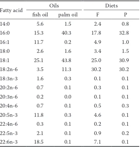
The fatty acid content in fish oil and palm oil, and in the F and P diet, is presented in Table 1.
The animals had free access to drinking water and were fed daily ad libitum; the daily ration was divided into two parts and fed twice daily (at 7.00 and 14.00 h). By subtracting leftovers, the net feed consumption was measured twice daily; due to the ad libitum access to feed, significant differ-ences in consumption between animals in a pen were not supposed. The animals were weighed in weekly intervals. The fattening lasted for 70 days.
Last day of fattening, all pigs were anesthetized by the intramuscular application of the TKX mix-ture (12.5 mg/ml of ketamine (Vétoquinol SA, France) + 12.5 mg/ml of xylazine (Bioveta, Czech Republic) + 12.5 mg/ml of tiletamine (Virbac, France) + 12.5 mg/ml of zolazepam (Virbac)) in the total volume of 0.2 ml/kg, and consequently sacrificed by bleeding.
Blood and tissue samples collection. Blood
samples were collected (from the aorta) to the heparin-coated test tubes and subsequently cen-trifuged at 200 g for 10 min at 4°C to obtain blood plasma. Liver (200 g), muscle (m. quadriceps femo-ris; 200 g), and visceral adipose tissue (VAT) (pel-vic; 200 g) samples were taken, aliquots (100 g of each tissue) were freeze-dried (Alpha 1-2 LDplus; Martin Christ Gefriertrocknungsanlagen GmbH, Germany; temperature programme: –45°C/27 h and –50°C/3 h) and stored at –20°C for subsequent fatty acid analyses. Total RNA was immediately iso-lated from another liver and VAT aliquots (50 and 50 mg, respectively).
Fatty acid analysis. Total lipid extraction, sample
derivatization, and fatty acid methyl ester sepa-ration were performed according to a procedure described in a paper of Komprda et al. (2013). The results were expressed as a percentage of the sum of all determined fatty acids and in mg/100 g of the fresh tissue.
Plasma lipids determination. TC, LDLC, HDLC,
and TAG values were determined by the enzymatic-colorimetric method using an automated chemical analyzer BS-200 (Mindray, China) and commercial kits (Greiner Diagnostic GmbH, Germany).
Quantification of cytokine gene expressions.
[image:3.595.305.532.504.742.2]Total RNA isolation, reverse transcription, quan-titative polymerase chain reaction (PCR), and calculation of the normalized relative quantity values were performed according to Komprda et al. (2016). The efficiency of the quantitative PCR for the target genes and the reference gene was determined using an external standard curve with serial dilutions of the cDNA plotted against cycle number to calculate the slope (Hellemans et al. 2007). TBP1 was used as a reference gene based on the data of Nygard et al. (2007) reporting good expression stability and expression level for low abundant transcripts across different pig tissues. Specific primers for the pig genes used in quan-titative PCR are characterized in Table 2.
Table 1. Fatty acid proportion in dietary oils and in the diets (% of the sum of all determined fatty acids)
Fatty acid Oils Diets
fish oil palm oil F P
14:0 5.6 1.5 2.4 0.8
16:0 15.3 40.3 17.8 32.8
16:1 11.7 0.2 4.9 1.0
18:0 2.6 1.6 3.4 1.5
18:1 25.1 43.8 25.0 30.9
18:2n-6 3.5 11.3 30.2 30.2
18:3n-3 1.6 0.3 0.1 0.1
20:2n-6 0.7 0.1 0.3 0.1
20:3n-6 0.2 0.0 0.1 0.1
20:4n-6 0.7 0.1 0.5 0.3
20:5n-3 11.8 0.3 4.6 0.1
22:4n-6 0.3 0.1 0.2 0.1
22:5n-3 2.1 0.1 0.9 0.2
22:6n-3 18.5 0.1 7.1 0.1
Determination of plasma cytokines. IL-1β, IL-4, IL-6, IL-10, IL-12, and TNFα concentrations in the pig blood plasma were measured by Milliplex®
MAP Porcine Cytokine/Chemokine Magnetic Bead Panel kit (Millipore Corp., USA) according to the producer’s recommendation.
Statistical evaluation. Normality of the data
distribution was tested by Kolmogorov-Smirnov test. The differences between dietary interventions were evaluated by one-way ANOVA including the post-hoc Tukey’s test and by the independent samples t-test (sets with a normal distribution, i.e. all data sets except gene expressions), and by the non-parametric Wilcoxon signed-rank test (data sets concerning relative expression of the liver and adipose tissue genes), respectively. For all evaluations, the STATISTICA Version 12 software package (StatSoft, USA) was used.
RESULTS
Feed intake, live weight, daily weight gain. No
significant differences (P > 0.05) between the F- and P-pigs were established either in feed intake (10.5 and 10.6 g/kg per day) or in daily weight gain (0.85 ± 0.05 kg/dayand 0.86 ± 0.04 kg/day) or in the final live weight (83.64 ± 1.82 kg and 84.06 ± 3.35 kg, respectively; data not presented in tables or figures).
Fatty acid deposition in the tissues.
Deposi-tion of the tested fatty acids in the three tissues relevant from the viewpoint of human nutrition was quantified either as percentages from the sum of all analyzed fatty acids (Table 3) or as absolute amounts in mg/100 g of the fresh tissue (not pre-sented in tables or figures).
As far as a comparison of the tissues is concerned, it follows from Table 3 that MUFA were deposited in the highest (P < 0.05) and PUFA in the lowest (P < 0.05) percentage in the muscle tissue. On the other hand, PUFA deposited preferentially in the liver: the highest (P < 0.05) PUFA percentage from all tested tissues was found in the liver not only of the F-pigs, but also of the pigs fed a diet with palm oil (38.5% of all fatty acids deposited in the P-livers were PUFA, contrary to e.g. only 22.3% in the VAT of the fish oil-fed pigs; P < 0.05; Table 3). As far as a comparison of the dietary oils from the viewpoint of PUFA is concerned, the F- and P-pigs did not differ (P > 0.05) in deposition of PUFA n-6 in either of the tested tissues. On the
Ta
ble 2. Pr
imers u se d f or qu an tit ative P C R ( TBP 1 u se d a
s a r
other hand, sum of PUFA n-3 was deposited more (P < 0.05) in the F-pigs as compared to the P-counterparts in all tested tissues. The F-pigs also had a more favourable (lower; P < 0.05) PUFA n-6/PUFA n-3 ratio in all tested tissues (data not explicitly presented in Table 3).
When expressed in the absolute values, the contents of nutritionally desirable PUFA n-3 in the liver, muscle tissue, and VAT of the F- and P-pigs were 530 and 129, 84 and 19, and 1365 and 191 mg/100 g of the fresh tissue, respectively (dif-ferences between dietary groups were significant at P < 0.01 in all tissues).
Plasma lipids. One of the objectives of the
pre-sent experiment was to use pigs as a model for evaluating the effect of dietary fish oil on plasma lipids as markers of the risk of CVD. Plasma levels of total cholesterol, high-density lipoprotein cho-lesterol, low-density lipoprotein chocho-lesterol, and
triacylglycerols are shown in Figure 1. Dietary fish oil in comparison with palm oil decreased (P < 0.05) TC. However, this decrease was apparently at the expense of the desirable HDL fraction, the level of which was also lower (P < 0.05) in plasma of the F-pigs. On the other hand, dietary fish oil had no effect (P > 0.05) either on LDL cholesterol or TAG.
Cytokines. Other hallmark of the CVD risk is a
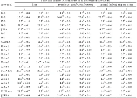
[image:5.595.61.533.407.744.2]chronic low inflammation in an organism. There-fore the next objective of the present experiment was to test dietary fish oil as a means for ame-lioration of an inflammatory status in a model organism; pro- and anti-inflammatory cytokines were used as pertinent markers. Expression of the genes coding for selected cytokines in the liver and VAT, and plasma levels of these cytokines (except of TGF-β1, whose antibody was not available in the kit used for the analysis) in plasma are presented in Figures 2 and 3.
Table 3. Fatty acid proportion in the liver, muscle tissue (m. quadriceps femoris), and visceral adipose tissue of pigs
fed 70 days a diet with 2.5 % of fish oil (F) and 2.5 % of palm oil (P), respectively (values are means ± standard error
of the means, n = 16)
Fatty acid
Fatty acid content (% of the sum of all determined fatty acids)
liver muscle (m. quadriceps femoris) visceral (pelvic) adipose tissue
F P F P F P
14:0 0.5A ± 0.0 0.5A ± 0.1 1.4B ± 0.0 1.3B ± 0.0 1.8C ± 0.0 1.4B ± 0.0
16:0 15.3A ± 0.6 17.4B ± 0.5 26.0CD ± 0.4 25.6C ± 0.3 27.5DE ± 0.4 25.8E ± 0.4
17:0 2.7A ± 1.8 0.5A ± 0.0 0.4A ± 0.0 0.3A ± 0.0 0.4A ± 0.0 0.5A ± 0.0
18:0 21.6C ± 0.1 21.1C ± 1.2 6.1A ± 0.7 7.5A ± 0.6 9.1AB ± 0.6 12.0B ± 0.8
∑SFA 40.1C ± 2.2 39.7C ± 0.9 33.8A ± 0.8 34.7AB ± 0.6 38.7BC ± 0.5 42.4C ± 0.5
16:1 1.0A ± 0.1 0.9A ± 0.1 3.0D ± 0.0 2.6C ± 0.1 2.8CD ± 0.1 1.6B ± 0.1
18:1 16.5A ± 0.5 20.2B ± 0.8 43.0D ± 0.5 45.9E ± 0.6 36.1C ± 0.8 36.6C ± 0.1
∑MUFA 17.6A ± 0.6 21.2B ± 0.8 46.1D ± 0.6 48.4D ± 0.6 38.9C ± 0.8 38.3C ± 0.5
18:2n-6 15.2A ± 0.2 16.5A ± 0.3 16.9A ± 3.6 12.9A ± 0.3 15.6A ± 0.5 16.3A ± 0.4
18:3n-6 2.9A ± 2.2 0.6A ± 0.0 1.0A ± 0.0 0.8A ± 0.02 1.3A ± 0.1 1.1A ± 0.0
20:2n-6 1.8A ± 1.4 0.7A ± 0.0 0.5A ± 0.0 0.6A ± 0.0 0.5A ± 0.0 0.5A ± 0.0
20:3n-6 2.2A ± 1.5 0.6A ± 0.0 0.2A ± 0.0 0.2A ± 0.0 0.1A ± 0.0 0.1A ± 0.0
20:4n-6 5.3B ± 0.1 13.7C ± 0.66 0.7A ± 0.1 1.3A ± 0.1 0.3A ± 0.0 0.3A ± 0.0
22:4n-6 1.6A ± 1.2 1.0A ± 0.0 0.2A ± 0.0 0.3A ± 0.0 0.1A ± 0.0 0.1A ± 0.0
PUFA n-6 29.0BC ± 6.3 33.0C ± 0.7 19.5AB ± 3.6 16.1A ± 0.4 17.9AB ± 0.5 18.4AB ± 0.4
18:3n-3 0.9A ± 0.6 0.3A ± 0.0 0.1A ± 0.0 0.1A ± 0.0 0.1A ± 0.0 0.2A ± 0.0
20:5n-3 10.0B ± 0.2 0.9A ± 0.1 1.1A ± 0.1 0.2A ± 0.0 1.0A ± 0.0 0.1A ± 0.0
22:5n-3 3.3D ± 0.13 2.2C ± 0.1 1.1B ± 0.0 0.4A ± 0.0 1.1B ± 0.0 0.2A ± 0.0
22:6n-3 7.4D ± 0.2 2.1BC ± 0.1 1.8B ± 0.1 0.3A ± 0.0 2.2C ± 0.1 0.2A ± 0.0
PUFA n-3 21.7D ± 0.7 5.5C ± 0.2 4.0BC ± 0.2 0.9A ± 0.1 4.4B ± 0.2 0.6A ± 0.1
∑PUFA 50.7B ± 6.9 38.5B ± 0.9 23.5A ± 3.54 17.0A ± 0.4 22.3A ± 0.7 19.0A ± 0.4
The most interesting finding following from Figure 2 is a tendency of fish oil to increase expres-sion of the genes coding for both anti- and pro-inflammatory cytokines. Due to a high variability between pigs, only difference in the IL6 gene was significant (P < 0.05) in the liver. However, in the VAT, dietary fish oil significantly increased expres-sion of the genes coding for anti-inflammatory cy-tokines IL-10 (P < 0.01) and TGF-β1 (P < 0.05), but also for pro-inflammatory cytokines IL-6 (P < 0.01), IL-12, and TNF-α (P < 0.05).
The plasma level of respective cytokines (Figure 3) did not correspond with the results of the genomic analysis (Figure 2). Due to the above-mentioned
high variability between pigs within both dietary groups, no significant differences in plasma cy-tokines between the F- and P-pigs were found out (P > 0.05). However, some interesting tendencies are worthy to mention. Dietary fish oil tended to increase plasma level of anti-inflammatory cytokine IL-4 (P = 0.07) on the one hand and to decrease plasma level of pro-inflammatory cytokines IL-1β (P = 0.12) and TNF-α (P = 0.16) on the other.
DISCUSSION
Weight, weight gain. Functional components of
fish oil, EPA and DHA, can probably contribute to weight loss in obese animals or humans (Howe et al. 2014). However, this is likely not the case
*
* NS
1 1.5 2 2.5
ncen
trati
on
(mmol/
l) P
F
NS
0 0.5
TC HDLC LDLC TAG
Pla
sma
co
n
Figure 1. Concentration of total cholesterol (TC), high-density lipoprotein cholesterol (HDLC), low-high-density lipoprotein cholesterol (LDLC), and triacylglycerols (TAG) in plasma of pigs fed 70 days a diet with 2.5% of fish oil (F) and 2.5% of palm oil (P), respectively. Values
are means ± SEM; n = 16
*P < 0.05, NS = not significant (independent samples
t-test)
NS NS NS NS * NS NS NS **
* **
** * *
100 150 200 250 300 350 400 450
pr
es
sio
n
(p
al
m
o
il
=
10
0
%
)
P F
0 50
IL
-4
IL
-1
0
TG
F-β1
IL
-1
β
IL
-6
IL
-1
2
TN
F-α
IL
-4
IL
-1
0
TG
F-β1
IL
-1
β
IL
-6
IL
-1
2
TN
F-α
T A V r
e v il
Re
lative
ex
p
Figure 2. Expression of the genes coding for selected pro- and anti-inflammatory cytokines in the liver and visceral adipose tissue (VAT) of pigs fed 70 days a diet sup-plemented with 2.5% of fish oil (F) relative to expression of these genes in the control pigs fed a diet with 2.5% of palm oil (P).
Values are means ± SEM; n = 8
*P < 0.05, **P < 0.01, NS = not significant
[image:6.595.303.531.99.256.2](Wilcoxon signed-rank test)
Figure 3. Concentration of selected cytokines in the plasma of pigs fed 70 days a diet with 2.5% of fish oil (F) and 2.5% of palm oil (P), respectively. Values are means ±
SEM; n = 8
NS = not significant (independent samples t-test)
NS
NS
NS
NS 0.3
0.4 0.5 0.6
ce
nt
ra
tio
n
(ng
/m
l)
P F
NS
NS
0 0.1 0.2
IL-4 IL-10 IL-1β IL-6 IL-12 TNFα
Plas
m
a c
[image:6.595.67.341.588.755.2]as far as normal-weight, non-obese animals are concerned. Chen et al. (2012) reported no signifi-cant effect of DHA oil in comparison with beef tallow on the body weight gain in weaned pigs, which agrees with the findings of the present ex-periment. Similarly, dietary intervention had no significant effect on either daily weight gain or the final live weight of rats fed a diet with fish oil and palm oil, respectively, in our previous experiment (Komprda et al. 2014).
Fatty acid deposition in the tissues. As follows
from Table 3, a relatively high proportion of the sum of total PUFA in all tested tissues was found out in the present experiment not only in the F-, but also in the P-pigs. This is likely a reflection of a high PUFA proportion both in the F-diet (44.0%) and in the P-diet (31.3%) (the data follow from individual fatty acid contents presented in Table 1); however, more than 30% of PUFA in both diets represented linolenic acid (n-6).
In an effort to enrich animal products (includ-ing pork) with desirable long-chain (LC)-PUFA n-3, supplementation of a diet with fish products (EPA + DHA) is more efficient in comparison with linseed (ALA); however, organoleptic proper-ties may be compromised (Moghadasian 2008). Unfortunately it was not possible to address this question in the present study; we were not able to perform sensory analysis, because the pigs were sacrificed after previous anesthesia by a mixture of ketamine, xylazine, tiletamine, and zolazepam.
Regarding PUFA deposition, it is interesting that Wojtasik et al. (2012), when fattening pigs with a diet containing 2.5% of linseed oil + 1% of fish oil, reported higher PUFA/SFA ratio in a subcutaneous fat than in a muscle tissue (m. lon-gissimus dorsi), which is contrary to our finding: the PUFA/SFA ratio was 0.58 in the VAT and 0.70 in the muscle tissue (m. quadriceps femoris) in the present experiment.
PUFA n-6/n-3 ratio established in the present experiment (5.6 in the muscle tissue, 4.2 in the VAT) is within the range of the results of the recent similar experiments in pigs fed diets containing fish oil: 6.39 in the outer shoulder fat layer (Hal-lenstvedt et al. 2012), and 3.57 and 3.11 in the muscle (m. triceps brachii) and backfat, respectively (Lisiak et al. 2013), or 3.51 and 3.99 in the muscle tissue (m. longissimus dorsi) and subcutaneous fat, respectively (Wojtasik et al. 2012). However, in the two last-mentioned experiments reporting
lower PUFA n-6/n-3 ratios (Wojtasik et al. 2012; Lisiak et al. 2013), in addition to fish oil the au-thors used also linseed oil, which has a very high content of α-linolenic acid; though a conversion efficiency of dietary ALA to the tissue LC-PUFA n-3 is very low (Komprda et al. 2013), ALA itself is efficiently transported from a diet to the animal (pig) tissues (Skiba et al. 2015) and therefore sub-stantially decreases the total PUFA n-6/n-3 ratio.
Nevertheless, from the viewpoint of human nutri-tion, the most valuable components of meat of pigs fed diets enriched with fish oil are LC-PUFA n-3, especially EPA + DHA. The EPA + DHA content found out in the muscle tissue of pigs fed a diet with 2.5% of fish oil in the present experiment (1.1 + 1.8 = 2.9% of the sum of all determined fatty acids; Table 3) is similar to the values reached using a diet with both a half (1.2%) and a double (5.0%) of fish oil content as reported by Haak et al. (2008) (EPA + DHA content was 2.4% of the sum of fatty acids in m. longissimus thoracis) and by Skiba et al. (2015) (EPA + DHA content was 2.4% after re-calculation: the authors reported kilograms of total fatty acids and grams of EPA and DHA in the whole body of pigs), respectively.
On the other hand, Wojtasik et al. (2012) and Lisiak et al. (2013) found out a lower EPA + DHA content (1.6% in m. triceps brachii and 0.8% in
m. longissimus dorsi, respectively) in pigs fed a diet with 3.5% of a mixture of fish oil and linseed oil.
Plasma lipids. The effect of fish oil on total
element-binding protein 1 (SREBP-1) signaling pathway that stimulates fatty acid β-oxidation and simultaneously inhibits fatty acid synthesis with the result of a decreased serum TAG (Jump 2008).
Unexpected results were found out in the present experiment regarding the plasma HDL-cholesterol fraction (Figure 1): significantly (P < 0.05) lower values in the F-pigs in comparison with the P-controls. This finding can be (rather counter-intuitively) interpreted as an ability of saturated fats to increase this favourable cholesterol fraction, which is, however, in agreement with the results of Puccinelli et al. (2015), who reported nearly four-times higher HDLC (2.28 vs 0.62 mmol/l) in plasma of pigs fed intermittently a diet with 20% of lard in comparison with control.
Cytokines. The findings of the present
experi-ment regarding the expression of cytokine genes (a tendency to increase and significant increase in the liver and VAT, respectively, of some genes coding for pro-inflammatory cytokines; Figure 2) are difficult to explain because they do not cor-respond with the presumptive effect of EPA/DHA as endogenous ligands of PPARγ, the ligation of which contributes to the inhibition of the signal-ing pathway of nuclear factor kappa B (NF-κB) (Komprda 2012); NF-κB is a transcription factor of (among others) the genes coding for the pro-inflammatory cytokines (Calder 2013).
Nevertheless, ligand induced activation of PPARγ did not ameliorate, but enhanced the pro-inflam-matory cytokine production in weaned pigs in an experiment of Liu et al. (2009); however, this finding can only partly explain the above-men-tioned results of the present experiment (Figure 2), because it does not concern gene expressions, but circulating proteins, and it was obtained in lipopolysaccharide (LPS)-challenged animals. Nevertheless, similarly to our results, Cormier et al. (2016) reported slightly over-expressed TNFα and IL6 genes in human subjects supplemented for six weeks with 5 g/day of fish oil.
As far as plasma cytokines are concerned, an in-significant effect of fish oil found out in the present experiment (Figure 3) was likely a consequence of the fact that there was no apparent inflammation in an organism of pigs and concentrations of cir-culating cytokines were therefore very low (only tenths of pg/ml). On the other hand, Upadhaya et al. (2015) reported significantly reduced serum TNF-α in the LPS-challenged pigs supplemented
with PUFA n-3 (from 50 to 44 pg/ml) − compare with only a tendency (P > 0.05) of fish oil to decrease plasma level of TNF-α in the present experiment (from 0.21 to 0.12 pg/ml) (Figure 3).
CONCLUSION
The hypothesis tested in the present experiment was confirmed only partially. On the one hand, fish oil fed at an amount of 2.5% of the feeding ration conspicuously increased the content of nutrition-ally valuable PUFA n-3 in all tested pig tissues (liver, muscle, fat). On the other hand, dietary fish oil decreased not only total plasma cholesterol, but also its favourable HDL fraction, and had no effect on plasma triacylglycerols, important risk factor of CVD. Moreover, the hypothesis concerning a favour-able effect of fish oil on the inflammatory status of a model organism was also not confirmed: fish oil increased expression not only of some genes coding for anti-inflammatory cytokines, but also of some genes coding for pro-inflammatory cytokines, and affected plasma cytokine profile only insignificantly.
In conclusion, as far as dietary fish oil is con-cerned, its effect on tissue fatty acid deposition is undeniable, but its effect on plasma markers related to the risk of chronic degenerative diseases requires further research.
REFERENCES
Alvarez-Rodriguez J., Villalba D., Cubilo D., Babot D., Tor M. (2016): Organic practices and gender are effec-tive strategies to provide healthy pork loin. Journal of Integrative Agriculture, 15, 608–617.
Bragt M.C.E., Mensink R.P. (2012): Comparison of the ef-fects of n-3 long-chain polyunsaturated fatty acids and fenofibrate on markers of inflammation and vascular function, and on the serum lipoprotein profile in over-weight and obese subjects. Nutrition, Metabolism and Cardiovascular Diseases, 22, 966–973.
Calder P.C. (2013): n-3 Fatty acids, inflammation and immu-nity: new mechanisms to explain old actions. Proceedings of the Nutrition Society, 72, 326–336.
Cormier H., Rudkowska I., Lemieux S., Couture P., Vohl M.-C. (2016): Expression and sequence variants of in-flammatory genes; effects on plasma inflammation bio-markers following a 6-week supplementation with fish oil. International Journal of Molecular Sciences, 17, 375. Haak L., De Smet S., Fremaut D., Van Walleghem K., Raes K.
(2008): Fatty acid profile and oxidative stability of pork as influenced by duration and time of dietary linseed or fish oil supplementation. Journal of Animal Science, 86, 1418–1425. Hallenstvedt E., Kjos N.P., Overland M., Thomassen M.
(2012): Changes in texture, colour and fatty acid composi-tion of male and female pig shoulder fat due to different dietary fat sources. Meat Science, 90, 519–527.
Hellemans J., Mortier G., De Paepe A., Speleman F., Vande-sompele J. (2007): qBase relative quantification framework and software for management and automated analysis of real-time quantitative PCR data. Genome Biology, 8: R19. Howe P.R.C., Buckley J.D., Murphy K.J., Pettman T., Milte C.,
Coates A.M. (2014): Relationship between erythrocyte omega-3 content and obesity is gender dependent. Nu-trients, 6, 1850–1860.
Jump D.B. (2008): n-3 Polyunsaturated fatty acid regula-tion of hepatic gene transcripregula-tion. Current Opinion in Lipidology, 19, 242–247.
Komprda T. (2012): Eicosapentaenoic and docosahexaenoic acids as inflammation-modulating and lipid homeostasis influencing nutraceuticals: a review. Journal of Functional Foods, 4, 25–38.
Komprda T., Zornikova G., Rozikova V., Borkovcova M., Przywarova A. (2013): The effect of dietary Salvia his-panica seed on the content of n-3 long-chain polyun-saturated fatty acids in tissues of selected animal species, including edible insects. Journal of Food Composition and Analysis, 32, 36–43.
Komprda T., Zornikova G., Knoll A., Vykoukalova Z., Ro-zikova V., Skultety O., Krobot R. (2014): Effect of dietary eicosapentaenoic and docosahexaenoic acid on expres-sion of rat liver genes controlling cholesterol homeostasis and on plasma cholesterol level. Czech Journal of Animal Science, 59, 391–398.
Komprda T., Sladek Z., Skultety O., Krizkova S., Rozikova V., Nemcova B., Sustrova T., Valova M. (2016): Effect of dietary Schizochytrium microalga oil on selected mark-ers of low-grade inflammation in rats. Journal of Animal Physiology and Animal Nutrition, 100, 1169–1178. Lisiak D., Grzeskowiak E., Borzuta K., Raj S., Janiszewski
P., Skiba G. (2013): Effects of supplementary vegetable and animal fats on the slaughter value of fatteners, meat quality, and fatty acid profile in pigs. Czech Journal of Animal Science, 58, 497–511.
Liu Y.L., Gong L.M., Li D.F., Feng Z.Y., Zhao L.D., Dong T. (2003): Effects of fish oil on lymphocyte prolifera-tion, cytokine production and intracellular signaling in weanling pigs. Archives of Animal Nutrition/Archiv für Tierernährung, 57, 151–165.
Liu Y.L., Shi J.X., Lu J., Meng G.Q., Zhu H.L., Hou Y.Q., Yin Y.L., Zhao S.J., Ding B.Y. (2009): Activation of peroxisome proliferator-activated receptor-gamma potentiates pro-inflammatory cytokine production, and adrenal and so-matotropic changes of weaned pigs after Escherichia coli lipopolysaccharide challenge. Innate Immunity, 15, 169–178. Moghadasian M.H. (2008): Advances in dietary enrichment with n-3 fatty acids. Critical Reviews in Food Science and Nutrition, 48, 402–410.
Nygard A.B., Jorgensen C.B., Cirena S., Fredholm M. (2007): Selection of reference genes for gene expression studies in pig tissues using SYBR green qPCR. BMC Molecular Biology, 8, 67.
Puccinelli E., Gervasi P.G., Trivella M.G., Vornoli A., Vigli-one F., Pelosi G., Parodi O., Sampietro T., Puntoni M. (2015): Modulation of lipid homeostasis in response to continuous or intermittent high-fat diet in pigs. Animal, 9, 1000–1007.
Skiba G., Polawska E., Sobol M., Raj S., Weremko D. (2015): Omega-6 and omega-3 fatty acids metabolism pathways in the body of pigs fed diets with different sources of fatty acids. Archives of Animal Nutrition, 69, 1–16.
Sobol M., Raj S., Skiba G. (2016): Effect of fat content in primal cuts of pigs fed diet enriched in n-3 polyunsatu-rated fatty acids on health-promoting properties of pork. Journal of Animal and Feed Sciences, 25, 20–28. Sopkova D., Hertelyova Z., Andrejcakova Z., Vlckova R.,
Gancarcikova S., Kresakova L. (2017): The application of probiotics and flaxseed promotes metabolism of n-3 polyunsaturated fatty acids in pigs. Journal of Applied Animal Research, 45, 93–98.
Upadhaya S.D., Kim J.C., Mullan B.P., Pluske J.R., Kim I.H. (2015): Vitamin E and omega-3 fatty acids indepen-dently attenuate plasma concentrations of
proinflamma-tory cytokines and prostaglandin E2 in Escherichia coli
lipopolysaccharide-challenged growing-finishing pigs. Journal of Animal Science, 93, 2926–2934.
Wojtasik M., Raj S., Skiba G., Weremko D., Czauderna M. (2012): The effects of diets enriched in omega-3 fatty acids on carcass characteristics and the fatty acid profile of intramuscular and subcutaneous fat in pigs. Journal of Animal and Feed Sciences, 21, 635–647.