http://www.scirp.org/journal/cc ISSN Online: 2332-5984
ISSN Print: 2332-5968
DOI: 10.4236/cc.2017.54012 Oct. 16, 2017 145 Computational Chemistry
Structural and Spectral (IR, NMR and
UV/Visible) Properties of Newly Designed
Boronic Acid Derivatives Containing
DO3A Sensitive to Uranyl Ion:
A DFT and TD-DFT Study
Kübra Sarikavak, Fatma Sevin
*Chemistry Department, Faculty of Science, Hacettepe University, Ankara, Turkey
Abstract
In this study, the structural, electronic and optical properties ofDO3A-based
boronic acid derivates with and without 2
2
UO + ion are studied by density
functional calculations with M062X/6 - 31 g + (d) method, in aqueous media.
The quantum chemical properties such as EHOMO, ELUMO, the energy gap (ΔE),
chemical potential (μ), hardness (η) are also performed. The theoretical
essen-tial UV-Vis. bands of DO3A-UO2(VI)-APB-o are at 392 and 687 nm.
DO3A-UO2 (VI)-ABB-o structure has two main bands at 393 and 650 nm. In
general, the bathochromic shift occurs and the HOMO-LUMO energy gap
decreases to about 2 eV, by binding 2
2
UO + ion in three different media. The
notable shifts in NMR spectrums have been found on α-carbon of carbonyl
group, ring carbons and amide protons. In IR-spectrums, the prominent peaks belong to BO-H and N-H of amide stretching vibrations of calculated structures.
Keywords
Uranium, DO3A, Benzimidazole, DFT
1. Introduction
Boronic acid derivatives have attracted attention from many applications due to
their unique properties and are widely used in Diels Alder reactions [1],
asym-metric synthesis [2], cross-coupling reactions [3], activation of carboxylic acids
How to cite this paper: Sarikavak, K. and Sevin, F. (2017) Structural and Spectral (IR, NMR and UV/Visible) Properties of Newly Designed Boronic Acid Derivatives Con-taining DO3A Sensitive to Uranyl Ion: A DFT and TD-DFT Study. Computational Chemistry, 5, 145-158.
https://doi.org/10.4236/cc.2017.54012
Received: September 10, 2017 Accepted: October 13, 2017 Published: October 16, 2017
Copyright © 2017 by authors and Scientific Research Publishing Inc. This work is licensed under the Creative Commons Attribution International License (CC BY 4.0).
http://creativecommons.org/licenses/by/4.0/
DOI: 10.4236/cc.2017.54012 146 Computational Chemistry
and pattern in synthesis [4]. Due to the binding properties of boronic acid
de-rivatives, they have been used in sensor studies [5]. Since using 2-arylboronic
acid as a chemosensor for polyols, it has been used as a fluorescence probe in
many studies [6]. Some organoboron compounds have been developed for
fluo-ride ion sensors [7]. Carotenoid derivatives containing B(OH)2 groups have been
used as potential sugar sensor [8].
On the other hand, DO3A (tetraazacyclododecane-1,4,7,-three acetic acid) molecules in the scope of this study is a compound known as a cyclin size macro and they can be seen as a chelating agent with higher affinity because of the
car-bonyl groups [9]. Since these molecules have been synthesized, they often used
in several ions complexes as Ni2+, Cu2+, Ca2+ and Gd3+ and also particularly
yt-trium complexes are investigated in a variety of research and applications like
therapeutic radiopharmaceuticals [10], positron emission tomography [11],
magnetic resonance imaging and medical diagnosis and treatment as high
ther-modynamic stability features [12].
One of the main applications of these molecules is the formation of the lan-thanide complexes clingy by DOTA (tetraazacyclododecane-1,4,7,10-tetra acetic
acid) lanthanides bound to the amino and carboxyl groups [13]. DOTA and
their complexes have been subjected to various experimental work and theoreti-cal studies in the literature. The study was conducted by Iglesias and different isomers of DOTA and related lanthanide complexes have been investigated in the solvent medium and stable geometries and energies specified with the DFT
method [14]. In another study, structural properties of Ln (III) complexes with a
tetrapyridine pendant-armed macrocyclic ligand have been investigated by DFT
method [15].
As can be seen from the above some mentioned studies, quick and versatile quantitative determination of molecular diagnostics of the mutated gene asso-ciated with human disease plays an important role in modern clinical treatment and genomic studies. On the other hand, radioactive waste, such as the uranyl ions consisting of the fuel nuclear power plant and ore beneficiation are carried to the living systems on through land, water and food chains. To solve the uranyl chemistry in living systems, specifically, understanding of interactions with biomolecules can be achieved by calculation studies at the molecular level.
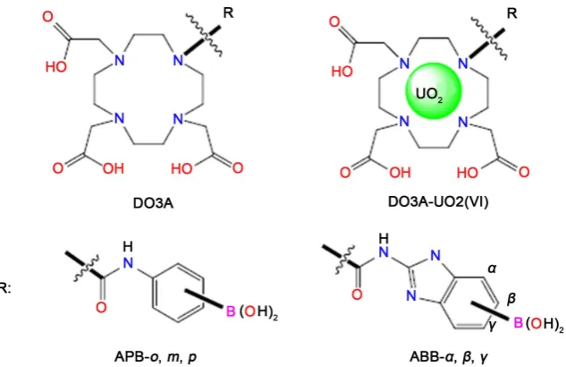
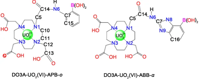
In this sense, new designed DO3A-based sensors containing benzimidazole
and phenyl group with boronic acid have been shown in Figure 1. Their stability
of uranyl ion and possible structures with DO3A have been calculated in water
media and also chemical properties such as EHOMO, ELUMO, the energy gap (ΔE),
chemical potential (μ), hardness (η), softness (S), the absolute electronegativity
(χ) and the electrophilicity index (ω) have been predicted. Moreover, detections of these complexes in water phase spectroscopic properties (UV-Vis, IR and NMR) have been given.
2. Method
DOI: 10.4236/cc.2017.54012 147 Computational Chemistry
Figure 1. Designed DO3A-based sensors containing benzimidazole and phenylgroup.
View 5.0.8 molecular modeling software [17]. Also, ChemCraft program [18]
was used to determine the diameter of the examined structures. M062X [19],
B3LYP [20] and B3PW91 [21] methods that can be suitable for calculations are
selected and compared with experimental absorption value of reference uranium
complex [22]. First of all, U (IV) DO3A complex was optimized with selected
three methods in the gas phase. Optimization in the solvent phase (methanol) was then carried out and the UV-Vis spectrum was obtained. The calculation results compared with experimental value and show that (Table S1 given in Supporting information) M062X functional was found better than the other se-lected methods. After determination of the appropriate method, all subsequent
calculations performed with M062X functional and 6 - 31 + G (d) [23] basis set.
The SARC ZORA basis set was used for the uranium atom [24]. The SMD
Solva-tion Model Density [25] method was chosen to reflect the effect of the water
phase on the calculations. The dielectric constant was chosen as the standard
value for water ε = 78.39). Calculations corresponding to acidic medium
imple-mented with nitrogen protonation. The basic medium is provided by the remov-al of hydrogen atoms in the carboxylic groups on DO3A.
3. Results and Discussion
3.1. DO3A-Based Aminophenol and Aminobenzo Imidazolyl
Boronic Acid Derivatives with and without
22
UO +
3.1.1. Stabilities
After selecting the appropriate method for the calculations, boronic ac-id-containing derivatives of ortho, para and meta positions of DO3A based
sen-sors have been investigated. Table 1 shows that energy values of DO3AAPB and
DO3A-ABB derivatives in gas and water phases.
DOI: 10.4236/cc.2017.54012 148 Computational Chemistry
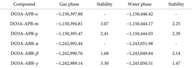
Table 1. Total energies of DO3A-APB and DO3A-ABB structures (kcal/mol).
Compound Gaz phase Stability Water phase Stability
DO3A-APB-o −1,150,397.88 - −1,150,446.42 -
DO3A-APB-m −1,150,394.81 3.07 −1,150,444.17 2.25
DO3A-APB-p −1,150,395.47 2.41 −1,150,444.03 2.39
DO3A-ABB-α −1,242,992.44 - −1,243,051.98 -
DO3A-ABB-β −1,242,990.76 1.68 −1,243,049.84 2.14
DO3A-ABB-γ −1,242,989.14 3.30 −1,243,050.51 1.47
is the more stable than the other positions in the gas phase, about of 2 - 3 kcal/mol energy values. In the water phase, the same trend was found about 2 kcal/mol. When the energy of structures in water and gas phases is compared, it has been found that the presence of water in the environment brings about the solvation stability.
Table 1 shows that DO3A-ABB-α structure is more stable than the other po-sitions in the gas and water phases, about of 1.5 - 3 kcal/mol energy values.
As a result, DO3A-APB-o and DO3A-ABB-α have been found the most stable
within calculated structures and so by choosing these compounds, further
calcu-lations have been continued. Optimized DO3A-APB-o and DO3A-ABB-α
structures have been presented in Table S2 and Table S3.
3.1.2. Structural Properties and Molecular Orbitals
Ortho structures with and without uranyl ion have been examined in terms of their structural, electronic and spectroscopic properties in acidic, basic and neu-tral media. While the acidic medium was formed by protonation of the nitrogen (N6), the basic medium was formed by the removal of the carboxylic proton lo-cated on DO3A. The numbered structures of some selected atoms of all calcu-lated of DO3A-based sensors with/without uranyl ion are presented, acidic,
neu-tral and basic media, in Figure 2 and Table 2.
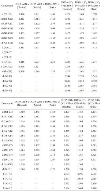
C-C bond lengths of the target compounds are in the range of 1.530 - 1.534 Å. This bond length with uranyl ion is changed from 1.564 to 1.576 Å. For com-pound without uranyl ion, the bond length between nitrogen and carbon in that fragment vary from 1.455 to 1.476 Å. For a compound with uranyl ion it in-creases to 1.464 - 1.528 Å.
The bond length between uranyl and nitrogen are between 2.246 and 1.2684 Å. For uranyl-free structures, the bond length of boron and hydroxide is be-tween 1.363 and 1.371 Å, while bond length with uranium slightly increased to range of 1.377 and 1.399 Å, within indicates three media. Diameters of related structures in the water medium have been determined theoretically and given in
Table S4. DO3A-APB-oand DO3A-ABB-α structures have been found to be
about 11 Å in three mediums. The diameter of the structures with uranyl ion is wider than the others, 1 Å.
DOI: 10.4236/cc.2017.54012 149 Computational Chemistry
Table 2. Optimized bond lengths (Å) of DO3A-APB-o and DO3A-ABB-α structures at
M062X/6 − 31 + G (d) level of theory.
Compound DO3A-APB-o (Neutral) DO3A-ABB-o (Acidic) DO3A-APB-o (Basic) DO3A-UO(VI) APB-o 2 (Neutral)
DO3A-UO2
(VI) APB-o (Acidic)
DO3A-UO2
(VI) APB-o (Basic)
d (N1-C5) 1.446 1.441 1.446 1.476 1.469 1.370
d (N1-C10) 1.463 1.464 1.463 1.506 1.521 1.511
d(C10-C11) 1.534 1.532 1.535 1.564 1.571 1.577
d (N2-C11) 1.475 1.474 1.468 1.522 1.524 1.527
d (N2-C12) 1.455 1.457 1.456 1.477 1.479 1.482
d (N6-C14) 1.353 1.517 1.353 1.355 1.506 1.357
d (C5-C14) 1.527 1.517 1.528 1.535 1.505 1.533
d (N6-C7) 1.411 1.471 1.409 1.414 1.488 1.413
d (N8-C7) - - - -
d (N9-C7) - - - -
d (O-C13) 1.218 1.217 1.258 1.226 1.226 1.271
d (OH-C13) 1.330 1.331 - 1.363 1.363 -
d (B-OH) 1.370 1.364 1.370 1.377 1.362 1.377
d (N1-U) - - - 2.544 2.570 2.543
d (N2-U) - - - 2.685 2.654 2.550
d (N3-U) - - - 2.549 2.507 2.484
d (N4-U) - - - 2.544 2.507 2.492
Compound DO3A-ABB-α (Neutral) DO3A-ABB-α (Acidic) DO3A-ABB-α (Basic) DO3A-UO(VI) ABB-α 2 (Neutral)
DO3A-UO2
(VI) ABB-α (Acidic)
DO3A-UO2
(VI) ABB-α (Basic)
d (N1-C5) 1.450 1.445 1.453 1.477 1.469 1.474
d (N1-C10) 1.463 1.467 1.465 1.515 1.522 1.514
d(C10-C11) 1.534 1.530 1.533 1.569 1.568 1.576
d (N2-C11) 1.476 1.473 1.469 1.521 1.514 1.528
d (N2-C12) 1.456 1.457 1.456 1.469 1.464 1.483
d (N6-C14) 1.364 1.554 1.365 1.373 1.577 1.375
d (C5-C14) 1.524 1.507 1.525 1.532 1.505 1.531
d (N6-C7) 1.385 1.437 1.388 1.384 1.445 1.383
d (N8-C7) 1.364 1.352 1.362 1.325 1.310 1.382
d (N9-C7) 1.310 1.298 1.310 1.382 1.367 1.325
d (O-C13) 1.218 1.214 1.267 1.226 1.225 1.271
d OH-C13) 1.330 1.347 - 1.365 1.365 -
d (B-OH) 1.368 1.371 1.371 1.390 1.390 1.378
d (N1-U) - - - 2.534 2.591 2.555
d (N2-U) - - - 2.677 2.658 2.555
d (N3-U) - - - 2.532 2.506 2.476
DOI: 10.4236/cc.2017.54012 150 Computational Chemistry
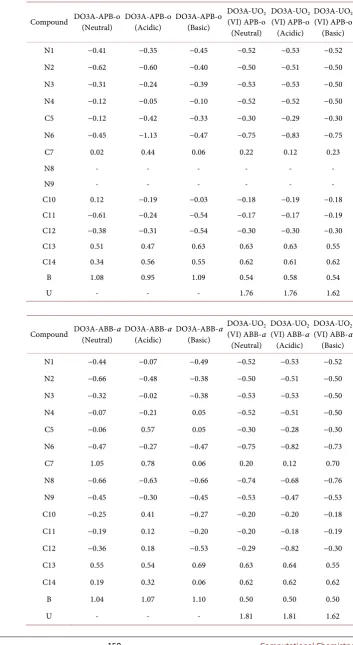
Table 3. Optimized Mulliken charges (Debye) of DO3A-APB-o and DO3A-ABB-α
structures.
Compound DO3A-APB-o (Neutral) DO3A-APB-o (Acidic) DO3A-APB-o (Basic) DO3A-UO(VI) APB-o 2 (Neutral)
DO3A-UO2
(VI) APB-o (Acidic)
DO3A-UO2
(VI) APB-o (Basic)
N1 −0.41 −0.35 −0.45 −0.52 −0.53 −0.52
N2 −0.62 −0.60 −0.40 −0.50 −0.51 −0.50
N3 −0.31 −0.24 −0.39 −0.53 −0.53 −0.50
N4 −0.12 −0.05 −0.10 −0.52 −0.52 −0.50
C5 −0.12 −0.42 −0.33 −0.30 −0.29 −0.30
N6 −0.45 −1.13 −0.47 −0.75 −0.83 −0.75
C7 0.02 0.44 0.06 0.22 0.12 0.23
N8 - - - -
N9 - - - -
C10 0.12 −0.19 −0.03 −0.18 −0.19 −0.18
C11 −0.61 −0.24 −0.54 −0.17 −0.17 −0.19
C12 −0.38 −0.31 −0.54 −0.30 −0.30 −0.30
C13 0.51 0.47 0.63 0.63 0.63 0.55
C14 0.34 0.56 0.55 0.62 0.61 0.62
B 1.08 0.95 1.09 0.54 0.58 0.54
U - - - 1.76 1.76 1.62
Compound DO3A-ABB-α (Neutral) DO3A-ABB-α (Acidic) DO3A-ABB-α (Basic) DO3A-UO(VI) ABB-α 2 (Neutral)
DO3A-UO2
(VI) ABB-α (Acidic)
DO3A-UO2
(VI) ABB-α (Basic)
N1 −0.44 −0.07 −0.49 −0.52 −0.53 −0.52
N2 −0.66 −0.48 −0.38 −0.50 −0.51 −0.50
N3 −0.32 −0.02 −0.38 −0.53 −0.53 −0.50
N4 −0.07 −0.21 0.05 −0.52 −0.51 −0.50
C5 −0.06 0.57 0.05 −0.30 −0.28 −0.30
N6 −0.47 −0.27 −0.47 −0.75 −0.82 −0.73
C7 1.05 0.78 0.06 0.20 0.12 0.70
N8 −0.66 −0.63 −0.66 −0.74 −0.68 −0.76
N9 −0.45 −0.30 −0.45 −0.53 −0.47 −0.53
C10 −0.25 0.41 −0.27 −0.20 −0.20 −0.18
C11 −0.19 0.12 −0.20 −0.20 −0.18 −0.19
C12 −0.36 0.18 −0.53 −0.29 −0.82 −0.30
C13 0.55 0.54 0.69 0.63 0.64 0.55
C14 0.19 0.32 0.06 0.62 0.62 0.62
B 1.04 1.07 1.10 0.50 0.50 0.50
DOI: 10.4236/cc.2017.54012 151 Computational Chemistry
Figure 2. Schematic structures of DO3A-based sensors with/without uranyl ion.
the charges of nitrogen atoms in theDO3A ring reduce in the acidic medium and are in the range of −0.60 to −0.05, when the nitrogen charges in the other medium are compared. For all three medium, these charges are close to reaching −0.50 after the bonded with the uranyl ion. The charge of amide nitrogen is about −0.40 in the neutral and basic medium while it increases to −1.13 in the acidic
medium. In the DO3A-UO2 (VI)-APB-o complexes, it increases to −0.75 in the
neutral and basic medium while it decreases to -0.85 in the acidic medium. The charge of C5 carbon atom linking DO3A to amide falls from -0.40 in the acidic
medium to −0.29 in the UO2 complexes. The charge of the boron atom in the
DO3A-APB-o structures decreases from −1.00 to −0.55 in DO3A-UO2
(VI)-APB-o structures. The uranium (VI) ion charge is in the range of 1.75 to
1.61. Similar trends and values have been found forDO3A-ABB-α and
DO3A-UO2 (VI)-ABB-α structures in all three mediums.
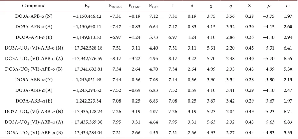
The highest occupied molecular orbital (HOMO) and lowest unoccupied mo-lecular orbital (LUMO) are very important parameters as a measure of relative
stability and reactivity. The energies of frontier orbitals, total energies (ET),
chemical hardness (η), electronic chemical potentials (μ), chemical softness (S)
and electrophilicity (ω) values are listed in Table 4.
When the total energy values of the DO3A-APB-o are examined,it can be seen that the energy stability decreases in these order DO3A-APB-o acidic > DO3A-APB-o
neutral > DO3A-APB-o basic media. The same trend has been for the DO3A-ABB-α,
DO3A-UO2 (VI)-ABB-α and DO3A-UO2 (VI)-APB-o series structures. The
most stable structures are in acidic media.
When the total energy values of the DO3A-APB-o are examined, it can be seen that the energy stability decreases in these order DO3A-APB-o acidic > DO3A-APB-o neutral > DO3A-APB-o basic media. The same trend has been
seen for the DO3A-ABB-α, DO3A-UO2 (VI)-ABB-α and DO3A-UO2 (VI)-APB-o
series structures. The most stable structures have been found in the acidic media. A large HOMO-LUMO energy gap has been associated with high stability of
structures. For DO3A-APB-o and DO3A-ABB-α series, the HOMO-LUMO
DOI: 10.4236/cc.2017.54012 152 Computational Chemistry
Table 4. Values of the global reactivity descriptors of the systems (eV).
Compound ET EHOMO ELUMO EGAP I A χ η S μ ω
DO3A-APB-o (N) −1,150,446.42 −7.31 −0.19 7.12 7.31 0.19 3.75 3.56 0.28 −3.75 1.97
DO3A-APB-o (A) −1,150,690.41 −7.47 −0.83 6.64 7.47 0.83 4.15 3.32 0.30 −4.15 2.60
DO3A-APB-o (B) −1,149,613.33 −6.97 −1.24 5.73 6.97 1.24 4.10 2.86 0.35 −4.10 2.94
DO3A-UO2 (VI)-APB-o (N) −17,342,528.18 −7.51 −3.11 4.40 7.51 3.11 5.31 2.20 0.45 −5.31 6.41
DO3A-UO2 (VI)-APB-o (A) −17,342,776.59 −8.17 −3.22 4.95 8.17 3.22 5.70 2.48 0.40 −5.70 6.55
DO3A-UO2 (VI)-APB-o (B) −17,341,682.81 −7.34 −2.64 4.70 7.34 2.64 4.99 2.35 0.43 −4.99 5.30
DO3A-ABB-α (N) −1,243,051.98 −7.44 −0.36 7.08 7.44 0.36 3.90 3.54 0.28 −3.90 2.15
DO3A-ABB-α (A) −1,243,294.62 −7.52 −0.69 6.83 7.52 0.69 4.10 3.41 0.29 −4.10 2.47
DO3A-ABB-α (B) −1,242,223.34 −7.08 −0.25 6.83 7.08 0.25 3.67 3.42 0.29 −3.67 1.97
DO3A-UO2 (VI)-ABB-α (N) −17,435,128.24 −7.26 −3.19 4.07 7.26 3.19 5.23 2.04 0.49 −5.23 6.71
DO3A-UO2 (VI)-ABB-α (A) −17,435,369.38 −7.95 −3.31 4.64 7.95 3.31 5.63 2.32 0.43 −5.63 6.83
DO3A-UO2 (VI)-ABB-α (B) −17,434,284.04 −7.21 −2.66 4.55 7.21 2.66 4.93 2.27 0.44 −4.93 5.35
lated chemical hardness (η), DO3A-APB-o in basic media, DO3A-ABB-α in
acidic media and both DO3A-UO2 (VI)-APB-o and DO3A-UO2 (VI)-ABB-α
structures in neutral media have lower values than the rest of the their series and these result indicating that they are kinetically more stable and less reactive than the other their structures.
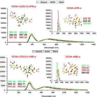
3.2. Absorption Spectra Analysis
The absorption spectrum of structures has been calculated using Time-Dependent DFT calculations same levels of theory in water phase three indicated media.
The calculated absorption bands are summarized in Figure 3.
For DO3A-APB-o compound in the indicated mediums absorption bands outside the visible region and are range of 205 nm - 230 nm. It can be seen from the Figure 3, when DO3A-APB-o bonded with uranyl ion, DO3A-UO2 (VI)-APB-o,
oscillator strength decreases and shift to long wavelength. At the beginning, a single peak has seen and two flattened peaks have formed at with the addition of uranyl ion. These peaks are in the range of 384 - 392 nm and 645 - 695 nm. For
DO3A-ABB-α, calculated absorption bands are range in 200 - 264 nm within
three indicated media. DO3A-UO2 (VI)-ABB-α gives the same trend and these
peaks are in the range of 374 - 393 nm and 645 - 688 nm. Consequently, when DO3A-APB-o structure bonds with uranyl ion, the maximum shift is in the
neu-tral medium, whereas maximum shifts of DO3A-ABB-α with uranyl ion are in
the basic medium.
3.3. Infrared and NMR Spectra Analysis
In this section, the changes in 13C-NMR and 1H-NMR chemical shift values of
the all the calculated complexes formed by the binding of uranyl ions have been
DOI: 10.4236/cc.2017.54012 153 Computational Chemistry
Table 5. 13C-NMR chemical shifts (ppm) of DO3A-APB-o and DO3A-ABB-α and their
structures with uranyl ion.
Compound DO3A-APB-o (Neutral) DO3A-APB-o (Acidic) DO3A-APB-o (Basic) DO3A-UO(VI) APB-o 2 (Neutral)
DO3A-UO2
(VI) APB-o (Acidic)
DO3A-UO2
(VI) APB-o (Basic)
C5 62.9 63.8 63.6 59.8 64.6 60.7
C7 158.6 145.2 158.3 158.7 152.3 159.7
C10 57.4 58.0 56.6 59.1 56.2 53.8
C11 46.7 48.7 45.9 64.6 65.1 59.6
C12 61.8 62.9 66.2 71.4 70.5 70.2
C13 191.3 189.7 182.6 189.3 189.2 191.4
C14 18.3 196.5 187.9 185.3 195.7 185.7
Compound DO3A-ABB-α (Neutral) DO3A-ABB-α (Acidic) DO3A-ABB-α (Basic) DO3A-UO(VI) ABB-α 2 (Neutral)
DO3A-UO2
(VI) ABB-α (Acidic)
DO3A-UO2
(VI) ABB-α (Basic)
C5 64.5 63.9 64.3 61.8 64.5 61.0
C7 149.2 142.4 154.5 61.8 144.6 155.3
C10 52.0 53.7 49.0 54.1 54.4 54.6
C11 63.7 62.6 61.6 54.7 56.8 60.6
C12 61.1 62.3 65.9 61.9 62.8 70.8
C13 190.1 189.9 192.1 188.0 187.7 191.2
C14 185.0 184.0 186.6 188.0 184.3 184.0
Table 6. 1H-NMR chemical shifts (ppm) of DO3A-APB-o and DO3A-ABB-α and their
structures with uranyl ion.
Compound DO3A-APB-o (Neutral) DO3A-APB-o (Acidic) DO3A-APB-o (Basic) DO3A-UO(VI) APB-o 2 (Neutral)
DO3A-UO2
(VI) APB-o (Acidic)
DO3A-UO2
(VI) APB-o (Basic)
BO-H 4.9 5.4 4.8 6.0 6.7 5.7
N6-H 11.0 6.4 11.2 12.4 8.1 11.8
N8-H - - - -
C14-H 7.0 7.5 - 8.3 8.3 -
C10-H 2.3 2.6 2.2 3.5 3.3 3.8
C11-H 3.1 2.9 3.3 3.9 4.1 3.9
C15-H 9.3 7.8 9.2 8.3 8.4 9.5
Compound DO3A-ABB-α (Neutral) DO3A-ABB-α (Acidic) DO3A-ABB-α (Basic) DO3A-UO(VI) ABB-α 2 (Neutral)
DO3A-UO2
(VI) ABB-α (Acidic)
DO3A-UO2
(VI) ABB-α (Basic)
BO-H 5.1 6.1 5.1 5.9 5.9 5.8
N6-H 9.4 11.1 9.5 8.4 8.3 8.0
N8-H 9.9 10.6 10.2 10.0 11.1 9.8
C14-H 9.4 11.5 - 8.3 8.3 -
C10-H 3.6 2.4 2.2 3.7 3.3 3.8
C11-H 3.0 2.5 3.3 3.3 3.9 2.7
[image:9.595.207.539.436.744.2]DOI: 10.4236/cc.2017.54012 154 Computational Chemistry
Figure 3. Computed UV-Vis absorption bands. The λmax values of structures also have
been indicated on the graphs.
Firstly, the chemical shift of TMS (tetramethylsilane) has been calculated with the same level of theory in order to carry out the NMR calculations. Subsequently, the NMR spectrum of the structures has been calculated and the difference
be-tween the two values gives the final result. In the calculated 13C-NMR spectra, a
chemical shift in the C5 atom ca. 0.5 - 3 ppm is observed in the high field while C11 carbon (ca. 14 - 18 ppm) has low field for DO3A-APB-o structures. The same trend exists for C12 (ca. 10 ppm). Similarly, C5 and C11 carbon atoms
be-long to DO3A-ABB-α structures have been found in the high field ca. 1 ppm and
ca. 5 - 9 ppm, respectively. There is no significant change in other carbon atoms,
but there is a low shift in C10 atom, ca. 2 - 5 ppm. In the calculated 1H-NMR
spectra, the corresponding proton peaks connected to C10, C11 (ca. 1 ppm) and N6 (ca. 2 ppm) of DO3A-APB-o structures have shifted to the low field. In the
DO3A-ABB-α structures, there are only high field shift in N6-H and C10-H
peaks, ca. 1 - 2 ppm, there is no big differences for the other peaks.
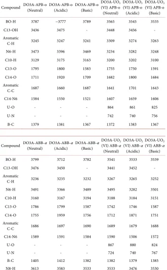
The calculated harmonic vibrations and most characteristic peaks of the
compounds are also included in this part and given in Table 7. The main effect
on DO3A-UO2 (VI)-APB-o on calculated IR-spectrum is seen at the BO-H and
N6-H stretching vibrations for all mediums. The bond stretching vibrations of
DOI: 10.4236/cc.2017.54012 155 Computational Chemistry
Table 7. Selected and calculated vibrational frequency (cm−1).
Compound DO3A-APB-o (Neutral) DO3A-APB-o (Acidic) DO3A-APB-o (Basic) DO3A-UO(VI) APB-o 2 (Neutral)
DO3A-UO2
(VI) APB-o (Acidic)
DO3A-UO2
(VI) APB-o (Basic)
BO-H 3787 −3777 3789 3565 3545 3535
C13-OH 3436 3475 - 3448 3456 -
Aromatic
C-H 3245 3247 3241 3309 3274 3263
N6-H 3473 3396 3469 3234 3282 3248
C10-H 3129 3175 3163 3200 3202 3100
C13-O 1795 1800 1583 1755 1750 1591
C14-O 1711 1920 1709 1682 1800 1684
Aromatic
C-C 1687 1660 1687 1641 1701 1643
C14-N6 1584 1550 1521 1607 1659 1606
U-O - - - 864 861 825
U-N - - - 742 740 756
B-C 1379 1381 1367 1372 1383 1367
Compound DO3A-ABB-α (Neutral) DO3A-ABB-α (Acidic) DO3A-ABB-α (Basic) DO3A-UO(VI) ABB-α 2 (Neutral)
DO3A-UO2
(VI) ABB-α (Acidic)
DO3A-UO2
(VI) ABB-α (Basic)
BO-H 3799 3712 3782 3541 3533 3539
C13-OH 3476 3450 - 3441 3452 -
Aromatic
C-H 3236 3235 3232 3267 3265 3252
N6-H 3491 3366 3489 3495 3282 3501
C10-H 3160 3167 3194 3188 3184 3151
C13-O 1786 1799 1587 1742 1746 1587
C14-O 1755 1959 1756 1712 1871 1751
Aromatic
C-C 1686 1697 1690 1689 1679 1688
C14-N6 1589 1591 1584 1590 1506 1572
U-O - - - 867 880 824
U-N - - - 724 740 767
B-C 1405 1412 1382 1382 1379 1385
N8-H 3613 3583 3533 3533 3476 3530
medium. The differences in between DO3A-APB-o and DO3A-UO2 (VI)-APB-o
of BO-H stretching modes are among 222 and 254 cm−1 and the N6-H one is in
between 114 - 239 cm−1 in neutral and acidic medium, appears in a lower
fre-quency. In DO3A-UO2 (VI)-ABB-α structures, the bond stretching vibrations of
DOI: 10.4236/cc.2017.54012 156 Computational Chemistry
medium. When the stretching mode of BO-H passing from DO3A-APB-o to
DO3A-UO2 (VI)-ABB-α structures have been shifted to lower frequency (180 - 258
cm−1). There is no significant difference in the basic medium at N8-H stretching
vibration frequency, whereas a shift has been found at about 80 - 100 cm−1 in the
neutral and acidic medium.
4. Conclusion
Our theoretical work represents structural and spectral properties of newly
de-signed DO3A-based structures sensitive to 2
2
UO +. The structures have been
op-timized in the gas phase and water phase at three levels of theory (M062X, B3LYP and B3PW9/6 - 31 g + (d)) for comparison with experimental value, after that M062X/6 - 31 g + (d) method has been chosen for all remaining calculations. The calculations have been shown that ortho position of boronic acid is the most stable within all calculated structures. By examining the diameter dimension of the structural features, it has been seen that the linking of the uranium causes the increase of the total diameter up to about 1 Å. The relative stability of the studied structures showed that all series are more stable in the acidic medium.
The calculated HOMO-LUMO gap results manifest that linking of 2
2
UO + ion
to designed structures has caused the decrease. The theoretical electronic
spec-trum of DO3A-UO2 (VI)-APB-o gives bands at 392 and 687 nm in neutral
me-dium. DO3A-UO2 (VI)-ABB-α structure has two main bands at 393 and 650 nm.
Both structures show red shift and are located within the visible region by
bind-ing 2
2
UO + ion. The notable shifts in NMR spectrums have been found on
α-carbon of carbonyl group, ring carbons and amide protons. In IR spectrum,
the prominent peaks belong to BO-H and N6-H stretching vibrations of calcu-lated structures.
Acknowledgements
This work was supported by The Scientific and Technological Research Council of Turkey (TÜBİTAK) (No: 115Z504). The calculations were performed on Tur-kish Academic Network and Information Center (ULAKBİM).
References
[1] Ishihara, K., and Yamamoto, H. (1999) Arylboron Compounds as Acid Catalysts in Organic Synthetic Transformations. European Journal of Organic Chemistry, 1999, 527-538,
https://doi.org/10.1002/(SICI)1099-0690(199903)1999:3<527::AID-EJOC527>3.0.C O;2-R
[2] Petasis, N.A. and Zavialov, I.A. (1997) A New and Practical Synthesis of A-Amino Acids From Alkenyl Boronic Acids. Journal of the American Chemical Society, 119, 445-446. https://doi.org/10.1021/ja963178n
[3] Miyaura, N. and Suzuki, A. (1997) Palladium-Catalyzed Cross-Coupling Reactions of Organoboron Compounds. Chemical Reviews, 95, 2457-2483.
DOI: 10.4236/cc.2017.54012 157 Computational Chemistry
[4] Latta, R., Springsteen, G. and Wang, B. (2001) Development and Synthesis of an Arylboronic Acid-Based Solid-Phase Amidation Catalyst. Synthesis, 2001, 1611-1613. https://doi.org/10.1055/s-2001-16758
[5] James, T.D., Sandanayake, S. and Shinkai, S. (1996) Saccharide Sensing with Mole-cular Receptors Based on Boronic Acid. Angewandte Chemie International Edition, 35, 1910-1922. https://doi.org/10.1002/anie.199619101
[6] Liua, S., Shia, F., Zhao, B., Chena, L. and Sua, X. (2013) 3-Aminophenyl Boronic Acid-Functionalized CuInS2 Quantum Dots as A Near Infrared Fluorescence Probe
for the Determination of Dopamine. Biosensors & Bioelectronics, 47, 379-384.
https://doi.org/10.1016/j.bios.2013.03.055
[7] DiCesare, N. and Lakowicz, J.R. (2002) New Sensitive and Selective Fluorescent Probes for Fluoride Using Boronic Acids. Analytical Biochemistry, 301, 111-116.
https://doi.org/10.1006/abio.2001.5476
[8] DiCesare, N. and Lakowicz, J.R. (2001) Spectral Properties of Fluorophores Com-bining the Boronic Acid Group with Electron Donor or Withdrawing Groups. Im-plication in the Development of Fluorescence Probes for Saccharides. Journal of
Physical Chemistry A, 105, 6834-6840. https://doi.org/10.1021/jp010076x
[9] Barge, A., Cappelletti, E., Cravotto, G., Ferrigato, A., Lattuada, L., Marinonib, F. and Tei, L. (2009) Synthesis of Functionalised HP-DO3A Chelating Agents for Conjuga-tion to Biomolecules. Organic & Biomolecular Chemistry, 7, 3810-3816.
https://doi.org/10.1039/b905369g
[10] Riesen, A., Zehnder, M. and Kaden, T.A. (1986) Metal Complexes of Macrocyclic Ligands. Part XXIII. Synthesis, Properties, and Structures of Mononuclear Com-plexes with 12- and 14-Membered Tetraazamacrocycle-N,N′,N″,N‴-Tetraacetic Acids. Helvetica Chimica Acta, 69, 2067-2073.
https://doi.org/10.1002/hlca.19860690830
[11] Rösch, F., Herzog, H., Stolz, B., Brockmann, J., Köhle, M., Mühlensiepen, H., Mar-bach, P. and Gärtner, H.W. (1999) Uptake Kinetics of the Somatostatin Receptor Ligand [86Y]DOTA-Dphe1-Tyr3-Octreotide ([86Y]SMT487) using Positron Emis-sion Tomography in Non-Human Primates and Calculation of Radiation Doses of The 90Y-Labelled Analogue. European Journal of Nuclear Medicine, 26, 358-366.
https://doi.org/10.1007/s002590050398
[12] Kumar, K., Chang, C.A., Francesconi, L.C., Dischino, D.D., Malley, M.F., Gougou-tas, J.Z. and Tweedle, M.F. (1994) Synthesis, Stability, and Structure of Gadoli-nium(III) and Yttrium(III) Macrocyclic Poly(Amino Carboxylates). Inorganic
Chemistry,33, 3567-3575.https://doi.org/10.1021/ic00094a021
[13] Loncin, M.F., Desreux, J.F. and Merciny, E. (1986) Coordination of Lanthanides by Two Polyamino Poly Carboxylic Macrocycles: Formation of Highly Stable Lantha-nide Complexes. Inorganic Chemistry, 25, 2646-2648.
[14] Iglesias, C.P. (2012) The Solution Structurea nd Dynamics of MR Probes Based on Lanthanide(III) Dota as Investigated by DFT and NMR Spectroscopy. European
Journal of Inorganic Chemistry,12,2023-2033.
https://doi.org/10.1002/ejic.201101164
[15] Fernandez, M., Bastida, R., Macıas, A., Perez-Lourido, P., Iglesias, C.P. and Valen-cia, L. (2006) Lanthanide(III) Complexes with a Tetrapyridine Pendant-Armed Ma-crocyclic Ligand: 1H NMR Structural Determination in Solution, X-Ray Diffraction,
and Density-Functional Theory Calculations. Inorganic Chemistry, 45, 4484-4496.
https://doi.org/10.1021/ic0603508
DOI: 10.4236/cc.2017.54012 158 Computational Chemistry
J.R., Scalmani, G., Barone, V., Mennucci, B., Petersson, G.A., Nakatsuji, H., Carica-to, M., Li, X., Hratchian, H.P., Izmaylov, A.F., Bloino, J., Zheng, G., Sonnenberg, J.L., Hada, M., Ehara, M., Toyota, K., Fukuda, R., Hasegawa, J., Ishida, M., Nakaji-ma, T., Honda, Y., Kitao, O., Nakai, H., Vreven, T., Montgomery, J.A., Peralta, J.E., Ogliaro, F., Bearpark, M., Heyd, J.J., Brothers, E., Kudin, K.N., Staroverov, V.N., Kobayashi, R., Normand, J., Raghavachari, K., Rendell, A., Burant, J.C., Iyengar, S.S., Tomasi, J., Cossi, M., Rega, N., Millam, J.M., Klene, M., Knox, J.E., Cross, J.B., Bakken, V., Adamo, C., Jaramillo, J., Gomperts, R., Stratmann, R.E., Yazyev, O., Austin, A.J., Cammi, R., Pomelli, C., Ochterski, J.W., Martin, R.L., Morokuma, K., Zakrzewski, V.G., Voth, G.A., Salvador, P., Dannenberg, J.J., Dapprich, S., Daniels, A.D., Farkas, Ö., Foresman, J.B., Ortiz, J.V., Cioslowski, J. and Fox, D.J. (2009) Gaussian 09, Revis. D.01.
[17] Dennington, R.D., Keith, T.A. and Millam, J.M. (2008) GaussView 5.0.8, Gaussian.
[18] https://www.chemcraftprog.com
[19] Walker, M., Harvey, A., Sen, A. and Dessent, C. (2013) Performance of M06, M06-2X, and M06-HF Density Functionals for Conformationally Flexible Anionic Clusters: M06 Functionals Perform Better than B3LYP for a Model System with Dispersion and Ionic Hydrogen-Bonding Interactions. The Journal of Physical
Chemistry A,117, 12590-12600.https://doi.org/10.1021/jp408166m
[20] Rives, T.J. and Jorgensen, W.L. (2008) Performance of B3LYP Density Functional Methods for a Large Set of Organic Molecules. Journal of Chemical Theory and
Computation,4, 297-306.https://doi.org/10.1021/ct700248k
[21] Govindarajana, M., Ganasanb, K., Periandyc, S. and Mohand, S. (2010) DFT (LSDA, B3LYP and B3PW91) Comparative Vibrational Spectroscopic Analysis of Aceto-naphthone. Spectrochimica Acta Part A, 76, 12-21.
[22] Natrajan, L.S. (2012) The First Structural and Spectroscopic Study of a Paramag-netic 5f DO3A Complex. Dalton Tran,41, 13167.
https://doi.org/10.1039/c2dt30573a
[23] Del Bene, J.E. (1995) Properties of Hydrogen-Bonded Complexes Obtained from the B3LYP Functional with 6-31G(d,p) and 6-31+G(d,p) Basis Sets: Comparison with MP2/6-31+G(d,p) Results and Experimental Data. The Journal of Physical
Chemistry, 99, 10705-1070.https://doi.org/10.1021/j100027a005
[24] Pantazis, D.A., Chen, X., Landi,s C.R. and Neese, F. (2008) All-Electron Scalar Rela-tivistic Basis Sets for Third-Row Transition Metal Atoms. Journal of Chemical
Theory and Computation,4, 908-919.https://doi.org/10.1021/ct800047t
[25] Marenich, A.V., Cramer, C.J. and Truhlar, D.G. (2009) Universal Solvation Model Based on Solute Electron Density and on Acontinuum Model of the Solvent Defined by the Bulk Dielectric Constant and Atomic Surface Tensions. The Journal of