AN AUTOMATIC PROBABILISTIC FRAMEWORK FOR BRAIN TUMOUR DETECTION USING
*,1
Nageswara Reddy,
1
Research Scholar, Jawaharlal Nehru Technological University, Kakinada
2Professor and Principal, Avanthi Institute of Engineering & Technology
3Professor, Department of
ARTICLE INFO ABSTRACT
Improvement of automated frameworks for detection of brain tumour is the real need of the clinical enhancement. A Moderate numbers of methods are introduced to analyse the biological symptoms and produce the report to be recognized by the trained professio
to errors due because of human interpretations. Also the computer aided reports leave huge scope for multiple further diagnoses. Thus the need of a novel algorithm for predictive analysis of diseases for brain di
mechanism based on an enhancement in accuracy of multilateral filter and EM multilateral filter enhances the image edges for better segmentation using sign
of the pixel. The final outcome of this paper produces the brain regions detected with anomalies and possible diseases, thus the number of possible further medical investigations are reduces.
Copyright©2016, Nageswara Reddy et al. This is an open access article distributed under the Creative Commons Att use, distribution, and reproduction in any medium, provided the original work is properly cited.
INTRODUCTION
The Detection of brain tumours are the key area of focus for many researchers. The process of detection is manual and prune to human errors. Thus, the researchers are bounds to carry out multiple autonomous framework based researches. Gliomas are the highly in-focus and malignant type
originated from the glial cells in the brain. Due to the malignant type, they grow fast in the surrounding cells and turns into high grade type of glioma. After the detection of these types of tumour, the life span of the patient is eventually r
to three years and prerequisites a gastric pre and post neurosurgical procedure. Nonetheless, the detection delay can reduce the life span further. Due to the criticalness of the type, patients need to undergo for extraordinary frequency of diagnosis and the client results in gigantic amount of datasets. Processing those datasets manually is extremely effort consuming. The collected dataset are eventually entailing with quantitative information and can be best captured in MRI methods.The MR Image method reproduces the tumour information in 3D and 4D structures with the quantified boundaries. Nevertheless, the process of representation and
*Corresponding author: Nageswara Reddy,
Research Scholar, Jawaharlal Nehru Technological University,
ISSN: 0975-833X
Article History:
Received 23rd September, 2016
Received in revised form 10th October, 2016
Accepted 29th November, 2016
Published online 30th December,2016
Key words:
Brain MR Images, T1, T2, T1C, Flair, EM-GM Method, Multilateral Filter, Disorder Detection.
Citation: Nageswara Reddy, P., Mohan Rao, C. P. V. N. J. and Ch.Satyanarayana
detection using MR images”, International Journal of Current Research,
RESEARCH ARTICLE
AN AUTOMATIC PROBABILISTIC FRAMEWORK FOR BRAIN TUMOUR DETECTION USING
MR IMAGES
P.,
2Mohan Rao, C. P. V. N. J. and
3Ch.Satyanarayana
Research Scholar, Jawaharlal Nehru Technological University, Kakinada
Professor and Principal, Avanthi Institute of Engineering & Technology
Professor, Department of Computer Science and Engineering, JNTU Kakinada
ABSTRACT
Improvement of automated frameworks for detection of brain tumour is the real need of the clinical enhancement. A Moderate numbers of methods are introduced to analyse the biological symptoms and produce the report to be recognized by the trained professions. Conversely, the final analysis is prone to errors due because of human interpretations. Also the computer aided reports leave huge scope for multiple further diagnoses. Thus the need of a novel algorithm for predictive analysis of diseases for brain disorder is much expected. This paper presents a fully reliable brain disease detection mechanism based on an enhancement in accuracy of multilateral filter and EM
multilateral filter enhances the image edges for better segmentation using sign
of the pixel. The final outcome of this paper produces the brain regions detected with anomalies and possible diseases, thus the number of possible further medical investigations are reduces.
is an open access article distributed under the Creative Commons Attribution License, which use, distribution, and reproduction in any medium, provided the original work is properly cited.
are the key area of focus for many researchers. The process of detection is manual and prune to human errors. Thus, the researchers are bounds to carry out multiple autonomous framework based researches. focus and malignant type of tumours originated from the glial cells in the brain. Due to the malignant type, they grow fast in the surrounding cells and turns into high grade type of glioma. After the detection of these types of tumour, the life span of the patient is eventually reduced to two to three years and prerequisites a gastric pre and post neurosurgical procedure. Nonetheless, the detection delay can reduce the life span further. Due to the criticalness of the type, patients need to undergo for extraordinary frequency of iagnosis and the client results in gigantic amount of datasets. Processing those datasets manually is extremely effort consuming. The collected dataset are eventually entailing with quantitative information and can be best captured in MRI mage method reproduces the tumour information in 3D and 4D structures with the quantified boundaries. Nevertheless, the process of representation and
Research Scholar, Jawaharlal Nehru Technological University, Kakinada
detection remains a difficult process as the glioma tumours preserve to grow (Menze et al
models of segmentation perform better in these situations. The framework, cited and proposed by many researchers, also includes a normalization phase, where the MR Image is being enhanced. Also the image modality can differ as MRI
MRI – FLAIR based on the segmentation algorithms used. This is proven that, various segmentation algorithms perform better on different types of MR Images. The recent researches also fail to achieve the unsurpassed accuracy
Henceforth the rest of the paper is furnished with the focus to exhibit the improvement in accuracy of disorder detection for T1, T2, T1C and FLAIR type MR Images.
Generic process of gliomas tumour types from
The following are the steps for detecting brain tumour in human using MRI scan.
A. Image Acquisition
In MRI scan we get images of brain and these scanned images will display in two dimensional matrices along with their pixels as its elements. Matrices will be dependent on size of the matrix and its field of view. The scanned images will be International Journal of Current Research
Vol. 8, Issue, 12, pp.43542-43547, December, 2016
INTERNATIONAL
Reddy, P., Mohan Rao, C. P. V. N. J. and Ch.Satyanarayana, 2016.“An automatic probabilistic framework for brain tumour
International Journal of Current Research, 8, (12), 43542-43547.
AN AUTOMATIC PROBABILISTIC FRAMEWORK FOR BRAIN TUMOUR DETECTION USING
Ch.Satyanarayana
Research Scholar, Jawaharlal Nehru Technological University, Kakinada
Professor and Principal, Avanthi Institute of Engineering & Technology
Science and Engineering, JNTU Kakinada
Improvement of automated frameworks for detection of brain tumour is the real need of the clinical enhancement. A Moderate numbers of methods are introduced to analyse the biological symptoms and ns. Conversely, the final analysis is prone to errors due because of human interpretations. Also the computer aided reports leave huge scope for multiple further diagnoses. Thus the need of a novel algorithm for predictive analysis of diseases for sorder is much expected. This paper presents a fully reliable brain disease detection mechanism based on an enhancement in accuracy of multilateral filter and EM-GM method. The multilateral filter enhances the image edges for better segmentation using signal amplitude moderation of the pixel. The final outcome of this paper produces the brain regions detected with anomalies and possible diseases, thus the number of possible further medical investigations are reduces.
ribution License, which permits unrestricted
detection remains a difficult process as the glioma tumours et al., 2014). Thus the predictive models of segmentation perform better in these situations. The framework, cited and proposed by many researchers, also des a normalization phase, where the MR Image is being enhanced. Also the image modality can differ as MRI – T2 or FLAIR based on the segmentation algorithms used. This is proven that, various segmentation algorithms perform better of MR Images. The recent researches also fail to achieve the unsurpassed accuracy (Bauer et al., 2013). Henceforth the rest of the paper is furnished with the focus to exhibit the improvement in accuracy of disorder detection for
e MR Images.
Generic process of gliomas tumour types from MR images
The following are the steps for detecting brain tumour in
In MRI scan we get images of brain and these scanned images will display in two dimensional matrices along with their pixels as its elements. Matrices will be dependent on size of the matrix and its field of view. The scanned images will be INTERNATIONAL JOURNAL OF CURRENT RESEARCH
stored in the file and will be displayed as a grey scale image which ranges from 0 to 255. Where 0 shows the total black colour and 255 shows the entire white colour in which ranges vary intensity between black and white
B. Pre-Processing Stage
In this stage noise reduction and enhancement techniques are implemented for obtaining the best result of an image. These results in more prominent edges and a sharpened image is obtained, noise will be reduced thus reducing the blurring effect from the image. In addition to this image segmentation (edge detection method) is applied which helps in detecting the edges of image for finding the exact location of the tumour.
1) Text Removal: In this stage all unwanted text-noise will be removed. As the MRI scanned image may contain some text in image
2) Noise Removal: In this stage different types Low pass filters are used to remove the noise from the images. Filters are used to remove salt and pepper noise from the image. This filter pixel’s value is replaced with its neighbourhood values.
3) Image Sharpening: After removing noise from the image the image is sharpened by using different high pass filters like Gaussian high pass filter that are widely used for sharpening the edges, which helps in detecting the boundaries of the tumour.
C. Skull Stripping
Skull striping is an important in medical image. It is done only to brain part, since it has to eliminate non brain tissues like extra cerebral tissues, fat, skin etc., it can be done by several techniques.
D. Processing Stage
In this stage segmentation of the image is done based on the division of the image of similar attributes into regions. These regions are grouped depending upon the similarity that helps in extracting the important features which information can be easily perceived.
E. Post-Processing Stage
In this processing stage segmentation of are done using different types of techniques or methods using algorithms which help for detecting accurate and effective location of tumour from brain. Some of the techniques or methods for segmentation are: watershed Segmentation, Threshold Segmentation, K-Means, Fuzzy C-Means… etc.
F. Morphological Operators
After conversion of image to binary format some morphological operations are performed to separate tumour part from the image. Only tumour part is visible in white in colour. The tumour portion contains highest intensity than other any regions of the image. This paper describes the applicability and improvement on tumour detection using EM-GM method. Thus the next section describes the functional parameters of EM-GM method.
EM – GM Method
In this section, the work explains the original EM – GM method and evaluates the applicability for image segmentation. Considering x is set of n independent sample of a mixture of multivalued distribution with dimension d.
1 2 3
( , , ,.... )
nx
x x x
x
(1)
and
Z is the connecting set of variable determines the component from which the result should be collected.
1 2 3
( , , ,.... )
nz
z z z
z
(2)
Thus,
1 1
| (
1)
( ,
)
i i d
X
Z
(3) and
2 2
| (
2)
(
,
)
i i d
X
Z
(4)
Where
1
(
i1)
P Z
(5)
and
2 1
(
i2)
1
P Z
(6)
Also the mixing value is calculated,
1 2 1 2
( ,
,
,
,
)
(7)
Thus the likelihood function can be represented as,
2
1 1
( ; )
( ;
,
),
n
j i j j
j i
L
f
x
x μ
(8)
Hence, the complete likelihood function is represented as,
2 1 1
( ; , )
( , | )
(
)
( ;
,
)
n
i i j j j
j i
L
p
z
j f
x z
x z
I
x μ
(9)
Henceforth, in the next section this work proposed the automatic probabilistic framework for tumour detection.
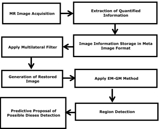
Proposed Framework
Fig.1. Proposed framework for Brain Anomaly Detection
A. Novel Multilateral Filter
The Proposed multilateral filter is based on the existing bilateral filter for improving the input image variance and standard deviation (Stilleet al., 1965; Subbannaet al., 2013).
The bilateral filter explained as
1 1
1
2 1 2 2 1
1 ( )
( )
( ). ( , ). ( ( ), ( ))
Co Px
IMG Co
N Co
IMG Co g Co Co P IMG Co IMG Co
(10)
Where,
IMG
, denotes the original imageIMG, denotes the filtered and noise removed image
1
Co
andCo2, denotes the spatial coordinates of the image Px, denotes the collection of pixels around the noise
1 ( )
N Co , denotes the normalization constant for each pixel to
restrict the value after normalization within geometric and photonic range denoted by Px
And g and p, denotes the geometric and photometric similarities of the image
Hence the enhancement of the image is proposed to regularize the local signal amplitude of every pixel value:
1
1 2 1 2
1
1
( ) ( ). ( , , )
( )Co Px
IMG Co IMG Co Co Co t
N Co
(11) As,
1 2
1 1 2
1
1 1 2 1 2 1 2
1
( , , )
(1 ( )). ( , )
( ). ( , ). ( ( ), ( )). ( , )
D
i i
Co Co t
a Co g Co Co
a Co g Co Co P IMG Co IMG Co d Co Co
(12) Where,
1 ( )
a Co
, denotes the regularized local signal amplitude of the pixel
di, denotes the image dimensions for during noise removal
B. Enhanced Expectation Maximization and Gaussian Mixture
The existing Gaussian mixture method (Gooyaet al., 2011; Weizman,2012; Avantset al., 2011) is applied for each pixel of the normalized image. After application of Gaussian mixture method, the expectation maximization needs to be applied. Thereafter, the Gaussian parameters are to be mapped into the score point and finally, the likelihood to be calculated to converge. Based on the region marks, this technique will predict the possible diseases (Table – 1)(Achantaet al.,2012; Hameeteman,2011; Ahmedet al., 2015). The predictions of diseases are demonstrated in results and discussion section of this paper.
Multilateral Filter Outcomes
The improvement in the input images are been recorded (Table – 2 to 5) and the improvement in variance and standard deviation is been observed. The improvements are visualized in this section (Fig. 2 to 5).
Apply EM-GM Method
MR Image Acquisition Extraction of Quantified Information
Apply Multilateral Filter
Region Detection Image Information Storage in Meta
Image Format
Generation of Restored Image
[image:3.595.64.519.593.647.2]Predictive Proposal of Possible Dieses Detection
Table 1. Brain Anomalous Area and Prediction of Diseases
Brain Region Predictable Diseases
Amygdala Memory Loss, Anxiety, Phobia, Post – Traumatic Disorder
Prefrontal Cortex Stress
Anterior Cingulate Cortex ADHD, Schizophrenia, Depression
Hippocampus Mood Disorder
Table 2. Improvement in the Input Data by Multilateral Filter – FLAIR
Image DatasetIn MHA
Actual Image Variance
Filtered Image
Variance Improvement
Actual Image Std. Deviation
Filtered Image Std.
Deviation Improvement
Dataset – 1 5060902.713 4915045.117 0.02882 69.797568 69.568505 0.003282
Dataset – 2 9554696.161 16036161.27 0.678354 77.144764 88.461004 0.146688
Dataset – 3 8616966.903 9325425.899 0.082217 73.134012 74.266098 0.01548
Dataset – 4 2898009.131 5441952.068 0.877824 58.438954 68.754658 0.176521
Dataset – 5 3297844.713 3310126.799 0.003724 60.892958 61.294396 0.006593
Dataset – 6 7956455.468 9209704.089 0.157513 61.032613 63.335792 0.037737
Dataset – 7 4355136.222 4923593.077 0.130526 68.444576 70.463172 0.029492
Dataset – 8 21368886.14 18066531.68 0.15454 63.106152 60.306925 0.044357
Dataset – 9 7298752.498 8231411.34 0.127783 71.921236 73.941303 0.028087
[image:3.595.51.555.682.799.2]Table 3. Improvement in the Input Data by Multilateral Filter – T1
Image Dataset In MHA
Actual Image Variance
Filtered Image
Variance Improvement
Actual Image Std. Deviation
Filtered Image Std.
Deviation Improvement
Dataset – 1 4426745.687 19162216.05 3.328737 72.648616 104.887534 0.443765
Dataset – 2 8406091.46 26021378.86 2.095538 74.846144 99.801705 0.333425
Dataset – 3 16958827.91 19501273.64 0.149919 100.156664 103.583045 0.03421
Dataset – 4 17772628.04 19802241.38 0.114199 99.038213 102.016712 0.030074
Dataset – 5 13768559.75 14906214.22 0.082627 93.611389 95.94429 0.024921
Dataset – 6 22898252.69 24819950.25 0.083923 93.447251 95.434675 0.021268
Dataset – 7 22003154 24379743.81 0.108011 104.394753 106.962605 0.024598
Dataset – 8 15379577.02 16119083.29 0.048084 103.211526 104.673618 0.014166
Dataset – 9 9672979.152 11018602.43 0.139112 78.361981 81.946585 0.045744
[image:4.595.49.548.228.343.2]Dataset – 10 9672979.152 11018602.43 0.139112 78.361981 81.946585 0.045744
Table 4. Improvement in the Input Data by Multilateral Filter – T2
Image Dataset In MHA
Actual Image Variance
Filtered Image
Variance Improvement
Actual Image Std. Deviation
Filtered Image
Std. Deviation Improvement
Dataset – 1 2893554.644 3752181.075 0.296738 60.258618 64.599329 0.072035
Dataset – 2 2406414.584 3177258.438 0.320329 50.748587 56.46155 0.112574
Dataset – 3 4859893.989 4779595.617 0.016523 66.462106 66.070983 0.005885
Dataset – 4 1722850.314 2062650.347 0.197231 47.485555 49.923533 0.051341
Dataset – 5 5053719.403 8311452.955 0.644621 63.456567 71.954974 0.133925
Dataset – 6 5053719.403 8311452.955 0.644621 63.456567 71.954974 0.133925
Dataset – 7 6116058.465 6181313.026 0.010669 56.694385 56.933726 0.004222
Dataset – 8 1655927.918 1632738.103 0.014004 50.417798 50.442167 0.000483
Dataset – 9 4688216.164 4697257.513 0.001929 68.233328 68.089125 0.002113
[image:4.595.54.547.375.493.2]Dataset – 10 3213676.027 3928247.099 0.222353 61.218723 65.484408 0.069679
Table 5. Improvement in the Input Data by Multilateral Filter – T1C
Image Dataset In MHA
Actual Image Variance
Filtered Image
Variance Improvement
Actual Image Std. Deviation
Filtered Image
Std. Deviation Improvement
Dataset – 1 7293520.405 17192522.9 1.357232 80.733374 100.264884 0.241926
Dataset – 2 1829464.97 10392524.45 4.680636 51.132055 79.382207 0.552494
Dataset – 3 752554.5295 8586590.521 10.409925 43.737097 81.375867 0.860569
Dataset – 4 1008211.942 6543428.922 5.490132 47.294959 76.133189 0.609753
Dataset – 5 1823443.794 5016480.102 1.751102 54.81342 71.403739 0.302669
Dataset – 6 3550669.136 9953151.433 1.803176 58.35015 75.674243 0.296899
Dataset – 7 596971.6746 4443766.514 6.443848 45.598477 76.271296 0.672672
Dataset – 8 596971.6746 4443766.514 6.443848 45.598477 76.271296 0.672672
Dataset – 9 1948743.064 3492151.849 0.792002 52.0609 60.615936 0.164327
Dataset – 10 1901077.531 7470530.45 2.92963 59.159467 84.129712 0.422084
Table 6. Accuracy Analysis over Expectation Maximization – Gaussian Mixture Method – FLAIR
Image DatasetIn MHA format EM – GM Novel Unification Technique Improvement (%)
Dataset – 1 97.78 99.01 1.26
Dataset – 2 98.11 99.11 1.02
Dataset – 3 97.02 98.02 1.03
Dataset – 4 93.71 94.71 1.07
Dataset – 5 95.87 96.87 1.04
Dataset – 6 94.3 95.3 1.06
Dataset – 7 92.48 93.48 1.08
Dataset – 8 95.1 96.1 1.05
Dataset – 9 89.28 90.28 1.12
Dataset – 10 98.08 99.08 1.02
Table 7. Accuracy Analysis over Expectation Maximization – Gaussian Mixture Method – T1
Image DatasetIn MHA format EM – GM Novel Unification Technique Improvement (%)
Dataset – 1 96.97 99.13 2.23
Dataset – 2 98.12 99.12 1.02
Dataset – 3 91.53 92.53 1.09
Dataset – 4 91.67 92.67 1.09
Dataset – 5 95.63 96.63 1.05
Dataset – 6 93.04 94.04 1.07
Dataset – 7 94.25 95.25 1.06
Dataset – 8 92.03 93.03 1.09
Dataset – 9 85.43 86.43 1.17
[image:4.595.59.540.526.634.2] [image:4.595.68.530.666.772.2]RESULTS AND DISCUSSION
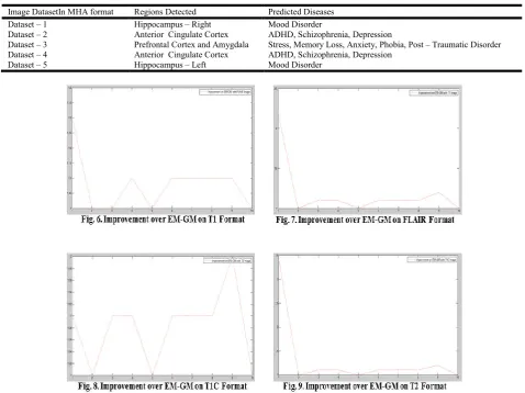
The optimal unification framework is been applied to FLAIR, T1, T2 and T1C image formats alongside with the EM-GM (Table – 6 to 9) techniques and a relative improvement in the result is observed. The improvements are visualized here (Fig. 6 to 9)
[image:5.595.79.514.166.273.2]Hence this work shows the improvement of accuracy for all the tested datasets for 10 Datasets’ dataset. The work also successfully predicts the diseases based on the anomalies detected on the brain regions. The outcomes of the predictive analysis is also demonstrated (Fig. 10) (Table – 10). This outcome makes the work unique in nature and reduced the span of further investigations.
Table 8. Accuracy Analysis over Expectation Maximization – Gaussian Mixture Method – T2
Image DatasetIn MHA format EM – GM Novel Unification Technique Improvement (%)
Dataset – 1 95.99 97.03 1.08
Dataset – 2 98.29 99.29 1.02
Dataset – 3 91.64 92.64 1.09
Dataset – 4 91.86 92.86 1.09
Dataset – 5 95.36 96.36 1.05
Dataset – 6 92.87 93.87 1.08
Dataset – 7 92.18 93.18 1.08
Dataset – 8 91.57 92.57 1.09
Dataset – 9 85.5 86.5 1.17
[image:5.595.79.517.307.413.2]Dataset – 10 97.46 98.46 1.03
Table 9. Accuracy Analysis over Expectation Maximization – Gaussian Mixture Method – T1C
Image DatasetIn MHA format EM – GM Novel Unification Technique Improvement (%)
Dataset – 1 95.86 99.09 3.37
Dataset – 2 98.29 99.29 1.02
Dataset – 3 91.75 92.75 1.09
Dataset – 4 91.99 92.99 1.09
Dataset – 5 95.61 96.61 1.05
Dataset – 6 93.27 94.27 1.07
Dataset – 7 93.15 94.15 1.07
Dataset – 8 91.62 92.62 1.09
Dataset – 9 85.85 86.85 1.16
Dataset – 10 97.86 98.86 1.02
Table 10. Region Based Disease Prediction Results
Image DatasetIn MHA format Regions Detected Predicted Diseases
Dataset – 1 Hippocampus – Right Mood Disorder
Dataset – 2 Anterior Cingulate Cortex ADHD, Schizophrenia, Depression
Dataset – 3 Prefrontal Cortex and Amygdala Stress, Memory Loss, Anxiety, Phobia, Post – Traumatic Disorder
Dataset – 4 Anterior Cingulate Cortex ADHD, Schizophrenia, Depression
[image:5.595.58.537.439.797.2]Dataset – 1 Dataset – 2 Dataset – 3
[image:6.595.57.269.48.204.2]Dataset – 4 Dataset – 5
Fig. 10. Disease Prediction for Various Datasets
Conclusion
Major Contributions of this work are the multilateral filter to normalize the image noises and the enhancement of EM-GM technique to improve the detection of brain diseases. Quantitative analysis of brain MR images allows a greater understanding of the nature of the diseases. The proposed algorithm in this work has been tested on BRATS 2012 (Nice), BRATS 2013 (Nagoya) and BRATS 2014 (Boston) challenge datasets and demonstrates higher accuracy. The work also concludes the optimal technique for medical image segmentation and detection of brain anomalies. Compared to the existing research outcomes, this work demonstrates the mapping of possible disease with the brain anomalous regions. With the final outcome of accuracy improvement for FLAIR, T1, T2 and T1C image data types on disease prediction, the work certainly and satisfyingly extends the possibilities of better medical image processing.
REFERENCES
Achanta R. et al., “SLICsuperpixels compared to state-of-the-art su- perpixel methods,” IEEE Trans. Pattern Anal. Mach. Intell., vol. 34, no. 11, pp. 2274–2282, Nov. 2012
Ahmed, S., K. M. Iftekharuddin and A. Vossough, 2015."Efficacy of texture, shape, and intensity feature fusion for posterior-fossa tumor segmentation in MRI", IEEE Trans. Inf. Technol. Biomed., vol. 15, no. 2, pp. 206-213.
Avants B. B. et al., “A reproducible evaluation of ANTs similarity metric performance in brain image registration,” Neuroimage, vol. 54, no. 3, pp. 2033–44, Feb. 2011 Bauer, S., R. Wiest, L.-P. Nolte and M. Reyes, "A survey of
MRI-based medical image analysis for brain tumor studies", Phys. Med. Biol., vol. 58, no. 13, pp. R97-R129, 2013
Gooya, A., G. Biros and C. Davatzikos, "Deformable registration of glioma images using EM algorithm and diffusion reaction modeling", IEEE Trans. Med. Imag., vol. 30, no. 2, pp. 375-390, 2011
Hameeteman, K. 2011. "Evaluation framework for carotid bifurcation lumen segmentation and stenosis grading", Med. Image Anal., vol. 15, no. 4, pp. 477-488.
Islam, A., S. M. S. Reza, and K. M. Iftekharuddin, “Multi-fractal texture estimation for detection and segmentation of brain tumors,” IEEE Trans. Biomed. Eng., vol. 60, no. 11, pp. 3204–3215, Nov. 2013.
Menze B. M. et al. “The multimodal brain tumor image segmentation benchmark (BRATS),” IEEE Trans. Med. Imag., vol. 33, no. 10, pp. 1993–2024, Oct. 2014.
Shattuck, D. W., G. Prasad, M. Mirza, K. L. Narr and A. W. Toga, "Online resource for validation of brain segmentation methods", Neuroimage, vol. 45, no. 2, pp. 431-439, 2009 Shin, H. C., M. R. Orton, D. J. Collins, S. J. Doran, and M. O.
Leach, “Stacked autoencoders for unsupervised feature learning and multiple organ detection in a pilot study using 4D Dataset data,” IEEE Trans. Pattern Anal. Mach. Intell., vol. 35, no. 8, pp. 1930–1943, Aug. 2013
Stille, M.,M. Kleine; J. Hagele; J. Barkhausen; T. M. Buzug, "Augmented Likelihood Image Reconstruction", IEEE Transactions on Medical Imaging, Volume:35, Issue:1 Subbanna, N., D. Precup, L. Collins, and T. Arbel,
“Hierarchical prob- abilistic Gabor and MRF segmentation of brain tumours in MRI vol- umes,” Proc. MICCAI, vol. 8149, pp. 751–758, 2013
Weizman, L. 2012. "Automatic segmentation, internal classification, and follow-up of optic pathway gliomas in MRI", Med. Image Anal., vol. 16, no. 1, pp. 177-188.