SWALLOWED FOREIGN BODIES IN CHILDREN: DIAGNOSTIC AND THERAPEUTIC IMPLICATIONS
*,1
Dr. Mohamed Ramadan Abdallah and
1
Pediatric Surgery Unit, Sohag Faculty of Medicine, Sohag, Egypt
2Department of Pediatric Surgery
ARTICLE INFO ABSTRACT
Backgrounds:
coins conti
outlining aspects of diagnosis and management.
Patients and Methods:
retrospectively reviewed f
Results:
FBs were
and 24 females. All were healthy with no prior medical problems except one who had primary repair of esophageal atresia during neonatal period. A variety of FBs were
commonest representing 71% of cases. Twenty seven FBs (43.5%) required either endoscopic or surgical removal (21 esophageal and 6 intestinal). The remaining 35 (56.5%) passed spontaneously.
Conclusions:
remain the commonest object to be swallowed. Impacted esophageal FBs require prompt removal.
esophagus, but early rec
Copyright © 2016, Dr. Mohamed Ramadan Abdallah and Dr. Ahmed H. Al
Attribution License, which permits unrestricted use, distribution,
INTRODUCTION
Swallowing of FBs by children is a common problem, with the greatest incidence among those aged 6 months to 4 years
(Kelley et al., 1993). Children often swallow usual household
objects such as coins, nails, pins, screws or toy parts
et al., 2013; Litovitz et al., 2010a; Conners 2008; Pavlidis
et al., 2008; Pryor et al., 2007). Fortunately, the majority of
these FBs will pass spontaneously without causing damage to the gastrointestinal tract. However, complications including bleeding, gastrointestinal perforation and obstructionare liable
to occur (Peters et al., 2015; Litovitz et al., 2010b; Dahiya and
Denton, 1999; Brayer, et al., 1998; Byard et al.,
FBs namely button batteries and magnets are increasingly swallowed by children and these require special attention as they carry particular risks and may cause serious life
threatening complications (Abbas et al., 2013, Litovitz
2010a; Pryor et al., 2007; Sheikh, 1993). This study is a review
of our experience with swallowed FBs in children with emphasis on aspects of diagnosis and management.
*Corresponding author:Dr. Mohamed Ramadan Abdallah Pediatric Surgery Unit, Sohag Faculty of Medicine, Sohag, Egypt
ISSN: 0975-833X
Article History:
Received 25th January, 2016
Received in revised form
07th February, 2016
Accepted 28th March, 2016
Published online 26th April,2016
Key words:
Swallowed foreign bodies, FBs, coins,
Magnets, Button battery.
Citation: Dr. Mohamed Ramadan Abdallah and Dr. Ahmed H. Al
implications”,International Journal of Current Research
RESEARCH ARTICLE
SWALLOWED FOREIGN BODIES IN CHILDREN: DIAGNOSTIC AND THERAPEUTIC IMPLICATIONS
Dr. Mohamed Ramadan Abdallah and
2Dr. Ahmed H. Al
Pediatric Surgery Unit, Sohag Faculty of Medicine, Sohag, Egypt
Pediatric Surgery, Maternity and children Hospital, Dammam, Saudi Arabia
ABSTRACT
Backgrounds: Children continue to swallow foreign bodies (FBs), some of which are bizarre but coins continue to be the most common. This review presents our experience with 62 swallowed FBs outlining aspects of diagnosis and management.
Patients and Methods: The medical records of all children with the diagnosis of swallowed FB were retrospectively reviewed for age, type of the swallowed FB, diagnosis and management.
Results: During a 7-year period, from June 2008 to July 2015, a total of 62 children with swallowed FBs were treated. Their age ranged from 8 months to 10 years (mean 5.2 years). There were 38 males and 24 females. All were healthy with no prior medical problems except one who had primary repair of esophageal atresia during neonatal period. A variety of FBs were
commonest representing 71% of cases. Twenty seven FBs (43.5%) required either endoscopic or surgical removal (21 esophageal and 6 intestinal). The remaining 35 (56.5%) passed spontaneously.
Conclusions: Swallowing of FBs by children continues to be a common medical problem. Coins remain the commonest object to be swallowed. Impacted esophageal FBs require prompt removal.Watchful waiting is preferred to deal with swallowed FBs once they passed beyond the esophagus, but early recognitison and management of any complication is necessary.
Dr. Mohamed Ramadan Abdallah and Dr. Ahmed H. Al-Salem. This is an open access article distributed under the Creative Commons use, distribution, and reproduction in any medium, provided the original work is properly cited.
Swallowing of FBs by children is a common problem, with the those aged 6 months to 4 years 1993). Children often swallow usual household objects such as coins, nails, pins, screws or toy parts (Abbas
2010a; Conners 2008; Pavlidis 2007). Fortunately, the majority of these FBs will pass spontaneously without causing damage to the gastrointestinal tract. However, complications including bleeding, gastrointestinal perforation and obstructionare liable 2010b; Dahiya and
et al., 1990). Certain
FBs namely button batteries and magnets are increasingly swallowed by children and these require special attention as sks and may cause serious life
2013, Litovitz et al.,
2007; Sheikh, 1993). This study is a review of our experience with swallowed FBs in children with
ement.
Dr. Mohamed Ramadan Abdallah Pediatric Surgery Unit, Sohag Faculty of Medicine, Sohag, Egypt
Patients and methods
The medical records of all children with the diagnosis of swallowed FBs were retrospectively reviewed for
diagnosis, type of the swallowed FB, diagnostic modalities and management.
RESULTS
During a7-year period, from June 2008 to July 2015, a total of 62 children with swallowed FBs were treated. Their age ranged from 8 months to 10 years (mean 5.2 ye
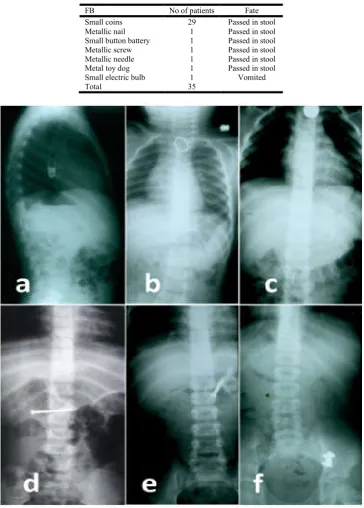
males and 24 females. All were healthy without preexisting medical problems except one who had repair of esophageal atresia with tracheoesophageal fistula. Coins were the commonest object swallowed and was found in 44 patients (71%). Small coins (n: 29; 46.8%) passed spontaneously to the stomach while large coins (n: 15; 24.2%) got impacted in the esophagus and required intervention to be removed (Figure 1, 2). Other objects and their frequencies are reported in table 1 and 2. The majority of the FBs (n: 58; 93.5%) were radiopaque and were readily visible on
Radiolucent FBs were found in 4 patients. Two of them presented with clinical picture of intestinal obstruction, and International Journal of Current Research
Vol. 8, Issue, 04, pp.29632-29637, April, 2016
INTERNATIONAL
Dr. Mohamed Ramadan Abdallah and Dr. Ahmed H. Al-Salem, 2016. “Swallowed foreign bodies in children: diagnostic and therapeutic
International Journal of Current Research, 8, (04), 29632-29637.
SWALLOWED FOREIGN BODIES IN CHILDREN: DIAGNOSTIC AND THERAPEUTIC IMPLICATIONS
H. Al-Salem
Pediatric Surgery Unit, Sohag Faculty of Medicine, Sohag, Egypt
, Maternity and children Hospital, Dammam, Saudi Arabia
Children continue to swallow foreign bodies (FBs), some of which are bizarre but nue to be the most common. This review presents our experience with 62 swallowed FBs
The medical records of all children with the diagnosis of swallowed FB were or age, type of the swallowed FB, diagnosis and management.
year period, from June 2008 to July 2015, a total of 62 children with swallowed treated. Their age ranged from 8 months to 10 years (mean 5.2 years). There were 38 males and 24 females. All were healthy with no prior medical problems except one who had primary repair of esophageal atresia during neonatal period. A variety of FBs were swallowed but coins were the commonest representing 71% of cases. Twenty seven FBs (43.5%) required either endoscopic or surgical removal (21 esophageal and 6 intestinal). The remaining 35 (56.5%) passed spontaneously.
ldren continues to be a common medical problem. Coins remain the commonest object to be swallowed. Impacted esophageal FBs require prompt Watchful waiting is preferred to deal with swallowed FBs once they passed beyond the
on and management of any complication is necessary.
is an open access article distributed under the Creative Commons reproduction in any medium, provided the original work is properly cited.
The medical records of all children with the diagnosis of swallowed FBs were retrospectively reviewed for age at diagnosis, type of the swallowed FB, diagnostic modalities and
year period, from June 2008 to July 2015, a total of 62 children with swallowed FBs were treated. Their age ranged from 8 months to 10 years (mean 5.2 years). There were 38 males and 24 females. All were healthy without preexisting medical problems except one who had repair of esophageal atresia with tracheoesophageal fistula. Coins were the commonest object swallowed and was found in 44 patients all coins (n: 29; 46.8%) passed spontaneously to the large coins (n: 15; 24.2%) got impacted in the esophagus and required intervention to be removed (Figure 1, Other objects and their frequencies are reported in table 1 ty of the FBs (n: 58; 93.5%) were radiopaque visible on plain x-rays (Figure 2). Radiolucent FBs were found in 4 patients. Two of them presented with clinical picture of intestinal obstruction, and
INTERNATIONAL JOURNAL OF CURRENT RESEARCH
although the presence of intraluminal obstructing FB had been suggested on gastrointestinal contrast studies, the nature of the FB was revealed only during laparotomy. These were a gelatinous type of sweets that did not dissolve and became impacted in the duodenum, and a soft rubber pacifier that became impacted in the ileum (Figure1 and figure 3). In the third patient, the FB was a large plant seed impacted on top of an esophageal stricture following repair of esophageal atresia. The nature of the FB was revealed during endoscopy (Figure 1). In the fourth patient, the FB was a radiolucent sharp plastic button that was impacted in the esophagus and caused esophageal perforation. The diagnosis was made on esophagogram and CT-scan (Figure 3).
[image:2.595.107.501.226.555.2]Out of 62 patients included in this study, 27patients (43.5%) required either endoscopic or surgical removal (21 esophageal and 6 intestinal). The remaining 35 FBs (56.5%) passed spontaneously (Table 1, 2). Twenty nine patients (46.7%) presented with esophageal FBs; 21 (33.8%) required intervention to be removed, 7 passed to the stomach prior to endoscopy and 1 (small electric bulb) was vomited spontaneously within 24 hours of swallowing. Twenty FBs (32%) were impacted in the esophagus and were removed endoscopically. These included 15 large coins (24%), 1 golden ring, 1 piece of sharp blade, 1 large button battery, 1 piece of lamb bone, and 1 large plant seed impacted on top of a stricture following repair of esophageal atresia (Table 1).
Figure 1. A variety of FBs swallowed by children: large plant seed (a), multiple magnets (b & c), small electric bulb (d), part of metallic necklace (e), rubber pacifier (f), piece of lamb bone (g), piece of sharp blade (h), coins (i) and metallic needle (j).
Table 1. Summary of patients with swallowed FBs that required intervention
FB No of patients Location Fate
Large coins 15 Esophagus Endoscopic removal
Golden ring 1 Esophagus Endoscopic removal
Piece of sharp blade 1 Esophagus Endoscopic removal
Large button battery 1 Esophagus Endoscopic removal
Piece of lamb bone 1 Esophagus Endoscopic removal
Large plant seed 1 Esophagus Endoscopic removal
Plastic sharp button 1 Esophagus Surgical removal
Gelatinous type of sweet 1 Duodenum Surgical removal
Metallic nail 1 Duodenum Surgical removal
Metallic necklace of several beads 1 Jejunum Surgical removal
Multiple magnets 2 Jejunum Surgical removal
Rubber pacifier 1 Ileum Surgical removal
[image:2.595.143.457.621.756.2]Table 2. Summary of patients with swallowed FBs treated conservatively
FB No of patients Fate
Small coins 29 Passed in stool
Metallic nail 1 Passed in stool
Small button battery 1 Passed in stool
Metallic screw 1 Passed in stool
Metallic needle 1 Passed in stool
Metal toy dog 1 Passed in stool
Small electric bulb 1 Vomited
[image:3.595.113.502.626.780.2]Total 35
Figure 2. Various radio-opaque FBs on plain x-rays: small electric bulb(a), golden ring (b), coin (c), nail (d), screw (e), and metallic dog toy (f)
Endoscopic removal was performed after a period that ranged from 6 hours to 4 days after swallowing. The majority of FBs were impacted in the upper third of the esophagus. Endoscopic removal was achieved using either rigid or flexible esophagoscope. In two patients a Foley’s catheter was used to remove coins from the upper esophagus. X-rays were repeated for all patients just prior to the planned endoscopic removal of the FB to confirm its location. The majority of these FBs were loosely impacted, except in one patient who was neglected for 4 days after swallowing a large coin which was deeply stuck and about to perforate the esophagus. Endoscopic retrieval was
difficult but successful. In the 21st patient, a neglected
swallowed plastic sharp button caused perforation of the upper esophagus that was diagnosed using esophagogram and CT-scan (Figure 3) and removed surgically.
Forty patients (64.5%) presented with swallowed FBs beyond the esophagus (including 7 patients presented initially with esophageal FBs that passed spontaneously to the stomach within a few hours from the initial presentation). Laparotomy was required in 6 patients (9.7%) who presented with pictures of complications. (Table 1; Figure 1, 2, 3). In 2 patients, the diagnosis was made intraoperatively because of the radiolucent nature of the FB. In one of them, the FB was a gelatinous type of sweet that did not dissolve and became impacted in the duodenum, and required a duodenotomy to be removed. In the other, the FB was a soft rubber pacifier that became impacted in the ileum. This was milked towards the colon and out of the anus, avoiding enterotomy. In 4 other patients, the FBs had been demonstrated preoperatively on plain x-rays. In one of them a metallic nail was lodged in the duodenal mucosa without causing perforation and endoscopic dislodgement was unsuccessful. This was removed via a duodenotomy. In another patient, a part of a metallic beaded necklace lodged in the jejunum and was deeply implanted into the bowel wall that necessitated bowel resection. In the remaining 2 patient, swallowed multiple magnets caused multiple jejunal perforations that were repaired by simple repair after removal of the magnets.
For the remaining 34 patients, a policy of watchful waiting was followed regardless of the shape or the size of the FB, including 29 small coins, 1 small button battery, 1 metallic screw, 1 metallic nail, 1 metallic needle, and 1 metallic dog toy (Table 2, Figure 1, 2). There were no attempts to remove FBs endoscopically once they reached the stomach except in one
patient who presented with an impacted duodenal FB. Our
protocol consisted of careful education of the family about the warning symptoms and signs of complications along with follow-up plain abdominal radiographs to monitor the progress of the FB until its passage in the stool. The frequency of these x-rays was variable but in general they were ordered every 48 hours and then weekly when there were no clinical manifestations of complications. The parents were also instructed to inspect the stools for the passage of the FB. No special diet was advised. Even the seemingly traumatic objects passed spontaneously with stools. The mean time for the passage of the FB in the stool ranged from 2 days to 12 days (mean: 4 days).
DISCUSSION
Children are known to ingest inadvertently or intentionally a variety of FBs. Hand-to-mouth activity is one of the primary mechanisms that infants use to connect with their surroundings and experience the world; hence the increased risk of FB ingestion in the younger age group (6 months to 4 years)
(Kirkham et al., 2015; McConnell, 2013; Paul et al., 2010;
Uyemura, 2005). Parents should be aware of the danger of
leaving small objects accessible to children (Peters et al., 2015;
Dahiya and Denton, 1999; Byard et al., 1990). Most ingested
FBs are usual household objects. Common items that get put in the mouth are those that are shiny and easy to grasp, including coins, button batteries, magnets, screws and small keys
(Kirkham et al., 2015). Coins continue to be the commonest
object swallowed by children. Forty-four (71%) of the swallowed FBs in our series were coins. Unusual objects reported in this study included a rubber baby pacifier ingested by a 10 years old boy because of jealousy from his younger brother, a gelatinous type of sweets that did not dissolve in the stomach and became impacted in the duodenum, a metal dog toy and a small electric bulb.
Ingestion of magnets is becoming a common problem among children and teens because magnets have become included in many kinds of modern toys and electronic devices. A single magnet most likely will pass through without causing any harm. However, if two or more magnets have been swallowed, attraction between them can result in pressure necrosis and
perforation of the intervening bowel (Abbas et al., 2013; Pryor
et al., 2007). This was the case in two of our patients who
presented with a picture of peritonitis and intestinal perforation due to neglected swallowed multiple magnets. Rapid evaluation of children with suspected swallowed magnets and prompt endoscopic removal of accessible ones is important to avoid these serious complications.
Swallowing of button (disk) batteries by children has also increased in proportion to the wide availability of electronic devices and remote controls that depend on these batteries as an energy source. Unlike other FBs which cause problems by mechanical action, batteries can lead to other kind of problems related to the discharge of electric current and leak of the chemical contents of the battery. Important factors that can help predict potential for complication include the battery size, its chemical composition, and its charge state. Small batteries can pass through the gastrointestinal tract without adverse effects. Problems can arise if the battery is of large size (>20 mm) and it becomes lodged (Litovitz 2010 a, b). When this occurs in the esophagus, serious complications can result including esophageal burns, perforations, fistula formation, hemorrhage and systemic poisoning with heavy metals
(McConnell, 2013; Chouhan et al., 2011; Vaishnav and Spitz,
1989; Mant et al., 1987). Therefore, rapid diagnosis and
emergency removal of esophageal button batteries is intuitive. On the other hand, button batteries in the stomach or intestines do not need to be removed immediately, as they generally pass through the gastrointestinal tract. Children can be managed at home with a protocol of family education, stool surveillance
However, batteries retained in the stomach or at a fixed spot in the intestines for 2-3 days should be removed either
endoscopically or surgically as appropriate (Volle et al., 1989).
Most ingested FBs are radiopaque and can be easily diagnosed and monitored by plain x-ray. Radiolucent FBs can be missed and often present with a picture of complications. In these situations, the use of CT scan and gastrointestinal contrast studies can be of help, but the nature of the FB is often revealed only at the time of endoscopy or surgery.
Fortunately, the majority of swallowed FBs pass
spontaneously without health consequences. It is estimated that up to 40% of these ingestions may go unnoticed (Uyemura, 2005). This however is not always the case and sometimes these objects can lead to serious complications including
gastrointestinal perforation or obstruction (Soprano et al.,
1999; Macpherson et al., 1996; Paul and Jaffe, 1988; Pellerin
et al., 1969). Complications from FB swallowing occurred in 7
out of 62 of our patients (11.3%). These included esophageal perforation in 1 patient, intestinal obstruction in 4 patientsand intestinal perforation in 2 patients.
The esophagus is a common site for FB impaction. In our series, esophageal FBs represented 46.7% of cases (29 out of 62 patients). The most common site of esophageal impaction is at the thoracic inlet and the cricopharyngeus level where about 70% of swallowed FBs become lodged. Another 15% become lodged at the mid esophagus and the remaining 15% become lodged at the gastroesophageal junction. FB impaction in the oesophagus can be associated with serious complications. Esophageal perforation by a neglected radiolucent sharp FB occurred in one of our patients. Therefore there is a general agreement that a FB impacted in the esophagus requires removal as soon as possible. This should be done within 24 hours from presentation and earlier if the impacted FB is a
button battery (Kirkham et al., 2015). Endoscopy
(esophagoscopy) is by far the most commonly used method to remove impacted esophageal FBs and is usually the procedure of choice. Blunt FBs may be removed by use of a Foley catheter or they may be advanced into the stomach with a
bougie (Conners, 2005; Waltzman et al., 2005; Vargas et al.,
2004; Dahshan and Kevin Donovan, 2007; Calkins et al.,
1999). The use of Magill forceps to remove upper esophageal FBs has also been shown to be safe and effective (Mahafza, 2002). Children with preexisting esophageal abnormalities such as congenital esophageal stenosis or stricture following repair of esophageal atresia are likely to have FB impaction at the site of the abnormality and these should be removed under vision using a rigid or flexible esophagoscopy.
Once they reach the stomach, the expected fate of most FBs is spontaneous passage with stool without causing complications. However, in our series, 6 out of 40 (15%) subdiaphragmatic FBs were associated with complications. Moreover, our findings came in contrary to the suggestion that sharp or pointed FBs carry an increased risk of complications (Paul and
Jaffe, 1988; Pellerin et al., 1969). It was suggested that these
traumatic FBs should be removed endoscopically from the stomach, and a "watchful waiting" policy should be followed only when they pass beyond the stomach. In our series, we adopted a policy of wait and follow-up for all FBs once they
crossed the esophagus irrespective of the size or the shape of the swallowed object. All objects their shapes implied an increased risk of complications passed smoothly without problems except one with a nail that was stuck in the wall of the duodenum. This was removed surgically after two failed attempts to remove it endoscopically. However, other seemingly traumatic objects were not associated with complications including a metallic screw, a metallic nail, a metallic needle, and a metallic dog toy. On the other hand, the majority of complicated abdominal FBs reported in our series (5 out of 6), were neither sharp nor pointed objects (a rubber pacifier, a gelatinous sweet, a beaded necklace, and multiple magnets). These findings may suggest that factors other than the shape of the FB e.g. its size, magnetic potential and chemical composition that may predict the risk of complications.
Conclusions
A variety of objects can be swallowed by children but coins continue to be the commonest. Impacted esophageal FBs require emergency removal to avoid serious complications. On the other hand, although watchful waiting is preferred to deal with the majority of swallowed FBs once they crossed the esophagus, complications are liable to occur and careful follow up is necessary for early recognition and prompt management of these complications.
REFERENCES
Abbas, M.I., Oliva-Hemker, M., Choi, J., Lustik, M., Gilger, M.A., Noel, R.A. 2013. Magnet ingestions in children presenting to US emergency departments, 2002-2011.
J.Pediatr.Gastroenterol.Nutr., 57(1):18-22.
Brayer, A.F., Conners, G.P., Ochsenschlager, D.W. 1998. Spontaneous passage of coins lodged in the upper
esophagus. Int. J.Pediatr.Otorhinolaryngol., 44(1):59-61.
Byard, R.W., Moore, L., Bourne, A.J. 1990. Sudden and unexpected death-a late effect of occult intraesophageal
foreign body. Pediatr.Pathol.,10(5):837-841.
Calkins, C.M., Christians, K.K., Sell, L.L. 1999. Cost analysis in the management of esophageal coins: endoscopy versus
bougienage.J.Pediatr. Surg.,34(3):412-414.
Chouhan, M., Yadav, J.S., Bakshi, J. 2011. Foreign body button battery in oesophagus - Time for intervention? Int. J.Pediatr.Otorhinolaryngol., 6:329-330.
Conners, G.P. 2005. Management of asymptomatic coin
ingestion. Pediatrics,116(3):752-753.
Conners, G.P. 2008. Esophageal coin ingestion: going low
tech. Ann.Emerg. Med., 51(4):373-374.
Dahiya, M. and Denton, J.S. 1999. Esophagoaortic perforation by foreign body (coin) causing sudden death in a 3-year-old
child. Am. J. Forensic Med.Pathol., 20(2):184-188.
Dahshan, A.H. and Kevin Donovan, G. 2007. Bougienage versus endoscopy for esophageal coin removal in children.
J.Clin.Gastroenterol., 41(5):454-456.
Kelley, J.E., Leech, M.H., Carr, M.G. 1993.A safe and cost-effective protocol for the management of esophageal coins in children.J.Pediatr. Surg., 28(7):898-900.
Litovitz, T., Whitaker, N., Clark, L. 2010. Preventing battery
ingestions: an analysis of 8648 cases. Pediatrics,
125(6):1178-1183.(b)
Litovitz, T., Whitaker, N., Clark, L., White, N.C., Marsolek, M. 2010. Emerging battery-ingestion hazard: clinical
implications. Pediatrics,125(6):1168-1177. (a)
Macpherson, R.I., Hill, J.G., Othersen, H.B., Tagge, E.P., Smith, C.D. 1996. Esophageal foreign bodies in children:
diagnosis, treatment, and complications. Am. J.Roentgenol.,
166(4):919-924.
Mahafza, T.M. 2002. Extracting coins from the upper end of
the esophagus using a Magill forceps technique. Int.
J.Pediatr.Otorhinolaryngol., 62(1):37-39.
Mant, T.G, Lewis, J.L., Mattoo, T.K., Rigden, SP., Volans, G.N., House, I.M., Wakefield, A.J., Cole, R.S. 1987.
Mercury poisoning after disc-battery ingestion.
Hum.Toxicol., 6(2):179-181.
McConnell, M.K. 2013. When button batteries become breakfast: The hidden dangers of button battery ingestion. J. Pediatr. Nurs., 28(6): 42-49
Paul, R.I. and Jaffe, D.M. 1988. Sharp object ingestions in
children: illustrative cases and literature review.
Pediatr.Emerg. Care, 4(4):245-248.
Paul, S.P., Hawes, D., Taylor, T.M. 2010. Foreign body ingestion in children: case series, review of the literature and guidelines on minimising accidental ingestions. J. Fam. Health Care, 20(6): 200-204
Pavlidis, T.E., Marakis, G.N., Triantafyllou, A., Psarras, K., Kontoulis, T.M., Sakantamis, A.K. 2008. Management of ingested foreign bodies. How justifiable is a waiting policy? Surg.Laparosc.Endosc.Percutan. Tech., 18(3):286-287.
Pellerin, D., Fortier-Beaulieu, M., Gueguen, J. 1969. The fate of swallowed foreign bodies. Experience of 1250 instances
of subdiaphragmatic foreign bodies in children. Prog.
Pediatr.Radiol., 2:286-302.
Peters, N.J., Mahajan, J.K., Bawa, M., Chabbra, A., Garg, R., Rao, K.L. 2015. Esophageal perforations due to foreign body impaction in children. J.Pediatr. Surg., 50(8):1260-1263.
Pryor, H.I. 2nd., Lange, P.A., Bader, A., Gilbert, J., Newman,
K. 2007. Multiple magnetic foreign body ingestion: a surgical problem. J. Am. Coll. Surg., 205(1):182-186. Sheikh, A. 1993. Button battery ingestions in children.
Pediatr.Emerg. Care, 9(4):224-229.
Soprano, J.V., Fleisher, G.R., Mandl, K.D. 1999. The spontaneous passage of esophageal coins in children. Arch.Pediatr.Adolesc. Med., 153(10):1073-1076.
Uyemura, M.C. 2005. Foreign body ingestion in children. Am. Fam. Physician, 72(2): 287-291.
Vaishnav, A. and Spitz, L. 1989. Alkaline battery-induced tracheo-oesophageal fistula. Br. J. Surg., 76(10):1045. Vargas, E.J., Mody, A.P., Kim, T.Y., Denmark, K., Moynihan,
J.A., Barcega, B.B. 2004. The removal of coins from the upper esophageal tract of children by emergency physicians: a pilot study. C.J.E.M.,6(6):434-440.
Volle, E., Beyer, P., Kaufmann, H.J. 1989. Therapeutic approach to ingested button-type batteries. Magnetic removal of ingested button-type batteries. Pediatr.Radiol., 19(2):114-118.
Waltzman, M.L., Baskin, M., Wypij, D., Mooney, D., Jones, D., Fleisher, G. 2005. A randomized clinical trial of the management of esophageal coins in children. Pediatrics, 116(3):614-619.