COMPARATIVE ANALYSIS OF POROUS HYDROXYAPATITE BONE GRAFT WITH AND WITHOUT
PLATELET RICH PLASMA IN THE TREATMENT OF PERIODONTAL INTRABONY OSSEOUS DEFECTS
1,*
Dr. Muzafar Ahmad Bhat,
1
Postgraduate student, Deptt., Of periodontics, Govt. Dental College, Srinagar Jammu & Kashmir
2
Senior Resident Department Of Dermatology,Kusturba Medical colleg
3
Assistant Professor Department of Pedodontics & Preventive Dentistry Manipal College Of Dental Science
ARTICLE INFO ABSTRACT
Background
introduction of a filler material into the defect inhope of inducing bone regeneration. The purpose of this study was to clinically and radiographically evaluate the use of porous hydroxyapatitebone graft with and without platelet
Methods:
depth
assigned to treatment with a combination of PRP + Hydroxyapatite (HA) (test sites) or HA alone (control sites). Theparameters were compared at baseline and 6 months postoperatively.
There was a statistically significa
the groups individually (more in experimental group); however, on comparing the two groups, the netreduction was not significant. Radiographic assessment showed a decrease in the defect s both the groups.
safe and shows improved defect fill as compared to the use of bone graft alone.
Copyright©2017, Muzafar Ahmad Bhat and Dr. Mirza Aumir Beg
which permits unrestricted use, distribution, and reproduction
INTRODUCTION
Periodontal therapy has always strived to control or eliminateperiodontal disease in an attempt to restore the structures,integrity, and the function of tissues that have been lost as aresult of inflammatory periodontal disease
2010) A well‑coordinatedsequence of a number of biologic events including cellmigration, adherence, multiplication, and differentiation hasincreased the predictability
regeneration (Arvidson et al., 2011). Treatment of intrabony defects has often focused on the bonydefect and this has led to the use of a number of graftingmaterials to stimulate bone repair (Carranza et al., 2002). Bone grafting
retained in the defect site provide a structural frameworkfor clot development, maturation, and remodeling that supportsbone formation in osseous defects
2010). A wide array of bonegraft substitutes is available today and has shown to producegreater clinical bone defect f flap debridement alone (Nasr, 1999). Bioceramicalloplasts primarily composed of calcium phosphateare available as tricalcium phosphate and hydroxyapatite.Hydroxyapatite became the ceramic of choice, producingpredictable short and long‑term results (Aichelmann‑Reidy, 1998
ISSN: 0975-833X
Vol.
Article History:
Received 24th September, 2017
Received in revised form
07th October, 2017
Accepted 23rd November, 2017
Published online 30th December, 2017
Citation: Dr. Muzafar Ahmad Bhat, Dr. Mirza Aumir Beg and Dr. Shafia Nisar Kakroo.
without Platelet‑Rich Plasma in the Treatment of Periodontal Intrabony Osseous Defects”
Key Words:
Bone graft, Periodontal Tissue Regeneration, Platelet‑Rich Plasma.
*Corresponding author:
RESEARCH ARTICLE
COMPARATIVE ANALYSIS OF POROUS HYDROXYAPATITE BONE GRAFT WITH AND WITHOUT
RICH PLASMA IN THE TREATMENT OF PERIODONTAL INTRABONY OSSEOUS DEFECTS
Dr. Muzafar Ahmad Bhat,
2Dr. Shafia Nisar Kakroo and
3Dr. Mirza Aumir Beg
Postgraduate student, Deptt., Of periodontics, Govt. Dental College, Srinagar Jammu & Kashmir
Senior Resident Department Of Dermatology,Kusturba Medical college Manipal
Assistant Professor Department of Pedodontics & Preventive Dentistry Manipal College Of Dental Science
Manipal
ABSTRACT
Background: Today, regenerative attempts for treatment of periodontal disease focus on the introduction of a filler material into the defect inhope of inducing bone regeneration. The purpose of this study was to clinically and radiographically evaluate the use of porous hydroxyapatitebone graft
ith and without platelet‑rich plasma (PRP) in the treatment of intrabony defects.
Methods: The study was carriedout in ten patients between 18 and 60 years. Patients with pocket depth ≥5 mm and radiographic evidence of vertical bone loss in t
assigned to treatment with a combination of PRP + Hydroxyapatite (HA) (test sites) or HA alone (control sites). Theparameters were compared at baseline and 6 months postoperatively.
There was a statistically significant reduction in probing depthand gain in clinical attachment in both the groups individually (more in experimental group); however, on comparing the two groups, the netreduction was not significant. Radiographic assessment showed a decrease in the defect s both the groups. Conclusion: PRP in additionto a bone graft in the treatment of intrabony defects is safe and shows improved defect fill as compared to the use of bone graft alone.
Mirza Aumir Beg. This is an open access article distributed under the reproduction in any medium, provided the original work is properly cited.
Periodontal therapy has always strived to control or eliminateperiodontal disease in an attempt to restore the of tissues that have been nflammatory periodontal disease (Sunitha, coordinatedsequence of a number of biologic events including cellmigration, adherence, multiplication, and ity of periodontal Treatment of intrabony defects has often focused on the bonydefect and this has led to the use of a number of graftingmaterials to stimulate bone Bone grafting materialswhen retained in the defect site provide a structural frameworkfor clot development, maturation, and remodeling that (Reynolds et al., A wide array of bonegraft substitutes is available today s shown to producegreater clinical bone defect fill than Bioceramicalloplasts primarily composed of calcium phosphateare available as tricalcium phosphate and hydroxyapatite.Hydroxyapatite producingpredictable short‑term
, 1998).
The graftmaterial acts as a biocompatible material within the gingivaltissue, and as it resorbs, it acts as a mineral reservoir and assistsbone formation through
mechanisms, resultingin clinically acceptable responses (Yukna, 2000; Wagner, 1989).
periodontal regeneration inthe present era is the use of growth factors that are a class ofnaturally occurring proteins which effectively stimulate theformation of mineralized as well as nonmineralized tissues (Plachokova
plasma (PRP) as introduced by Marx is definedas an autologous concentration of platelets in a small volumeof plasmaand is considered to be a rich source of autologousgrowth factors (Marx
dentistry, PRP has been used indifferent clinical procedures (i.e., sinus floor elevation, alveolarridge augmentation, mandibular reconstruction, maxillary cleftrepair, treatm periodontal defects, gingival recession,and treatment of extraction sockets), where it has been appliedalone or in addition to bone grafts (Carlson
2010; Boyapati, 2006; Rutkowski
Kumar, 2012). PRP once grafted intothe defect site begins to release alpha granules within 10 minof clot development and secrete over 90% of their prepackagedgrowth factors within 1
International Journal of Current Research
Vol. 09, Issue, 12, pp.62154-62159, December, 2017
Ahmad Bhat, Dr. Mirza Aumir Beg and Dr. Shafia Nisar Kakroo. 2017. “Comparative analysis of Porous Hydroxyapatite Bone Graft with and
‑Rich Plasma in the Treatment of Periodontal Intrabony Osseous Defects”, International Journal of Current Research
COMPARATIVE ANALYSIS OF POROUS HYDROXYAPATITE BONE GRAFT WITH AND WITHOUT
RICH PLASMA IN THE TREATMENT OF PERIODONTAL INTRABONY OSSEOUS DEFECTS
Dr. Mirza Aumir Beg
Postgraduate student, Deptt., Of periodontics, Govt. Dental College, Srinagar Jammu & Kashmir
e Manipal
Assistant Professor Department of Pedodontics & Preventive Dentistry Manipal College Of Dental Science
for treatment of periodontal disease focus on the introduction of a filler material into the defect inhope of inducing bone regeneration. The purpose of this study was to clinically and radiographically evaluate the use of porous hydroxyapatitebone graft rich plasma (PRP) in the treatment of intrabony defects. Materials and
The study was carriedout in ten patients between 18 and 60 years. Patients with pocket 5 mm and radiographic evidence of vertical bone loss in the affectedsite were randomly assigned to treatment with a combination of PRP + Hydroxyapatite (HA) (test sites) or HA alone (control sites). Theparameters were compared at baseline and 6 months postoperatively. Results:
nt reduction in probing depthand gain in clinical attachment in both the groups individually (more in experimental group); however, on comparing the two groups, the netreduction was not significant. Radiographic assessment showed a decrease in the defect size in : PRP in additionto a bone graft in the treatment of intrabony defects is safe and shows improved defect fill as compared to the use of bone graft alone.
the Creative Commons Attribution License, cited.
The graftmaterial acts as a biocompatible material within the gingivaltissue, and as it resorbs, it acts as a mineral reservoir and assistsbone formation through osteoconductive mechanisms, resultingin clinically acceptable responses . A different approach used for periodontal regeneration inthe present era is the use of growth factors that are a class ofnaturally occurring proteins which effectively stimulate theformation of mineralized as well as Plachokova et al., 2008). Platelet‑rich plasma (PRP) as introduced by Marx is definedas an autologous concentration of platelets in a small volumeof ed to be a rich source of Marx, 1998). In the field of dentistry, PRP has been used indifferent clinical procedures (i.e., sinus floor elevation, alveolarridge augmentation, mandibular reconstruction, maxillary cleftrepair, treatment of periodontal defects, gingival recession,and treatment of extraction sockets), where it has been appliedalone or in Carlson et al., 2002; Alissa et al., Rutkowski, 2010; Wojtowicz, 2007; . PRP once grafted intothe defect site begins to release alpha granules within 10 minof clot development and secrete over 90% of their prepackagedgrowth factors within 1
INTERNATIONAL JOURNAL OF CURRENT RESEARCH
Comparative analysis of Porous Hydroxyapatite Bone Graft with and
h, thereby initiating a greater andfaster initial cellular response than a normal blood clot. Platelet‑rich‑derived fibrin clot formation stimulates collagen synthesis in the periodontium and effectively promotes woundhealing at sites of injury in periodontal tissue (Lacoste, 2003; Rodrigues, 2012). Thus, the purpose of this study was to clinically andradiographically evaluate the use of porous hydroxyapatitebone graft with and without PRP in the treatment of periodontalintrabony osseous defects.
MATERIALS AND METHODS
Patient selection: This randomized controlled study was
carried out in the Department of Periodontics Govt Dental College Srinagar after approval by the Ethical Committee. The criteria for inclusion were systemically healthy individuals between age groups 18 and 60 years of either sex,no history of any medication affecting the periodontium in the past 6 months, and those who had not undergone anyperiodontal treatment in the past 6 months. Patients who hada clinical evidence of an intrabony defect with probing pocket depth (PD) ≥5 mm and radiographic evidence of angular boneloss in the affected site were included in the study. Patients excluded were individuals with systemic diseases (diabetes mellitus and platelet deficiencies), pregnant and lactating females, individuals with a present history of tobacco usage, and individuals on anticoagulant or immunosuppressive therapy.
Initial therapy: The patients were subjected to oral
prophylactic procedures, occlusal equilibration, if required, and routine laboratory investigations before surgery. Patients oral hygiene status was evaluated by plaque Index (Silness and Loe) (Löe, 1967) and gingival index (Loe and Silness) (Loe, 1962) On reevaluation of Phase I therapy, only those patients who had attained a score of ≤1 were selected for the surgical phase.
Clinical parameters: To standardize the reproducibility of clinical measurements, occlusal acrylic stents for positioning the periodontal probe were fabricated on a cast obtained from an alginate impression (Camargo et al., 2002). The following clinical parameters were recorded
Radiographic parameters: Preoperative radiographs were
obtained at baseline and then at 3 and 6 months postsurgery. The size of the defect or defect fill was measured using depth of the infrabonycomponent, and the bony defect width as described by Eickholz et al. (2004)
Group allocation: Before the commencement of the surgical procedure, the site to be treated was randomly allocated into experimental (PRP +Hydroxyapatite (HA)) or control (HAalone) study groups.
Plateletrich plasma procurement: Just before the surgery, 10
ml of blood was withdrawn from the antecubital vein of the patients and collected in tubes containing sodium citrate anticoagulant. The test tube was placed into the automated centrifuge machine always ensuring that the tubes were counterbalanced, as per the centrifuge manual. The first cycle of 2400 rpm for 10 min separated the whole blood into a platelet‑poor plasma layer at the top, a white buffy coat in the middle, and a layer of red blood corpuscles (RBC) at the bottom. The upper two layers andthe top 1–2 mm of RBC layer were expressed into another tube (without anticoagulant) and
centrifuged at 3600 rpm for 15 min that resulted in an upper portion of clear yellow supernatant with a very low concentration of platelets and are d‑tinged bottom layer with highly concentrated platelets. At the time of application, PRP was combined with an equal volume of a sterile saline solution containing 10% calcium chloride (a citrate inhibitor) and human thrombin (an activator) which resulted in a sticky gel that was relatively easy to apply in surgical defects (Okuda, 2005; Tözüm, 2003)
Surgical procedure: A standardized conventional periodontal
flap surgery wasperformed by a single operator. The site was anesthetized usingadequate local anesthesia (2% lidocaine hydrochloride withadrenaline 1:80,000). Intracrevicular buccal and palatal incisionswere given and full thickness mucoperiosteal flaps were elevatedto expose the defect. A thorough debridement was carried out toensure a clean site followed by thorough root planing. For thecontrol site, adequate quantity of the graft (Biograft HA) wasmixed with a few drops of saline to obtain a workable mass andthe defect was filled (Figure 1). At the experimental site, HA graftwas mixed with PRP gel in a proportion of 1:1 and was insertedup to the vertical height of the corresponding adjacent bonelevel (Figure 2). Flap was repositioned and sutured with 3‑0 silksuture material (Ethicon) followed by a periodontal dressing. Postoperative instructions and medications were prescribed tothe patients and were recalled after 10 days for suture removal.Postoperative care included reinforcement of oral hygiene andscaling when necessary. Patients were periodically monitoredand the clinical and radiographic parameters were recorded at3 and 6 months postsurgery.
Statistical analysis: The collected data were assessed for both the control and the experimental groups individually as well as compared with each other using SPSS v19. Baseline, 3 months and 6 months postoperative data were tabulated and analyzed statistically.
RESULTS
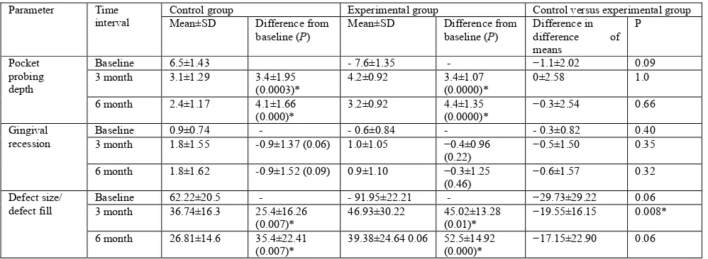
baseline to6 months). The difference between the groups was also not statistically significant (P = 0.34) (Table 3).
In the control group, the amount of defect fill from base line to 6 months post treatment was 56.91% (P = 0.0007). For the experimental group, greater defect fill was observed from baseline to 6 months (57.16%; P = 0.00, respectively). When a comparison was made between the groups, statistically significant difference (P = 0.008) was seen in the differences of means between the groups from baseline to 3 months. However, from baseline to 6 months, the difference was not significant (P = 0.06) (Table 3).
DISCUSSION
The management of periodontal osseous defects including the destruction of the periodontium has always been a challenge in clinical periodontics. A plethora of literature is available substantiating the use of bone graft materials along with growth factors with an aim to optimize the outcome of periodontal regeneration by assisting the proliferation, migration, and differentiation of periodontal ligament cells, cement oblasts, and osteoblasts (Pradeep, 2009) This study combined PRP with poroushydroxyapatite bone graft (Biograft HA®) to enhance the regenerative potential of the graft used. Various bone grafting materials have been used to fill periodontal intrabony defects with particle size between 300and 500 μm in diameter, which has resulted in clinicallyacceptable responses (Yukna, 2000; Mellonig, 1992)
Hence, the particle size of poroushydroxyapatite bone graft (Biograft HA) used in this studywas 350–500 μm.
It has been seen that porous HA bone graftshave excellent bone conductive properties which permitoutgrowth of osteogenic cells from existing bone surfacesinto adjacent bone graft material (Stahl, 1987) Since there are no organiccomponents contained in HA, this bone graft material does not induce any allergic reaction; however, true periodontalregeneration is not achieved because the healing which occursis a connective tissue encapsulation of the graft with a longjunctional epithelium (Meffert, 1985; Sculean, 2004). Different techniques of PRP preparation have been known toyield substantially different amounts of cells, i.e., platelets and leukocytes as well as different levels of growth factors (Weibrich, 2002). As periodontal defects are small in size, only 8–10 ml of venous blood was withdrawn with a preparation time of about 30 min that is performed simultaneously during the surgery, there by not increasing the chair‑side time. The method of procurement of PRP used in this study was similar to that used in the study performed by Okuda et al. (2005) According to de Obarrioet al. (2000) PRP preparation assumes a sticky consistency, due to highfibrin content, making it a hemostatic and stabilizing agentthat aid bone graft immobilization and has been suggested asan important event in wound healing. PRP is an auto genouspreparation and is inherently safe and free from concerns over transmissible diseases (Pradeep, 2009). In the present study also, the lack of adverse reactions, abscesses, or
Table 1. Distribution of intrabony defect in relation to tooth type and treatment modality
Group Arch Anterior Bicuspid Molar total
Test (10) Maxilla mandible 31 11 22 64
Control (10) Maxilla mandible 50 01 22 73
Table 2. Comparison of mean values of oral hygiene status within and between control and experimental groups
Parameter Time
interval
Control group Experimental group Control versus experimental group
Mean±SD Difference from
baseline (P)
Mean±SD Difference from
baseline (P)
Difference in difference of means
P
Plaque index Baseline 0.53±0.22 - 0.7±0.37 - -0.17±0.50 0.21
3 month 0.45±0.26 0.07±0.26 (0.39) 0.5±0.31 0.2±0.23 (0.02)* −0.125±0.33 0.27
6 month 0.25±0.20 0.27±0.28 (0.01)* 0.2±0.19 0.5±0.26 (0.00) * −0.225±0.38 0.07
Gingival index Baseline 0.85±0.27 - 0.725±0.34 - 0.125±0.27 0.37
3 month 0.42±0.26 0.425±0.43 (0.01)* 0.425±0.21 0.30±0.28 (0.00)* 0.125±0.42 0.45
6 month 0.22±0.20 0.625±0.39 (0.00)* 0.2±0.19 0.525± 0.29 (0.00)* 0.1±0.26 0.53
[image:3.595.43.542.300.483.2]*Significant P<0.05. Value for difference between groups indicates that experimental group value is higher than control group. SD: Standard deviation
Table 3. Comparison of mean values of various parameters within and between control and experimental groups
Parameter Time
interval
Control group Experimental group Control versus experimental group
Mean±SD Difference from
baseline (P)
Mean±SD Difference from
baseline (P)
Difference in
difference of
means
P
Pocket probing depth
Baseline 6.5±1.43 ‑ 7.6±1.35 - −1.1±2.02 0.09
3 month 3.1±1.29 3.4±1.95
(0.0003)*
4.2±0.92 3.4±1.07
(0.0000)*
0±2.58 1.0
6 month 2.4±1.17 4.1±1.66
(0.000)*
3.2±0.92 4.4±1.35
(0.0000)*
−0.3±2.54 0.66
Gingival recession
Baseline 0.9±0.74 - ‑ 0.6±0.84 - ‑ 0.3±0.82 0.40
3 month 1.8±1.55 -0.9±1.37 (0.06) 1.0±1.05 −0.4±0.96
(0.22)
−0.5±1.50 0.35
6 month 1.8±1.62 -0.9±1.52 (0.09) 0.9±1.10 −0.3±1.25
(0.46)
−0.6±1.57 0.32
Defect size/ defect fill
Baseline 62.22±20.5 - ‑ 91.95±22.21 - −29.73±29.22 0.06
3 month 36.74±16.3 25.4±16.26
(0.007)*
46.93±30.22 45.02±13.28
(0.01)*
−19.55±16.15 0.008*
6 month 26.81±14.6 35.4±22.41
(0.007)*
39.38±24.64 0.06 52.5±14.92
(0.000)*
−17.15±22.90 0.06
rejection of implanted materials suggested that HA and PRP used were well tolerated and failed to show any foreign body reaction during the entire study period. Improvement in plaque and gingival scores in the present study can be attributed to the fact that only those patients who showed maintenance of optimal oral hygiene were included in the study, and this level was maintained throughout the study period by reinforcement of plaque control measures and oral hygiene instructions at various recall periods. These results are in accordance with the results of Hanna et al. (2004) and Okuda et al. (2005) who reported that all patients enrolled for the study maintained very low mean plaque and gingival index scores at baseline and 6 months demonstrating high compliance with oral hygiene instructions. The change in probing depth and clinical attachment level could not be attributed to any significant difference in the levels of oral hygiene between both the groups. Periodontal pocket is considered as a pathognomonic sign of periodontal disease and reduction in PD is one of the requisites for successful periodontal therapy. When both experimental and control groups were assessed individually, the mean reduction in the probing depth from baseline to 6 months showed statistically significant results. This reduction can be attributed to the decrease in inflammation, shrinkage of the pocket wall, change in the tissue tone and the placement of graft material into defect, that may modify the gingival tissue consistency thereby impeding the penetration of periodontal probe.(34,35).
Results of the present study are in conformity with the triple combination therapy including PRP, bovine porous bone mineral (BPBM), and guided tissue regeneration (GTR) carried out by Lekovic et al., (2002) who reported a slightly greater reduction of PD (4.19 ± 0.81) in the test group (PRP + BPBM + GTR) when compared to 3.98 ± 1.02 in the control group (PRP + BPBM)implying that GTR adds no clinical benefit to PRP + BPBM. Okuda et al. (2005) reported that the test group (PRP + HA) exhibited statistically significant changes compared to the control sites (HA alone) which differ from the results of the present study due to a longer duration of evaluation, although the mean PD reduction of both groups was comparable to the present study. The mean gain in the clinical attachment levels was greater in the experimental group than the control group 6 months Post treatment. On comparing the two groups, the results were found insignificant during any of the time intervals. The explanation for slightly higher mean gain in CAL for PRP + HA could be the potential of PRP to contribute in tissue healing. Arikan et al. (2007) and Cáceres et al. (38) suggested the ability of PRP to stimulate gingival fibroblast and to modulate several cell responses potentially involved in wound healing such as cell adhesion, cell migration, and my ofibroblastic differentiation. The results of this study are comparable with the studies of Yilmaz et al. (2010) and Demir et al.(2007). The amount of gingival recession increased with time in boththe groups; however, it was higher in the control group andwas statistically not significant between the two groups at 6 months (P = 0.34). The results of the present study are inconformity with the results of the study carried out by Kaushicket al., (2011) who reported no significant change in the levels of gingival margin between the groups at the end of 6 months. Following periodontal therapy, the reduction in the probing depth was due to a combination of gingival recession andgain in the attachment levels. Hence, the levels of the gingival margins were not significant. The defect fill from baseline to 3 months was
greater in the experimental group than in the control group with a significant difference on the intergroup comparison. This can be interpreted as an increased remodeling of the graft due to addition of PRP which delivers a highly concentrated source of autologous platelets containing a variety of biological mediators and improves the handling properties of the graft material with which it is combined, facilitating graft placement and stability (Kaushick et al., 2011). The bone gain by PRP + HA observed in this study is in accordance with that of Marx et al.,(10) who reported that the addition of PRP to grafts evidenced a radiographic maturation rate 1.62–2.16 times that of grafts without PRP. In the present study, there was no significant difference between the groups at the end of 6 months. The results are in accordance with the results of Okuda et al.,(2005) who observed no statistical difference in the mean radiographic intrabony defect gain between the PRP + HA group and saline + HAgroup at 12 months although better results were seen in thetest group (PRP + HA was 70% and saline + HA was 56%).
These findings were explained by the fact that both treatments have the ability to retain HA granules in intrabony defects fora period of 12 months or longer, suggesting a longer period of monitoring to determine whether the end result is true regeneration rather than repair. Defect resolution following bone grafting can be a result of connective tissue encapsulation of the graft and the long junctional epithelium formation or because of remodeling of the graft and replacement by host bone.( Kaushick, 2011) The varying results from different studies may bederived from the use of different graft materials, the varying morphology of the initial defects, and/or study designs. The explanations of the reason for the lack of additive effect of PRP will be speculative due to the limitations of the present study which were small sample size, shorter follow‑ up period, absence of re‑entry, and histological examination. Furthermore, the potential mechanisms of PRP for bone formation were not tested. No blood parameters were evaluated which might have led to the production of PRP with low platelet counts.
Conclusion
Both treatment modalities demonstrated a significant improvement in the probing depth, clinical attachment level, and radiographic size of the defect at 6 months. Within the limitations of the current study, it can be concluded that PRP
addition to a bone graft in the treatment of
periodontalintrabony osseous defects shows improved defect fill as compared to the use of bone graft alone. The synergistic effect of PRP is safe and effective in treatment of periodontalintrabony osseous defects. However, long‑term clinical trials with larger sample size are needed to evaluate the individual role of PRP along with the regenerative potential when used in combination with bone substitutes.
REFERENCES
Aichelmann‑Reidy ME., Yukna RA. 1998. Bone replacement grafts. The bone substitutes. Dent Clin North Am., 42:491‑503.
Arikan F., Becerik S., Sonmez S., Gurhan I. 2007. Effect of platelet‑rich plasmaon gingival and periodontal ligament fibroblasts: New in‑vitro growthassay. Braz J Oral Sci., 6:1432‑7.
Arvidson K., Abdallah BM., Applegate LA., Baldini N., Cenni E., Gomez‑Barrena E. et al. 2011. Bone regeneration and stem cells. J Cell Mol Med., 15:718‑46.
Blumenthal NM. 1988. The effect of supracrestaltricalcium phosphateceramic‑microfibrillar collagen grafting on postsurgical soft tissuelevels. J Periodontol., 59:18‑22. Boyapati L., Wang HL. 2006. The role of platelet‑rich plasma
in sinusaugmentation: A critical review. Implant Dent., 15:160‑70.
Cáceres M., Hidalgo R., Sanz A., Martínez J., Riera P., Smith PC. et al. 2008. Effect of platelet‑rich plasma on cell adhesion, cell migration,and myofibroblastic differentiation in human gingival fibroblasts. J Periodontol., 79:714‑20. Camargo PM., Lekovic V., Weinlaender M., Vasilic N.,
Madzarevic M., Kenney EB. et al., 2002. Platelet‑rich plasma and bovine porous bone mineralcombined with guided tissue regeneration in the treatment of intrabonydefects in humans. J Periodontal Res., 37:300‑6. Carlson NE., Roach RB. Jr. 2002. Platelet‑rich plasma:
Clinical applications indentistry. J Am Dent Assoc., 133:1383‑6.
Carranza FA., Camargo PM. 2002. The periodontal pocket. In: Newman MG,Takei HH, Carranza FA, editors. Carranza’s Clinical Periodontology.9th ed. Philadelphia: W.B. Saunders and Co. p. 336‑53.
Demir B., Sengün D., Berberoğlu A. 2007. Clinical evaluation of platelet‑richplasma and bioactive glass in the treatment of intra‑bony defects. J Clin Periodontol., 34:709‑15. deObarrio JJ., Araúz‑Dutari JI., Chamberlain TM., Croston A.
2000. The useof autologous growth factors in periodontal surgical therapy: Plateletgel biotechnology – case reports. Int J Periodontics Restorative Den., 20:486‑97.
Eickholz P., Hörr T., Klein F., Hassfeld S., Kim TS. 2004. Radiographic parametersfor prognosis of periodontal healing of infrabony defects: Two differentdefinitions of defect depth. J Periodontol., 75:399‑407.
Hanna R., Trejo PM., Weltman RL. 2004. Treatment of intrabony defects withbovine‑derived xenograft alone and in combination with platelet‑richplasma: A randomized clinical trial. J Periodontol., 75:1668‑77.
Kaushick BT., Jayakumar ND., Padmalatha O., Varghese S. 2011. Treatment ofhuman periodontal infrabony defects with hydroxyapatite + β tricalciumphosphate bone graft alone and in combination with platelet rich plasma:A randomized clinical trial. Indian J Dent Res., 22:505‑10. Kumar A., Triveni MG., Mehta DS. 2012. Subepithelial
connective tissue graftused with platelet‑rich plasma in treatment of gingival recession. Dent Update., 39:218‑20. Lacoste E., Martineau I., Gagnon G. 2003. Platelet
concentrates: Effects ofcalcium and thrombin on endothelial cell proliferation and growth factorrelease. J Periodontol., 74:1498‑507.
Lekovic V., Camargo PM., Weinlaender M., Vasilic N., Kenney EB. 2002. Comparison of platelet‑rich plasma, bovine porous bone mineral, andguided tissue regeneration versus platelet‑rich plasma and bovineporous bone mineral
in the treatment of intrabony defects: A reentrystudy. J Periodontol., 73:198‑205.
Löe H. 1967. The gingival index, the plaque index and the retention indexsystems. J Periodontol., 38:610-6.
Loe H., Silness J. 1963. Periodontal disease in pregnancy. I. Prevalence andseverity. Acta Odontol Scand., 21:533‑51. Marx RE., Carlson ER., Eichstaedt RM., Schimmele SR.,
Strauss JE. Georgeff KR. et al., 1998. Platelet‑rich plasma: Growth factor enhancementfor bone grafts. Oral Surg Oral Med Oral Pathol Oral Radiol Endod., 85:638‑46.
Meffert RM., Thomas JR., Hamilton KM., Brownstein CN. 1985. Hydroxylapatite as an alloplastic graft in the treatment of humanperiodontal osseous defects. J Periodontol., 56:63‑73.
Mellonig JT. 1992. Autogenous and allogenic grafts in periodontal therapy Crit Rev Oral Biol Med., 3:333‑52. Nasr HF., Aichelmann‑Reidy ME., Yukna RA. 1999. Bone
and bone substitutes.Periodontol 2000. 19:74‑86.
Okuda K., Tai H., Tanabe K., Suzuki H., Sato T., Kawase T. et al. 2005. Platelet‑rich plasma combined with a porous hydroxyapatite graft forthe treatment of intrabony periodontal defects in humans: A comparativecontrolled clinical study. J Periodontol., 76:890‑8.
Plachokova AS., Nikolidakis D., Mulder J., Jansen JA., Creugers NH. 2008. Effect of platelet‑rich plasma on bone regeneration in dentistry:A systematic review. Clin Oral Implants Res., 19:539‑45.
Pradeep AR., Pai S., Garg G., Devi P., Shetty SK. 2009. A randomized clinicaltrial of autologous platelet‑rich plasma in the treatment of mandibulardegree II furcation defects. J Clin Periodontol., 36:581‑8.
Reynolds MA., Aichelmann‑Reidy ME., Branch‑Mays GL.
2010. Regenerationof periodontal tissue: Bone
replacement grafts. Dent Clin North Am., 54:55‑71. Rodrigues SV., Acharya AB., Thakur SL. 2012. Platelet‑rich
plasma. A review. N Y State Dent J., 78:26‑30
Rutkowski JL., Johnson DA., Radio NM., Fennell JW. 2010. Platelet rich plasmato facilitate wound healing following tooth extraction. J Oral Implantol., 36:11‑23.
Sculean A., Jepsen S. 2004. Biomaterials for the reconstructive treatment ofperiodontal intrabony defects. Perio., 1:5‑15. Stahl SS., Froum SJ. 1987. Histologic and clinical responses
to poroushydroxylapatite implants in human periodontal defects. Three to twelvemonths postimplantation. J Periodontol., 58:689‑95.
Strub JR., Gaberthüel TW., Firestone AR. 1979. Comparison of tricalciumphosphate and frozen allogenic bone implants in man. J Periodontol., 50:624‑9.
Subbaiah R., Thomas B. 2011. Efficacy of a bioactive alloplast, in the treatmentof human periodontal osseous defects‑a clinical study. Med Oral PatolOral Cir Bucal., 16:e239‑44.
Sunitha J., Manjunath K. 2010. A combination of platelet rich plasma andhydroxyapatite (osteogen) bone graft in the treatment of intrabonydefects – A case report: A preliminary study. J Clin Diagn Res., 15:2984‑8.
Tözüm TF., Demiralp B. 2003. Platelet‑rich plasma: A promising innovation indentistry. J Can Dent Assoc 2003;69:664.
prior to endosseousimplant surgery. J Oral Implantol., 15:186‑92.
Weibrich G., Kleis WK. 2002. Curasan PRP kit vs. PCCS PRP system.Collection efficiency and platelet counts of two different methodsfor the preparation of platelet‑rich plasma. Clin Oral Implants Res., 13:437‑43.
Wojtowicz A., Chaberek S., Urbanowska E., Ostrowski K. 2007. Comparisonof efficiency of platelet rich plasma, hematopoieic stem cells and bonemarrow in augmentation of mandibular bone defects. N Y State Dent J., 73:41‑5.
Yilmaz S., Cakar G., Ipci SD., Kuru B., Yildirim B. 2010. Regenerative treatmentwith platelet‑rich plasma combined with a bovine‑derived xenograft insmokers and non‑smokers: 12‑month clinical and radiographic results. J ClinPeriodontol., 37:80‑7.
Yukna RA. 1993. Synthetic bone grafts in periodontics. Periodontol2000. 1:92‑9.