doi:10.4236/ijcm.2011.23059 Published Online July 2011 (http://www.SciRP.org/journal/ijcm)
Prevalence of Anti-Cardiolipin and Anti-β2
Glycoprotein Antibodies in Indian Systemic Lupus
Erythematosus Patients
Vandana Pradhan1, Anjali Rajadhyaksha1, Pranaya Joshi1, Manisha Patwardhan1, Shruti Dighe1, Kanjaksha Ghosh2
1Department of Autoimmune Disorders, National Institute of Immunohaematology, Indian Council of Medical Research, King
Ed-ward Memorial Hospital, Mumbai, India. 2Department of Medicine, King Edward Memorial Hospital, Mumbai, India.
Email: pradhanv69@rediffmail.com
Received March 15th, 2011; revised April 26th, 2011; accepted May 27th, 2011.
ABSTRACT
Anti-phospholipid antibodies (APA) like anti-cardiolipin antibodies (ACA) and anti-β2glycoprotien (anti-β2 GP) are important cause of venous and arterial thrombosis and other occlusive vascular diseases. The prevalence of these anti-bodies in SLE patients at the time of diagnosis is not known in Indian SLE patients. This study was conducted to evalu-ate the prevalence of ACA and anti-β2 GP autoantibodies in SLE patients and to correlate them with disease activity and immune parameters such as C3, C4 and CRP levels, where 85 SLE patients referred from Rheumatology Depart-ment, KEM hospital, Mumbai were studied. SLE disease activity was evaluated by SLE Disease Activity Index (SLEDAI) score at the time of evaluation. All patients studied were in an active stage of disease of which 37.6% patients had renal disorders, which were categorized as Lupus Nephritis (LN) and 62.3% patients did not show any renal manifestations (non-LN). ACA and anti-β2 GP autoantibodies, to IgG and IgM subclasses were tested by ELISA. C3, C4 and CRP lev-els were detected by nephelometer. It was observed that 12.9% patients were IgG-ACA and IgM-ACA positive and ACA positivity was noted more among LN group Anti-β2 GP autoantibody positivity was 27.1% for IgG and 31.8% for IgM., IgG-anti-β2 GP antibodies were slightly higher in non-LN patients, whereas a higher incidence of IgM-anti-β2 GP an-tibodies were detected in LN patients. Hence detection both ACA and anti-β2 GP anan-tibodies along with associated im-mune parameters were helpful to evaluate their possible association with disease severity in SLE patients. A long term follow up of patients having ACA and anti-β2 GP antibodies without thrombotic event is also needed to detect their pos-sible thrombotic event in future along with their clinical presentation.
Keywords: Systemic Lupus Erythematosus (SLE), Anti-Cardiolipin Antibodies (ACA), Anti-β2 Glycoprotein Antibodies (anti-β2 GP), Lupus Nephritis (LN), SLE without Nephritis (Non-LN)
1. Introduction
Antiphospholipid antibody syndrome (APS) is perhaps one of the most confounding immunologic disorders. It's an acquired autoimmune disorder defined by the pres-ence of antibodies against phospholipids. Anti-phos-pholipid antibodies (APA), namely the lupus anticoagu-lant (LAC) and the anti-cardiolipin antibodies (ACA) are a group of antibodies directed against negatively-charged phospholipid antigens (phosphatidylserine), on endothe-lial cell membranes and platelets. Previously these anti-bodies were thought to be recognizing epitopes on ani-onic phospholipids and a complex of lipid-bound human prothrombin [1].Several components including high ti- tres of APA, β2 GP-I and activation of endothelium or
platelets, are now suspected to encompass the APS [2]. It seems likely that binding of APA on endothelial cells is mediated through the cofactor β2 GP-I [3]. The target antigen for could be a complex of β2 GP-I and anionic phosphoator lipids.
most patients with SLE with the involvement of varying degree of renal disease. Autoimmunity plays a major role in the pathogenesis of LN where autoantibodies form pathogenic immune complexes that deposit in kidneys. Glomerular thrombosis is another mechanism that may play a role in pathogenesis of LN, mainly in patients with APS and is believed to be the result of antibodies di-rected against negatively charged phospholipid-protein complexes [6,7]. APS is classified as primary or secon-dary depending on its association with other autoimmune disorders. Primary APS is diagnosed in patients demon-strating the clinical and laboratory criteria without other recognized autoimmune disease. Secondary APS is di-agnosed in patients with other autoimmune disorders such as SLE. One-third of patients with SLE also have antiphospholipid antibodies, and approximately one-third of those with antibodies have clinical signs of antiphos-pholipid antibody syndrome [8].
Phospholipids such as cardiolipin, β2 glycoprotein and LAC are responsible for prevention of blood clotting. In patients with SLE who have bad obstetric history (BOH) or recurrent pregnancy loss (RPL), both cardiolipin and lupus anticoagulant antibodies are often present in high titre [9]. ACA may belong to both IgG and IgM subtypes. The IgG antibodies seem to be better predictors of fetal outcome. More recent studies suggest that the antibodies that really matter are those to β2 GP, the cofactor by which ACA binds to phospholipid and usually are pre-sent with ACA [10]. Earlier studies have confirmed that patients’ positive for ACA are at risk of repeated epi-sodes of thrombosis, fetal loss and thrombocytopenia. APA occurs in up to 60% of patients with SLE and may be of pathogenic significance in LN where the presence of intra glomerular capillary thrombosis has also been described [11-14].
Present study was designed to evaluate the prevalence of ACA and anti-β2 GP autoantibodies in SLE patients and to correlate them with disease activity and immune parameters.
2. Materials and Methods
This study was conducted in 85 SLE patients from Rheumatology department of KEM hospital, Mumbai, India over the period of 2 years (2008-2010). SLE pa-tients were diagnosed according to the American College of Rheumatology (ACR) criteria [15]. This study was carried out after obtaining the requisite Ethics Committee approval and a written consent from the patient. The dis-ease activity was assessed at the time of evaluation using the Systemic Lupus Erythematosus Disease Activity In-dex (SLEDAI) [16]. Pregnant and post menapausal women, smokers, patients with diabetes and patients with significant hyperlipemia were excluded. After blood
col-lection, sera were stored in aliquots at –80˚C until tested. Renal biopsies of Lupus Nephritis (LN) cases were ex-amined by light microscopy using hematoxylin, eosin, periodic Schiff (PAS) staining. Immunofluorescence microscopy was done using anti-IgG, anti-IgM, anti-IgA, anti-C3, anti-C4 and anti-fibrinogen fluorescein isothio-cyanate conjugate (FITC). In LN patients the renal his-tology was classified according to WHO criteria [17]. Anti-Cardiolipin antibodies (ACA) to IgG and IgM iso-types and anti-β2 GP autoantibodies to IgG as well as IgM isotypes were detected by ELISA using commer-cially available kits (Euroimmune, Lubeck). C3, C4 and CRP levels were detected using a Nephelometer (BN ProSpec, Dade Behring, Germany). The Laboratory was blinded to the disease status of patients and their visceral involvement and a double blinded study was conducted on the autoantibody positive samples.
3. Results
A total 85 SLE patients were included in the study of which 80 were females and remaining 5 were males. The ages ranged between 16 yrs and 36 yrs with mean age being 26.8 ± 9.9 yrs. According to the ACR criteria, clinical manifestation of patients indicated that majority of these patients (60%) had arthritis followed by rash (Malar and Discoid) among 38.8% patients. It was ob-served that renal disorders were seen among 32/85 (37.6%) patients and these patients were categorized as LN. Remaining 53 patients (62.4%) did not showed renal manifestation and was categorized as non-LN. Other manifestations such as photosensitivity (20%), serositis (24.7%) and neurological disorder (16.5%) were noted among the study group. Laboratory findings indicated that all these patients were in active stage of disease where ANA positivity was noted among all. Hemato-logical disorders showed anemia (Hb < 7.0 g/dl) and/or autoimmune hemolytic anemia (AIHA) in 27.1%, leuco-penia (WBC count < 4.0 × 10³/μl) in 16.5%,
lym-phopenia (total lymphocyte count < 2.0 × 10³/μl) in
11.8% and thrombocytopenia (platelet count < 150 × 103/μl) in 25.9% patients. Immunological disorders
showed raised CRP levels (>5 mg/L) in 51.8% patients where 61.4% patients had previous history of bacteria, viral or parasitic infections and remaining 38.6% patients had no history of previous infections. Decreased C3 lev-els alone (<90 - 180 mg/μl) were detected in 22.4% pa-tients whereas in 13% papa-tients, a decreased C4 levels alone (<10 - 40 mg/μl) were noted. Both C3 and C4 lev-els were reduced in 27.1% patients where as 37.7% pa-tients had normal C3 as well as C4 levels (Table 1).
Table 1. Baseline characteristics of SLE patients according to ACR criteria (n = 85).
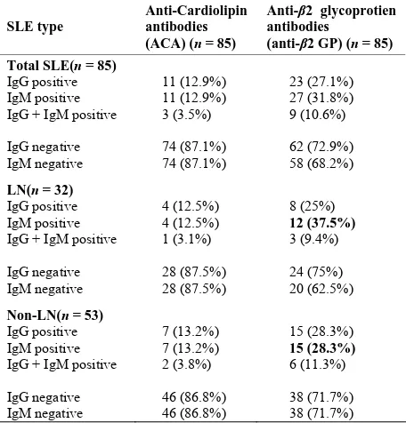
Table 2. Anti-Cardiolipin (ACA) and anti-2 glycoprotein (anti--2 GP) antibodies in (n = 85).
SLE type
Anti-Cardiolipin antibodies (ACA) (n = 85)
Anti-β2 glycoprotien antibodies
(anti-β2 GP) (n = 85)
Total SLE(n = 85) IgG positive IgM positive IgG + IgM positive
IgG negative IgM negative
11 (12.9%) 11 (12.9%) 3 (3.5%)
74 (87.1%) 74 (87.1%)
23 (27.1%) 27 (31.8%) 9 (10.6%)
62 (72.9%) 58 (68.2%)
LN(n = 32) IgG positive IgM positive IgG + IgM positive
IgG negative IgM negative
4 (12.5%) 4 (12.5%) 1 (3.1%)
28 (87.5%) 28 (87.5%)
8 (25%) 12 (37.5%) 3 (9.4%)
24 (75%) 20 (62.5%)
Non-LN(n = 53) IgG positive IgM positive IgG + IgM positive
IgG negative IgM negative
7 (13.2%) 7 (13.2%) 2 (3.8%)
46 (86.8%) 46 (86.8%)
15 (28.3%) 15 (28.3%) 6 (11.3%)
38 (71.7%) 38 (71.7%)
IgG and IgM antibodies to ACA. When these patients were categorized further into LN and non-LN groups, ACA positivity for both IgG and IgM autoantibodies was slightly higher in non-LN group (13.2%) as compared to 12.5% in LN group. Anti-β2 GP positivity was 27.1% for IgG-β2 GP and 31.8% for IgM-β2 GP where as 10.6% of the patients developed anti-β2 GP antibodies to both IgG and IgM subclasses. Among LN and non-LN groups,
IgG-β2 GP positivity revealed a slightly higher incidence (28.3%) in non-LN patients as compared to LN patients (25%) where as LN patients showed a higher incidence for IgM-β2 GP positivity (37.5%) as compared to 28.3% of patients in non-LN group. The cut off levels for IgG-ACA was 185 u/ml, IgM-ACA is 186 u/ml, IgG-β2 GP is 156 u/ml and IgM-β2 GP is 290 u/ml as per the normal individuals tested.
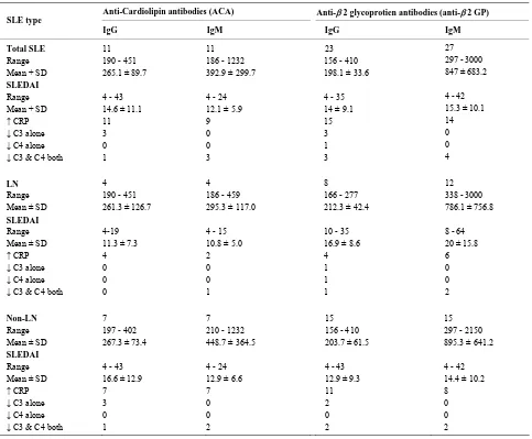
Table 3 gives the correlation of ACA and anti-β2 GP levels with SLEDAI and other immunological parame-ters such as CRP, C3 and C4. IgM-β2 GPpositive LN patients had higher SLEDAI scores (mean ± SD; 20 ± 15.8) Out of 23 patients with IgG-anti-β2 GP positivity, 15 (65.2%) patients showed raised CRP levels where non-LN patients 11/15 (73.3%) had higher CRP levels as compared to 4/8 (50%) in LN group. Among IgM anti-β2 GP positive patients 17/27 patients (63%) showed re-duced C3 and C4 levels, where in LN group 10/12 pa-tients (83.3%) had reduced C3 and C4 levels as com-pared to 7/15 patients (46.7%) in non-LN group.
The distribution of clinical manifestations according to the ACR criteria among ACA and anti-β2 GP positive patients at the time of evaluation was as shown in Table 4. ACA positive patients showed a higher incidence for clinical manifestations such as malar and discoid rash, photosensitivity, oral ulcers, vasculitis, alopecia, fever, arthritis and myositis. A slightly higher incidence for clinical manifestations such as renal disorders, serositis, neurological and hematological manifestations such as leucopenia, thrombocytopenia and autoimmune hemo-lytic anemia (AIHA) were noted among anti-β2 GP pa-tients. It was observed that among ACA positive patients, none of the patient had leucoepenia where as among anti-β2 GP positive patients having leucopenia, WBC counts ranged between (2.9 - 3.7) × 103/μl with mean ±
SD value of 3.3 ± 0.6. In a group of ACA positive pa-tients having thrombocytopenia, platelet counts ranged between (39 - 71) × 103/μL with a mean ± SD value 5.5 ±
22.6 where as anti-β2 GP patients having thrombocyto-penia showed platelet counts ranged between (12 – 141) × 103/μl with a slightly higher mean ± SD value (58.1 ±
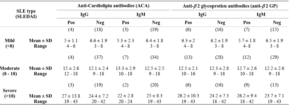
48.3). Table 5 shows distribution of clinical severity categorized into mild, moderate and severe based on the SLEDAI scores.
4. Discussion
Anti-phospholipid antibodies (APA) are a distinct group of autoantibodies that appear in a variety of autoimmune diseases, particularly Systemic Lupus Erythematosus (SLE). They are associated with clinical events such as arterial and/or venous thrombosis, and obstetric compli-cations with a strong association of ACA with thrombo- sis, thrombocytopenia, recurrent fetal losses and Coombs’
Characteristics:
Sex Ratio (F:M) 16:1
Mean Age (Years ± SD) 26.8 + 9.9
Clinical Manifestations (%) Rash (Malar or Discoid) 33 (38.8%)
Photosensitivity 17 (20%)
Oral Ulcers 24 (28.2%)
Arthritis 51 (60%)
Serositis 21 (24.7%)
Renal Disorders 32 (37.6 %)
Neurological Disorders 14 (16.5%)
Laboratory Characteristics Hematological Disorders
Anemia/ AIHA Leucopenia Lymphopenia Thrombocytopenia
23 (27.1%) 14 (16.5%) 10 (11.8%) 22 (25.9%) Immunological Disorders
↑ CRP
↓ C3 alone
↓ C4 alone
↓ C3 & C4 both
[image:3.595.59.287.375.613.2]Table 3. Correlation of ACA and anti-2 GP levels with SLEDAI and other immunological parameters.
Anti-Cardiolipin antibodies (ACA) Anti-2 glycoprotien antibodies (anti-2 GP) SLE type
IgG IgM IgG IgM
Total SLE Range Mean + SD SLEDAI Range Mean + SD
↑ CRP
↓ C3 alone
↓ C4 alone
↓ C3 & C4 both
11 190 - 451 265.1 ± 89.7
4 - 43 14.6 ± 11.1 11 3 0 1
11 186 - 1232 392.9 ± 299.7
4 - 24 12.1 ± 5.9 9 0 0 3
23 156 - 410 198.1 ± 33.6
4 - 35 14 ± 9.1 15 3 1 3
27 297 - 3000 847 ± 683.2
4 - 42 15.3 ± 10.1 14 0 0 4
LN Range Mean ± SD SLEDAI Range Mean ± SD
↑ CRP
↓ C3 alone
↓ C4 alone
↓ C3 & C4 both
4 190 - 451 261.3 ± 126.7
4-19 11.3 ± 7.3 4 0 0 0
4 186 - 459 295.3 ± 117.0
4 - 15 10.8 ± 5.0 2 0 0 1
8 166 - 277 212.3 ± 42.4
10 - 35 16.9 ± 8.6 4 1 1 1
12 338 - 3000 786.1 ± 756.8
8 - 64 20 ± 15.8 6 0 0 2
Non-LN Range Mean ± SD SLEDAI Range Mean ± SD
↑ CRP
↓ C3 alone
↓ C4 alone
↓ C3 & C4 both
7 197 - 402 267.3 ± 73.4
4 - 43 16.6 ± 12.9 7 3 0 1
7 210 - 1232 448.7 ± 364.5
4 - 24 12.9 ± 6.6 7 0 0 2
15 156 - 410 203.7 ± 61.5
4 - 43 12.9 ± 9.3 11 2 0 2
15 297 - 2150 895.3 ± 641.2
4 - 42 14.4 ± 10.2 8 0 0 2
Table 4. Distribution of clinical manifestations according to ACR criteria among ACA and anti-2 GP positive patients.
Clinical presentation ACA positives
(IgG and/or IgM) (n = 19) Anti-2 GP positives (IgG and/or IgM) (n = 41)
Malar Rash &/
Discoid Rash 9 (43.4%) 14 (34.1%)
Photosensitivity 4 (21.1%) 8 (19.5%)
Oral ulcers 7 (36.8%) 11 (26.8 %)
Arthritis 14 (73.7%) 24 (58.5%)
Serositis 6 (31.6%) 14 (34.1%)
Renal Disorders 3 (15.8 %) 8 (19.5%)
Neurological Disorders 2 (10.5%) 8 (19.5%)
Hematological Disorders 1 (5.3%) 5 (12.1%)
Systemic vascular thrombosis 4 (21.0%) 10 (24.4%)
Myositis 7 (36.8%) 10 (24.4%)
Alopecia 8 (42.1%) 16 (39.0%)
[image:4.595.65.538.527.719.2]Table 5. Distribution of clinical severity based on the SLEDAI scores.
Anti-Cardiolipin antibodies (ACA) Anti-2 glycoprotien antibodies (anti-2 GP)
IgG IgM IgG IgM
SLE type (SLEDAI)
Pos Neg Pos Neg Pos Neg Pos Neg
Mild (<8)
Mean ± SD Range
(4)
5 ± 1.1 4 - 6
(18)
6.6 ± 1.9 3 - 8
(3)
5.3 ± 2.3 4 - 8
(19)
6.4 ± 1.8 3 - 8
(6)
6.3 ± 2 4 - 8
(16)
6.2 ± 1.9 3 - 8
(7)
5.7 ± 1.8 4- 8
(15)
6.5 ± 1.9 3 - 8
Moderate (8 - 18)
Mean ± SD Range
(4)
15 ± 2.6 12 - 18
(37)
12.1 ± 2.4 9 - 18
(7)
13.3 ± 2.9 10 - 18
(34)
12.5 ± 2.5 9 - 18
(13)
12.5 ± 2.1 10 - 16
(28)
12.3 ± 2.8 9 - 18
(12)
12.7 ± 2.6 10 - 18
(29)
12.2 ± 2.6 9 - 18
Severe
(>18) Mean ± SD Range
(3)
27 ± 13.8 19 - 43
(19)
24.4 ± 7.2 20 - 42
(2)
22 ± 2.8 20 - 24
(20)
25 ± 8.3 19 - 43
(6)
26.2 ± 10.3 19 - 43
(16)
24.2 ± 7.3 18 - 42
(9)
26.2 ± 9.4 18 - 42
(13)
23.7 ± 7.1 19 - 43
positivity in SLE and related autoimmune disorders [18-22].
Recently Mostafa et al., 2010 had reported an inci-dence of 16.7% for ACA among SLE patients [23]. Similar incidence was found in our study where IgG-ACA and IgM-ACA positivity was 12.9% each which was lower than anti-β2 GP autoantibody positivity (IgG-β2GP: 27.1% and IgM-β2 GP: 31.8%). Other stud-ies such as Petri et al., 2010 reported 47% ACA and 32.5% anti-β2 GP autoantibodies in SLE, Biggioggero et al., 2010 had reported 16.5% IgG-ACA and 9.4% of IgM-ACA and an incidence of 4.7% IgG-β2 GP and 5.9% for IgM-β2 GP antibodies, Jallouli et al., 2008 had reported 71.6% for ACA and Descloux et al., 2008 had reported an incidence of 49% ACA [24,25,13,14]. In a study on South African SLE patients, Gould et al., 2006 had reported a very high incidence of 53% and 84% for ACA and anti-β2 GP antibodies where as Al Arfaj et al., 2009 had reported an incidence of 49.7% and 33.5% for IgG-ACA and IgM-ACA respectively among Saudi Ara-bian SLE patients [26,27]. Recently Woo et al., 2010 had reported an incidence of 18.2% and 31.8% for IgG-ACA and IgM-ACA respectively and 5.7% for anti-β2 GP to IgG and IgM isotypes in Korean SLE patients. Shrivastav et al., 2001 had reported 51% IgG-ACA and 44.7% IgG-β2 GP autoantibodies [28,29].
Thrombosis varies in SLE patients from 7.2 to 12%. Sarabi et al., 2005 reported that the most frequent causes of death in active SLE are infection and thrombosis [30]. The risk of thrombosis for SLE patients reported to be significantly higher and due to the increased incidence of traditional cardiovascular and nontraditional lupus-related thrombosis risk factors, SLE patients are at significantly increased risk of premature atherosclerosis and/or throm- bosis. The prevalence of vascular events in SLE patients ranges between 10% and 30%, for symptomatic coronary
artery disease 6% - 20%, stroke 2% - 15%, and subclini-cal coronary artery disease 30% - 40% [31].Our study showed a higher incidence of systemic vascular throm-bosis in anti-β2 GP positive patients as compared to ACA positive patients with an equal distribution for ve-nous and arterial thrombosis in both the groups.
Recurrent pregnancy loss (RPL) has been associated with APA including ACA and lupus anticoagulant. It had been reported that the risk of fetal loss is found to be increased in patients with hypertension, active SLE, LN, or abnormally low complement levels. Risk also in-creased for patients with APA: from 6% to 24% of pa-tients with SLE are positive for LAC and 40% are posi-tive for ACA [32-34].In our study two patients had RPL and other two had BOH, but they did not show the pres-ence of ACA or anti-β2 GP antibodies. Shrivastav et al., 2001 reported that incidence of neurological disorders such as seizures were noted in 9.4% SLE patients which was significantly associated with the presence of ACA and anti-β2 GP antibodies [29]. Neurological disorders were seen more in anti-β2 GP positive patients than in ACA positive patients in our study.
anti-β2 GP positive patients than ACA positives. Hence detection both ACA and anti-β2 GP antibodies along with associated immune parameters were found to be helpful parameters to evaluate their possible association with disease severity in SLE patients. A long term follow up of patients having ACA and anti-β2 GP antibodies without thrombotic event is required to detect their pos-sible thrombotic event in future along with their clinical presentation.
5. Acknowledgements
We are grateful to Prof. Yehuda Shoenfeld, MD, Head, Department of Medicine at the Tel Aviv University, and Ramat Aviv, Israel for his inputs in correcting this manu-script and giving his valuable suggestions to modify it.
REFERENCES
[1] Y. Sherer, M. Blank and Y. Shoenfeld, “Anti-Phosphol-ipid Syndrome (APS): Where Does It Come from?” Best
Practice and Research Clinical Rehumatology, Vol. 21,
No. 6, 2007, pp. 1071-1078.
doi:10.1016/j.berh.2007.09.005
[2] E. M. Bevers, M. Galli, T. Barbui, et al., “Lupus Antico-agulant IgG’s (LA) Are Not Directed to Phospholipids Only, but to a Complex of Lipid-Bound Human Proth- rombin,” Journal of Thrombosis and Haemostasis, Vol. 66, No. 6, 1991, pp. 629-632.
[3] S. Miyakis, M. D. Lockshin, T. Atsumi, D. W. Branch, R. L. Brey and R. Cervera, “International Consensus State-ment on an Update of the Classification Criteria for Defi-nite Antiphospholipid Syndrome (APS),” Journal of
Thrombosis and Haemostasis, Vol. 4, No. 2, 2006, pp.
295-306. doi:10.1111/j.1538-7836.2006.01753.x
[4] H. P. McNeil, R. J. Simpson, C. N. Chesterman and S. A. Krilis, “Antiphospholipid Antibodies Are Directed agai- nst a Complex Antigen That Includes a Lipid-Binding In-hibitor of Coagulation: Beta 2-Glycoprotein I (Apolipo-protein H),” Proceedings of National Academy of
Sci-ences of the United States of America, Vol. 87, No. 1,
1990, pp. 4120-4125. doi:10.1073/pnas.87.11.4120
[5] P. L. Meroni, N. D. Papa, D. Gambini, A. Tincami and G. Balesterari, “Antiphospholipid Antibodies and Endothe-lial Cells: An Unending Story,” Lupus, Vol. 4, No. 3, 1995, pp. 169-171. doi:10.1177/096120339500400301
[6] D. S. Pisetsky, G. Gilkeson and E. W. St. Clair, “Sys-temic Lupus Erythematosus. Diagnosis and Treatment,”
Medical Clinics of North America,Vol. 81, No. 1, 1997,
pp. 113-128. doi:10.1016/S0025-7125(05)70507-1
[7] G. S. Cooper, M. A. Dooley, E. L. Treadwell, E. W. St. Clair, C. G. Parks and G.S. Gilkeson, “Hormonal, Envi-ronmental, and Infectious Risk Factors for Developing Systemic Lupus Erythematosus,” Arthritis and Rheuma-tism, Vol. 41, No. 10, 1998, pp. 1714-1724.
doi:10.1002/1529-0131(199810)41:10<1714::AID-ART3 >3.0.CO;2-U
[8] V. D. D’Agati and G. B Appel, “Lupus Nephritis:
Pa-thology and Pathogenesis,” In: D. J .Wallace and B. H. Hahn, Eds., Dubois’ Lupus Erythematosus, 7th Edition,
Lippincott Williams & Wilkins, Philadelphia, 2007, pp. 1094-1111.
[9] H. Zheng, Y. Chen, W. Ao, Y. Shen, X. W. Chen, M. Dai, X. D. Wang, Y. C. Yan and C. D. Yang, “Antiphosphol-ipid Antibody Profiles in Lupus Nephritis with Glomeru-lar Microthrombosis: A Prospective Study of 124 Cases,”
Arthritis Research and Therapy, Vol. 11, No. 3, 2009, p.
R93. doi:10.1186/ar2736
[10] J. P. Grande, “Mechanisms of Progression of Renal Dam-age in Lupus Nephritis: Pathogenesis of Enal Scarring,”
Lupus, Vol. 7, No. 9, 1998, pp. 604-610.
doi:10.1191/096120398678920721
[11] J. S. Levine, D. W. Branch and J. Rauch, “The An-tiphospholipid Syndrome,” The New England Journal of
Medicine, Vol. 346, No. 2002, pp. 752-763.
doi:10.1056/NEJMra002974
[12] M. L. Davies, S. P. Young, K. Welsh, M. Bunce, B. P. Wordsworth, et al., “Immune Responses to Native β2-Glycoprotein I in Patients with Systemic Lupus Ery-thematosus and the Anti-Phospholipid Syndrome,”
Rheu-matology,Vol.41, No. 4, 2002, pp. 395-400.
doi:10.1093/rheumatology/41.4.395
[13] M. Jallouli, M. Frigui, M. B. Hmida, S. Marzouk, N. Kaddour and Z. Bahloul, “Clinical and Immunological Manifestations of Systemic Lupus Erythematosus: Study on 146 South Tunisian Patients,” Saudi Journal of Kidney
Diseases and Transplantation, Vol. 19, No. 6, 2008, pp.
1001-1008.
[14] E. Descloux, I. Durieu, P. Cochat, D. V. Durand, J Ninet, N. Fabien and R. Cimaz, “Pediatric Systemic Lupus Ery-thematosus: Prognostic Impact of Antiphospholipid An-tibodies,” Rheumatology, Vol. 47, No. 2, 2008, pp. 183- 187. doi:10.1093/rheumatology/kem335
[15] M. C. Hockberg, “Updating the American College of Rheumatology Revised Criteria for the Classification of Systemic Lupus Erythematosus,” Arthritis and Rheuma-tism, Vol. 40, No. 9, 1997, p. 1725.
doi:10.1002/art.1780400928
[16] C. Bombardier, F. F. Gladmsn, M. B. Urowit, D. Caron and C. H. Chang, “Derivation of SLEDAI: A Disease Ac-tivity Index for Lupus Patients. The Committee on Prog-nosis Studies in SLE,” Arthritis and Rheumatism, Vol. 35, No. 6, 1995, pp. 630-640. doi:10.1002/art.1780350606
[17] J. J. Weening, V. D. Agati, M. M. Schwartz, S. V. Seshan, C. E. Alpers and G. B. Appel, “The Classification of Glomerulonephritis in Systemic Lupus Erythematosus Re visited,” Journal of American Society of Nephrology, Vol. 15, No. 2, 2004, pp. 241-250.
doi:10.1097/01.ASN.0000108969.21691.5D
[18] J. R. Mueh, K. D. Herbst and S. I. Rapaport, “Thrombosis in Patients with the Lupus Anticoagulant,” Annals of
In-ternal Medicine, Vol. 92, No. 2, 1980, pp. 156-159.
Growth Retardation and Intrauterine Death,” British
Journal of Obstetrics and Gynaecology, Vol. 88, No. 9,
1981, pp. 890-894.
doi:10.1111/j.1471-0528.1981.tb02224.x
[20] M. L. Boey, C. B. Colaco, A. E. Gharavi, K. B. Elkon, S. Loizou and G. R. Hughes, “Thrombosis in Systemic Lu-pus Erythematosus: Striking Association with the Pres-ence of Circulating Lupus Anticoagulant,” British
Medi-cal Journal (Clinical Research Edition), Vol. 287, No.
6398, 1983, pp. 1021-1023.
[21] E. N. Harris, A. E. Gharavi and M. L. Boey, “Anti Cardi-olipin Antibody Detection by Radioimmunoassay and Association with Thrombosis in Systemic Lupus Erythe-matosus,” The Lancet, Vol. 332, No. 8361, 1983, pp. 1211-1214. doi:10.1016/S0140-6736(83)91267-9
[22] M. D. Lockshin and L. R. Sammaritano, “Lupus Preg-nancy,” Autoimmunity, Vol. 36, No. 1, 2011, pp. 33-40.
doi:10.1080/0891693031000067313
[23] G. A. Mostafa, D. H. Ibrahim, A. A. Shehab and A. K. Mohammed, “The Role of Measurement of Serum Auto- antibodies in Prediction of Pediatric Neuropsychiatric Systemic Lupus Erythematosus,” Journal of
Neuroim-munology, Vol. 227, No. 1-2, 2010, pp. 195-201.
[24] M. Petri, “Update on Anti-Phospholipid Antibodies in SLE: The Hopkins’ Lupus Cohort,” Lupus, Vol. 19, No. 4, 2010, pp. 419-423. doi:10.1177/0961203309360541
[25] M. Biggioggero and P. L. Meroni, “The Geoepidemiol-ogy of the Antiphospholipid Antibody Syndrome,”
Auto-immun Reviews, Vol. 9, No. 5, 2010, pp. A299-A304.
doi:10.1016/j.autrev.2009.11.013
[26] T. Gould, M. Tikly, R. Asherson, S. Loizou and S. Singh, “Prevalence and Clinical Correlates of Anti-Phospholipid Antibodies in South Africans with Systemic Lupus Ery-thematosus,” Scandinavian Journal of Rheumatology, Vol. 35, No. 1, 2006, pp. 29-34.
doi:10.1080/03009740510026913
[27] A. S. Al Arfaj and N. Khalil, “Clinical and Immunologi-cal Manifestations in 624 SLE Patients in Saudi Arabia,”
Lupus, Vol. 18, No. 5, 2009, pp. 465-473.
doi:10.1177/0961203308100660
[28] K. S. Woo, K. E. Kim, J. M. Kim, J. Y. Han, W. T. Chung and K. H. Kim, “Prevalence and Clinical Associa-tions of Lupus Anticoagulant, Anticardiolipin Antibodies, and Anti-Beta2-Glycoprotein I Antibodies in Patients with SLE,” The Korean Journal of Laboratory Medicine, Vol. 30, No. 1, 2010, pp. 38-44.
doi:10.3343/kjlm.2010.30.1.38
[29] A. Shrivastava, S. Dwivedi, A. Aggarwal and R. Misra, “Cardiolipin and Beta2 Glycoprotein I Anti-bodies in Indian Patients with Systemic Lupus Erythe-matosus: Association with the Presence of Seizures,” Lu-pus, Vol. 10, No. 1, 2001, pp. 45-50.
doi:10.1191/096120301671577528
[30] Z. S. Sarabi, E. Chang, R. Bobba, D. Ibanez, D. Gladman, M. Urowitz, et al., “Incidence Rates of Arterial and Ve-nous Thrombosis after Diagnosis and Systemic Lupus Erythematosus,” Arthritis and Rheumatism, Vol. 53, No. 4, 2005, pp. 609-612. doi:10.1002/art.21314
[31] D. Erkan, “Lupus and Thrombosis,” The Journal of
Rheumatology, Vol. 32, No. 9, 2006, pp.1715-1717.
[32] M. Petri, M. Golbus, R. Anderson, Q. Whiting-O’Keefe, L. Corash and D. Hellmann, “Antinuclear Antibody, Lu-pus Anticoagulant, and Anticardiolipin Antibody in Women with Idiopathic Habitual Abortion. A Controlled Prospective Study of Forty-Four Women,” Arthritis and
Rheumatism, Vol. 30, No. 6, 1987, pp. 601-606.
doi:10.1002/art.1780300601
[33] M. Petri and J. Allbritton, “Fetal Outcome of Lupus Preg-nancy: A Retrospective Case-Control Study of the Hop-kins Lupus Cohort,” TheJournal of Rheumatology, Vol. 20, No. 4, 1993, pp. 650-656.
[34] E. N. Harris, A. E. Gharavi and G. R. Hughes, “Anti- Phospholipid Antibodies,” Clinics in Rheumatic Diseases, Vol. 11, No. 3, 1985, pp. 591-609.